5359
Observation of the Cerebral Perforating Arteries around Circle of Willis Using Compressed Sensing Time-of-Flight at 7T1MR Collaboration, Siemens Healthineers Ltd., Beijing, China, 2Tiantan Neuroimaging Center of Excellence, Beijing, China, 3Beijing Tiantan Hospital, Capital Medical University, China National Clinical Research Centre for Neurological Diseases, Beijing, China, 4Beijing Tiantan Hospital, Capital Medical University, Neurology Department, Beijing, China
Synopsis
Keywords: Atherosclerosis, Vessels, perforating arteries, ultra high resolution
The impairment of microvessels can lead to neurologic diseases such as stroke and vascular dementia. Perforating arteries imaging requires an extremely high resolution due to their small caliber size. In this study, for the first time, the feasibility of noninvasive visualization of perforators around Circle of Willis in vivo was demonstrated by using 7T compressed sensing TOF. The number of stems and branches of the perforators was then calculated. The origin and anatomical distribution of the perforating arteries are described. This work revealed that ultra-high-resolution CS TOF might be a promising method for detecting microvasculopathies of cerebral vascular diseases.Introduction
The perforating arteries (PA), originate from the main cerebral arteries and mainly supply the paramedian part of the brain stem and the diencephalon, as well as the basal ganglia and the internal capsule[1, 2]. Perforator anatomy is essential for understanding the pathogenesis and consequences of ischemic and hemorrhagic stroke. However, the lack of effective PA imaging technology significantly hinders related research. 7T TOF MRA is demonstrated to visualize lenticulostriate arteries (LSAs)[3, 4], and basilar artery perforators[5], due to the increased SNRs and improved contrast. However, these techniques are still insufficient to bring into view the complete cerebral perforators due to their limited resolution. It was recently demonstrated that the novel compressed sensing (CS) acceleration technique could be implemented in 7T TOF while preserving the image quality [6]. Our previous work has demonstrated that more LSA stems and branches can be delineated with CS TOF than conventional TOF due to the higher resolution (0.2mm-iso) [7, 8]. In this study, for the first time, the feasibility of using 7T CS TOF to display the complete cerebral PA around the circle of Willis noninvasively was demonstrated.Methods
All measurements were performed on a 7T MR system (MAGNETOM Terra, Siemens Healthcare, Erlangen, Germany) using a 32- channel Rx/8Tx head-coil (Nova Medical, Wilmington, Massachusetts, USA). Fifteen healthy volunteers (6 males; aged 24–50 years; mean age 35.3 ± 8.7 years) were recruited in this study. All measurements were carried out with the approval of the local Ethics Committee. A prototype CS TOF MRA was used to achieve 0.2 mm isotropic LSA imaging. The imaging parameters were consistent with our previous studies [7, 8]. These perforators are divided into several groups according to their parent artery, the anterior cerebral artery (ACA) perforators, the middle cerebral arteries (MCA) perforators, the anterior communicating artery (AComA) perforators, the posterior cerebral artery (PCA) perforators, the posterior communicating artery (PComA) perforators, the basilar artery (BA) perforators, and the internal carotid artery (ICA) perforators. The numbers of stems and branches of the perforators were then calculated in each category for all participants. The origin and anatomical distribution of the PA are briefly described.Results
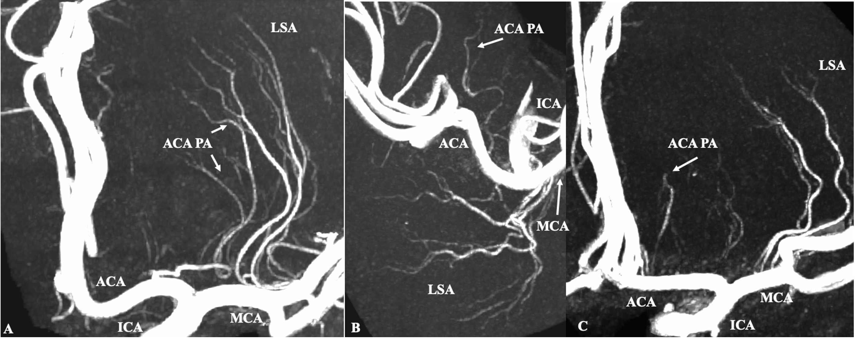
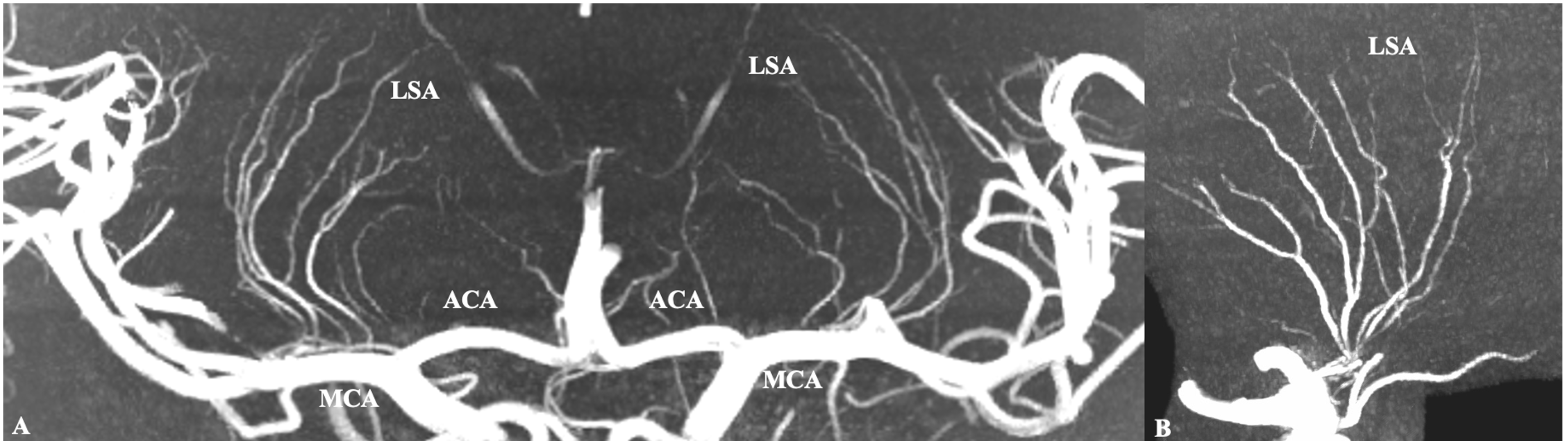
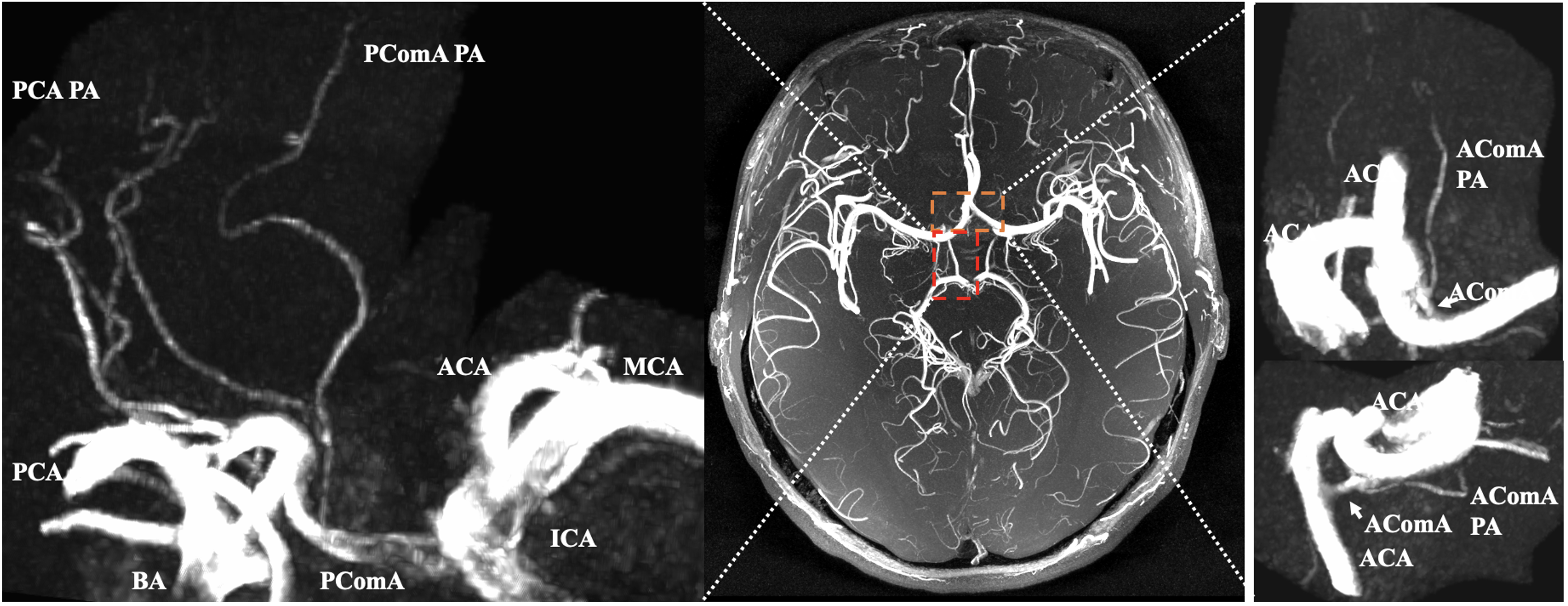
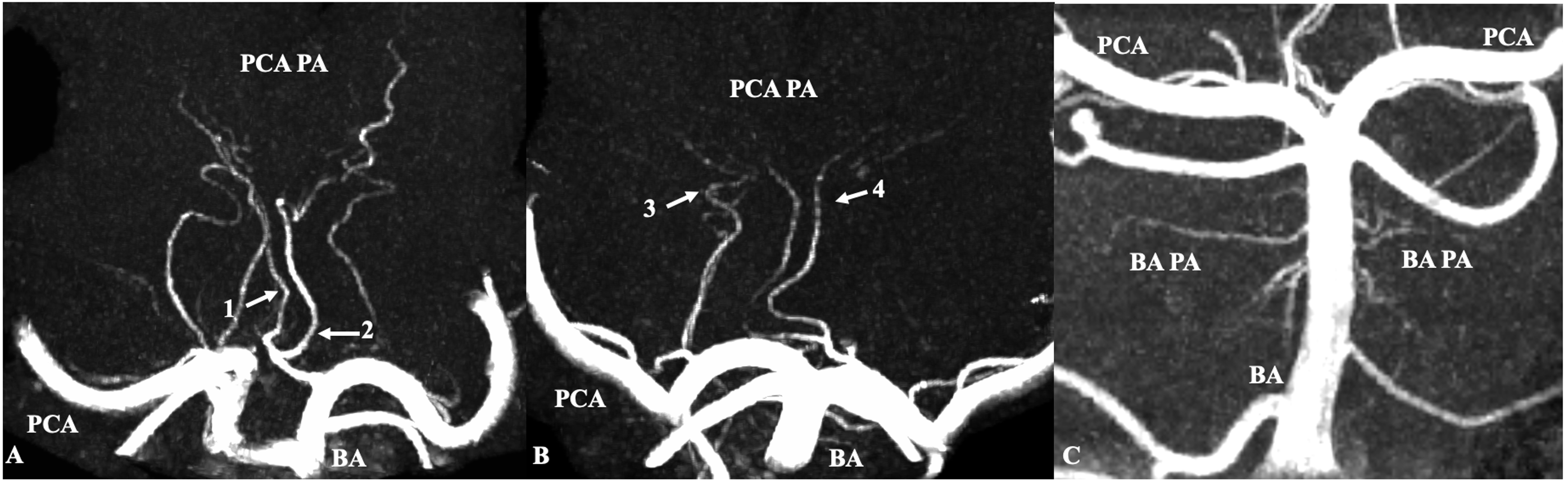
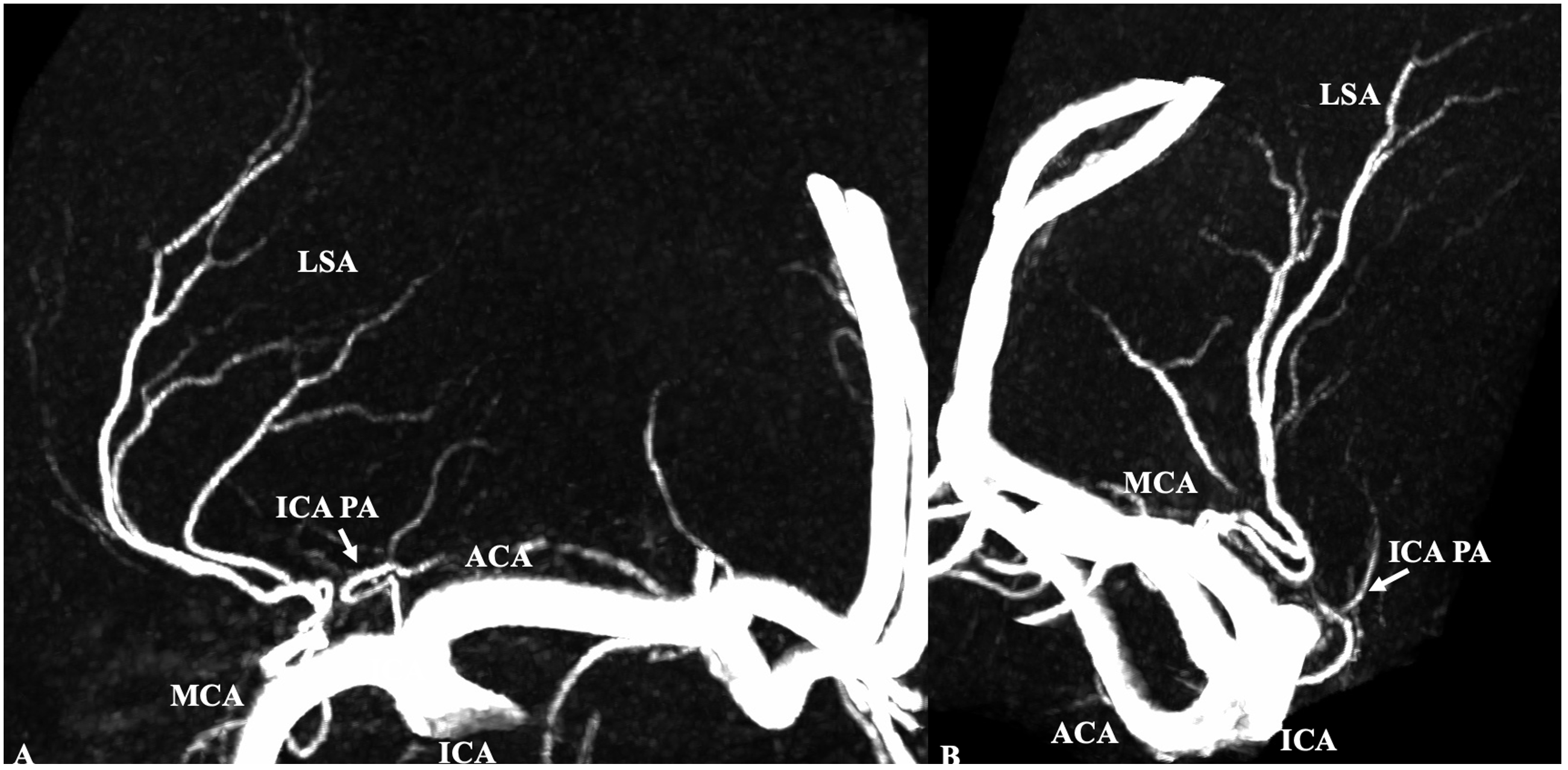
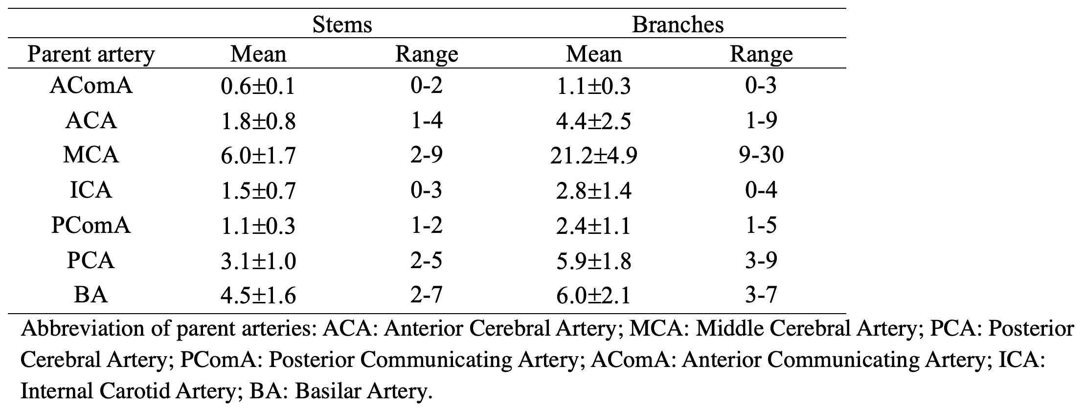
The identified mean number and range of perforating arteries were summarized in Table 1. The ACA perforators comprise the recurrent artery of Heubner and the typical perforating vessels (Figs. 1). Heubner’s artery, originates from the ACA (Figs. 1. A). The ACA PAs originate most frequently from the A1 segment (Fig. 1. BC), and commonly arise as individual vessels. The MCA perforators, known as the lenticulostriate arteries, average 6.0±1.7 in stem number and 21.2±4.9 in branch number (Figs. 2). The short AComA (Fig. 3), varies in form considerably. It was absent in 5 hemispheres with a fusion of the two ACAs, and in 3 hemispheres with a hypoplastic A1 segment of the ACA which directly joined a large opposite ACA. The PComA, most often possesses only one perforating branch (Figs. xx) known as the premamillary artery. It was absent in 11 hemispheres. The PAs from PCA can be grouped into two perforators: the thalamogeniculate and the thalamoperforating vessels. The thalamogeniculate arteries are located within the ambient cistern (Fig 4B). The thalamoperforating arteries most often originate from the P1 segment of the PCA (Figs. 4). The constant basilar perforators arise from both the right and left sides of the BA (Figs. 4C). The perforators of the ICA arise from its choroidal segment, i.e., between its bifurcation site and the AChA origin (Figs. 5).Discussion
This is the first time that all cerebral PA imaging around the circle of Willis has been demonstrated. This is thanks to the ultra-high resolution and improved artery-tissue contrast enabled by the use of CS TOF. Anatomy ex vivo studies have suggested that the perforator around the circle of Willis are very abundant [9], but perforators are challenging to observe in routine clinical practice due to limited SNR and resolution. Improved SNR and increased T1 relaxation time of 7T TOF MRA result in better visualization of small arteries. Still, conventional 7T TOF MRA (around 300um) might only describe the larger perforators, such as lenticustrate artery, whereas the small perforators can't delineate, such as the AComA perforators (mean diameter: 175um), ICA perforators (mean diameter: 237um), and thalamoperforating vessels (mean diameter: 255um) [9]. The compressed sensing technique improves the SNR efficiency while preserving imaging quality due to its novel acquisition trajectory and reconstruction. Together with the signal sparsity nature of TOF MRA, it is possible to achieve higher resolution using CS under clinically acceptable acquisition times. Thus making it possible to visualize the whole perforators around the Circle of Willis.In our future research, more subjects would be enrolled to verify the repeatability of CS TOF.Also, many parameters to quantify perforators, such as the length, the distance, and the tortuosity, should be adopted to explore population differences. Furthermore, patients with cerebral vascular diseases should be evaluated.Conclusion
This work demonstrates the feasibility of noninvasive visualization of perforators around the Circle of Willis in vivo. Ultra-high-resolution CS TOF may be a promising tool for the evaluation of cerebral small vessel diseases.Acknowledgements
No acknowledgement found.References
1. Tatu L, Moulin T, Bogousslavsky J, Duvernoy H (1996) Arterial territories of human brain: brainstem and cerebellum. Neurology 47:1125–1135. https://doi.org/10.1212/wnl.47.5.1125.
2. Ghika JA, Bogousslavsky J, Regli F (1990) Deep Perforators From the Carotid System: Template of the Vascular Territories. Archives of Neurology 47:1097–1100. https://doi.org/10.1001/archneur.1990.00530100063014.
3. Kang C-K, Park C-W, Han J-Y, et al (2009) Imaging and analysis of lenticulostriate arteries using 7.0-Tesla magnetic resonance angiography. Magn Reson Med 61:136–144. https://doi.org/10.1002/mrm.21786.
4. Cho Z-H, Kang C-K, Han J-Y, et al (2008) Observation of the Lenticulostriate Arteries in the Human Brain In Vivo Using 7.0T MR Angiography. Stroke 39:1604–1606. https://doi.org/10.1161/STROKEAHA.107.508002.
5. Kang C-K, Park C-A, Kim K-N, et al (2010) Non-invasive visualization of basilar artery perforators with 7T MR angiography. J Magn Reson Imaging 32:544–550. https://doi.org/10.1002/jmri.22250.
6. Meixner CR, Liebig P, Speier P, et al (2019) High resolution time-of-flight MR-angiography at 7 T exploiting VERSE saturation, compressed sensing and segmentation. Magnetic Resonance Imaging 63:193–204. https://doi.org/10.1016/j.mri.2019.08.014.
7. Zhe Zhang, Qingle Kong, Jing jing, et al. 0.20 mm isotropic intracranial perforating arteries imaging using compressed sensing TOF-MRA at 7T. In Proceedings of the 31st Annual Meeting of ISMRM, London, England, UK, 2022. Abstract 41428.
8. Zhe Zhang, Qingle Kong, Yingkui Zhang, et al. Improved Characterization of Lenticulostriate Arteries using Compressed Sensing Time-of-Flight at 7T. European radiology. Under Review.
9. Vogels V, Dammers R, van Bilsen M, Volovici V (2021) Deep Cerebral Perforators: Anatomical Distribution and Clinical Symptoms: An Overview. Stroke 52:. https://doi.org/10.1161/STROKEAHA.120.034096
Figures