5358
Preliminary Evaluation of the Tumor Vessels Based on Ultrafast DCE MRI in Differential Diagnosis of Breast Tumors (BI-RADS 4)1First Affiliated Hospital of Dalian Medical University, Dalian, CHINA, Dalian, China, 2zhongshan Hospital of Fudan University, Shanghai, CHINA., Shanghai, China, 3Clinical & Technical Support, Philips Healthcare, Beijing, China, Beijing, China
Synopsis
Keywords: Breast, Blood vessels
The occurrence and development of the breast cancer are closely related to the growth and infiltration of blood vessels.This study mainly analyzed the correlation between the maximum diameter of benign and malignant breast tumors, the number of peripheral blood vessels and the first appearance of vascular phase. It is concluded that the number of blood vessels had a certain value in distinguishing benign and malignant breast tumors. There existed a certain correlation between the maximum diameter of malignant tumor and the number of blood vessels.Prospective of Main Findings
In this study, we prospectively analyzed the maximum diameter of benign and malignant tumors, the number of peripheral blood vessels and the appearance period of blood vessels. Results showed that the peripheral blood vessel lesions in the malignant group were significantly higher than those in the benign group, with a distinct difference (P=0.007). There was no obvious difference in the first appearance of the vascular phase between two groups of benign and malignant breast tumors (P=0.121). The maximum diameter of the breast cancer was positively correlated with the number of blood vessels and the scatter plot showed a linear distribution. There was no significant difference in the maximum diameter of breast benign tumor and the number of blood vessels (P=0.18).Introduction
DCE-MRI can dynamically observe and evaluate the new blood vessels, which has been widely used in the early diagnosis of the breast cancer and has been considered as the most commonly used vascular functional MR Imaging method at present. By tracking the pharmacokinetics characteristics of paramagnetic T1WI contrast agents, microvascular structures and functions can be quantitatively and semi-quantitatively analyzed in a non-invasive manner[1-4]. 3D-MIP plays an important role in the diagnosis of the breast cancer and provides an important basis for clinical preoperative evaluation, surgical plan, treatment and prognosis [5].Previous studies have found that when >3 ipsilateral blood vessels are used as the diagnostic criteria for malignant breast lesions, the number of blood vessels in benign and malignant tumors is statistically significant, and the diagnostic efficiency is relatively high, with sensitivity and specificity of 81.6% and 80.8%, respectively [6]. Herein, we measured the number of blood vessels in benign and malignant tumors based on DCE and further explored whether the number of blood vessels had certain value in distinguishing benign and malignant breast tumors.Methods
In this prospective study, a total of 20 female patients (46.2±8.92, range: 31-67 years) were enrolled and informed consent was acquired from each patient. All patients were diagnosed as MR BI-RADS 4 and performed MR scan using a 3.0T whole-body MR scanner (Philips Ingenia CX, Philips Healthcare, the Netherlands). Based on the histopathology results, Group 1 consisted of 10 patients with malignant breast tumors, including 7 Invasive Ductal Carcinoma (IDC) II and 3 IDC III; Group 2 consisted of 10 patients with 10 benign breast tumors, including 8 fibroadenomas and 2 intraductal papillomas. The Mann-Whitney U test was used to analyze the distribution differences between two groups. Image parameters (the maximum diameter of the lesion, the number of blood vessels around the lesion, the first appearance of vascular phase images during lesion enhancement) measured by those two groups were compared, and the diagnostic performance based on these parameters was quantified by ROC curve.The relationship between the maximum diameter of the lesion, the number of blood vessels around the lesion and the first appearance of the vascular phase in Group 1 and Group 2 was explored.At the same time, the maximum diameter of the lesion was measured on the dynamic enhanced image,and the number of tumor blood vessels within 3cm around the tumor was calculated.Results
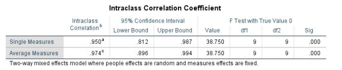
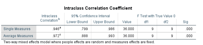
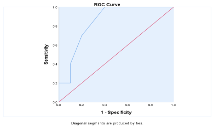
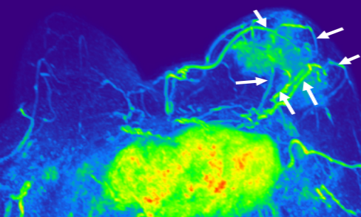
No significant difference was found in age between two groups (p = 0.063). The intraclass correlation coefficients (ICC, 0.94 and 0.95 for group 1 and 2, respectively) indicated a good inter-observer agreement of the measured image values. There was no significant difference between the two groups in the maximum diameter of the lesions, the early appearance of the lesions at the time of enhancement and the phase images, respectively (P = 0.121, P = 0.177) (Table 1). The number of blood vessels around the lesions in the malignant group was significantly higher than that in the benign group, with an obvious difference (P = 0.007). The best threshold for differentiating benign and malignant breast tumors was 1.5, and the sensitivity、 specificity and AUC were 100%, 60%, and 0.845, respectively (Fig. 1). The breast cancer malignancy had a sufficient blood supply and therefore, 6 vessels could be clearly observed on dynamic enhanced magnetic resonance imaging in Fig. 2. There was no significant difference in the maximum diameter and the number of blood .vessels of benign breast tumors (P=0.237, P=0.28). The scatter plot showed a linear distribution, indicating that there existed remarkable difference between the maximum diameter of breast malignant tumor and the number of blood vessels. There was a positive correlation between the maximum diameter of breast malignant tumors and the number of blood vessels (R=0.888, P =0.001).DISCUSSION AND CONCLUSION
The application of MRI in breast blood vessels is becoming more and more mature and extensive. According to the needs of clinical diagnosis and treatment purposes, the reasonable use of MR Functional imaging technology can improve the clinical application efficiency and provide more abundant reference information for the diagnosis and postoperative follow-up of the breast cancer. Our results may indicate that the number of blood vessels in benign and malignant breast tumors has a certain value in differentiating BI-RADS 4 breast lesions.Acknowledgements
No acknowledgement found.References
[1] Honda M, Kataoka M, Iima M, et al. Background parenchymal enhancement and its effect on lesion detectability in ultrafast dynamic contrast-enhanced MRI. Eur J Radiol 2020; 129:108984. 10.1016/j.ejrad.2020.108984
[2] Kim SY, Cho N, Choi Y, et al. Ultrafast dynamic contrast-enhanced breast MRI: Lesion conspicuity and size assessment according to background parenchymal enhancement. Korean J Radiol 2020; 21:561–571. 10.3348/kjr.2019.0567
[3] Onishi N, Kataoka M, Kanao S, et al. Ultrafast dynamic contrast‐enhanced mri of the breast using compressed sensing: breast cancer diagnosis based on separate visualization of breast arteries and veins. J Magn Reson Imaging 2018; 47:97–104. 10.1002/jmri.25747
[4] Mus RD, Borelli C, Bult P, et al. Time to enhancement derived from ultrafast breast MRI as a novel parameter to discriminate benign from malignant breast lesions. Eur J Radiol 2017; 89:90–96.[5] Çetinkaya E, Yıldız Ş, Otçu H, Sharifov R, Çelik Yabul F, Alkan A. The value of adjacent vessel sign in malignant breast tumors. Diagn Interv Radiol. 2022 Aug 23. doi: 10.5152/dir.2022.211228. Epub ahead of print. PMID: 35997479.
[6] Ao F, Yan Y, Zhang ZL, Li S, Li WJ, Chen GB. The value of dynamic contrast-enhanced magnetic resonance imaging combined with apparent diffusion coefficient in the differentiation of benign and malignant diseases of the breast. Acta Radiol. 2022 Jul;63(7):891-900.
Figures