5343
The value of Synthetic MRI combined with histogram analysis in predicting treatment response to chemoradiotherapy in nasopharyngeal carcinoma
Fan Yang1, Haoran Wei1, Yujie Li1, Xiaoduo Yu1, Lizhi Xie2, and Meng Lin1
1Department of Diagnostic Radiology, National Cancer Center/National Clinical Research Center for Cancer/Cancer Hospital, Chinese Academy of Medical Sciences and Peking Union Medical College, Beijing, 100021, China, Beijing, China, 2GE Healthcare, MR Research China, Beijing, Beijing, China
1Department of Diagnostic Radiology, National Cancer Center/National Clinical Research Center for Cancer/Cancer Hospital, Chinese Academy of Medical Sciences and Peking Union Medical College, Beijing, 100021, China, Beijing, China, 2GE Healthcare, MR Research China, Beijing, Beijing, China
Synopsis
Keywords: Head & Neck/ENT, MR Fingerprinting, Nasopharyngeal carcinoma; treatment response; Synthetic MRI
Almost 80% nasopharyngeal carcinoma patients are diagnosed with locoregionally-advanced stage at the time of initial diagnosis. Early detection of treatment response is important for adjusting therapy regimens. SyMRI could not only generate multi-contrast images (including T1WI, T2WI and PDWI) in a single scan, but also generate quantitative T1, T2 and PD maps. Our study demonstrated that SyMRI combined with histogram analysis could non-invasive predict early treatment response. The predict performance of combined SyMRI and clinical factors was significantly higher than clinical only or TNM stage only.Introduction
Nasopharyngeal carcinoma (NPC) is an aggressive head and neck cancer and almost 80% patients tend to present with locoregionally-advanced stage cancer(1). Although concurrent chemoradiotherapy (CCRT) have contributed to significant survival benefit, locoregional residue and relapse after CCRT remain the important clinical challenges for locoregionally-advanced nasopharyngeal carcinoma (LA-NPC)(2). Therefore, early detection of treatment response is important for guiding clinicians in timely and appropriate adjustments of therapy regimens and for predicting the survival of NPC. Synthetic MRI (SyMRI), using a multiple-delay multiple-echo (MDME) sequence, could generate multiple contrast images in single scan without contrast agent, including T1-weighted imaging (T1WI), T2-weighted imaging (T2WI), proton density weighted imaging (PDWI) and so on. Besides, SyMRI simultaneously generate the longitudinal and transverse relaxation times (T1 and T2) and proton density (PD) of tissues, providing clinicians an objective assessment method. The diagnostic value of SyMRI has been demonstrated in NPC, breast cancer, prostate cancer and so on(3-5). To our knowledge, this is the first study to explore the predictive value of SyMRI combined with histogram analysis in treatment response evaluation in NPC.Materials and Methods
This prospective study was approved by the Ethics Committee of our hospital, and informed consent was obtained from all patients before MRI examination. The detailed information of SyMRI (Discovery MR 750, GE Healthcare, Milwaukee, WI, USA) is as follows: repetition time, 6200; echo time, 18.9/94.7; the field of vision, 26; acquisition matrix, 256 × 320; slice thickness/gap, 4.0/0.4; number of excitations, 1; acquisition time, 7.02. A total of 42 patients with LA-NPC between August 2018 and May 2019 were included in this study (Table 1).All patients underwent CCRT with or with out induction chemotherapy (IC). 12 weeks after the completion of CCRT was chosen as evaluation point(6). 16 patients were divided into the residual group and 26 patients were divided into the non-residual group according to the standard of Response Evaluation Criteria in Solid Tumors (RECIST, version 1.1)(7). The representative patients are shown in Fig.1. Two radiologists (18 and 3 years of tumor-imaging experience) manually delineated volumes of interest (VOIs) of primary tumor on SyT2WI imaging using ITK-SNAP software (version 2.2.0, www.itksnap.org), excluding any visible necrosis and hemorrhage. Then, the VOIs were automatically copied to the T1 map, T2 map and PD map, and histogram parameters (90th Percentile, Mean, Entropy and Mean Absolute Deviation (MAD)) were extracted using open-source Pyradiomics (http://www.radiomics.io/pyradiomics.html). The independent sample-t test, Mann-Whitney U test, or chi-square test was used, as appropriate. Besides, multivariate logistic regression analysis was used to construct models (SyMRI model, Clinical model, SyMRI + Clinical model and TNM stage model). A nomogram was then constructed and calibration curves were plotted via bootstrapping with 1000 resamples. The Delong test was used to compared the predictive performance.
Results
All histogram parameters derived from T1 map, T2 map and PD map showed excellent interclass agreement (all ICC > 0.965, Table 2). In univariate analysis, non-residual group has higher T2_90th, T2_Mean, PD_90th, PD_Mean and PD_Entropy than residual group (all P ≤ 0.020, Table 2). After multivariate logistic regression analysis, PD_Mean and PD_Entropy were included to construct SyMRI model (AUC: 0.834), and two clinical factors (EBV DNA level and Age) were included to construct Clinical model (AUC: 0.745). As for combined model (AUC: 0.894), PD_Mean, PD_Entropy, EBV DNA level and Age were included and the nomogram and calibration curve were shown in Fig.2. The combined model showed a significantly higher performance compared with Clinical model (P = 0.041), while there is no significant difference when compared with SyMRI model (P = 0.165). Interestingly, the combined model showed higher predictive performance than TNM stage model (AUC: 0.666, P = 0.026). The AUC, sensitivity, specificity and accuracy of models are shown in Table 3.Conclusions
Pre-treatment SyMRI parameters provide a non-invasive approach for early treatment response assessment of NPC. The combined model that included SyMRI and clinical factors may provide an effective reference for individual treatment regimen and prognostic prediction.Acknowledgements
Not applicableReferences
1. Liu LT, Chen QY, Tang LQ, et al. Advanced-Stage Nasopharyngeal Carcinoma: Restaging System after Neoadjuvant Chemotherapy on the Basis of MR Imaging Determines Survival. Radiology 2017;282(1):171-181.2. Xiao WW, Huang SM, Han F, et al. Local control, survival, and late toxicities of locally advanced nasopharyngeal carcinoma treated by simultaneous modulated accelerated radiotherapy combined with cisplatin concurrent chemotherapy: long-term results of a phase 2 study. Cancer 2011;117(9):1874-1883.
3. Wang P, Hu S, Wang X, et al. Synthetic MRI in differentiating benign from metastatic retropharyngeal lymph node: combination with diffusion-weighted imaging. Eur Radiol 2022.
4. Li Q, Xiao Q, Yang M, et al. Histogram analysis of quantitative parameters from synthetic MRI: Correlations with prognostic factors and molecular subtypes in invasive ductal breast cancer. Eur J Radiol 2021;139:109697.
5. Cui Y, Han S, Liu M, et al. Diagnosis and Grading of Prostate Cancer by Relaxation Maps From Synthetic MRI. J Magn Reson Imaging 2020;52(2):552-564.
6. Chen YP, Chan ATC, Le QT, Blanchard P, Sun Y, Ma J. Nasopharyngeal carcinoma. Lancet 2019;394(10192):64-80.
7. Eisenhauer EA, Therasse P, Bogaerts J, et al. New response evaluation criteria in solid tumours: revised RECIST guideline (version 1.1). Eur J Cancer 2009;45(2):228-247.
Figures
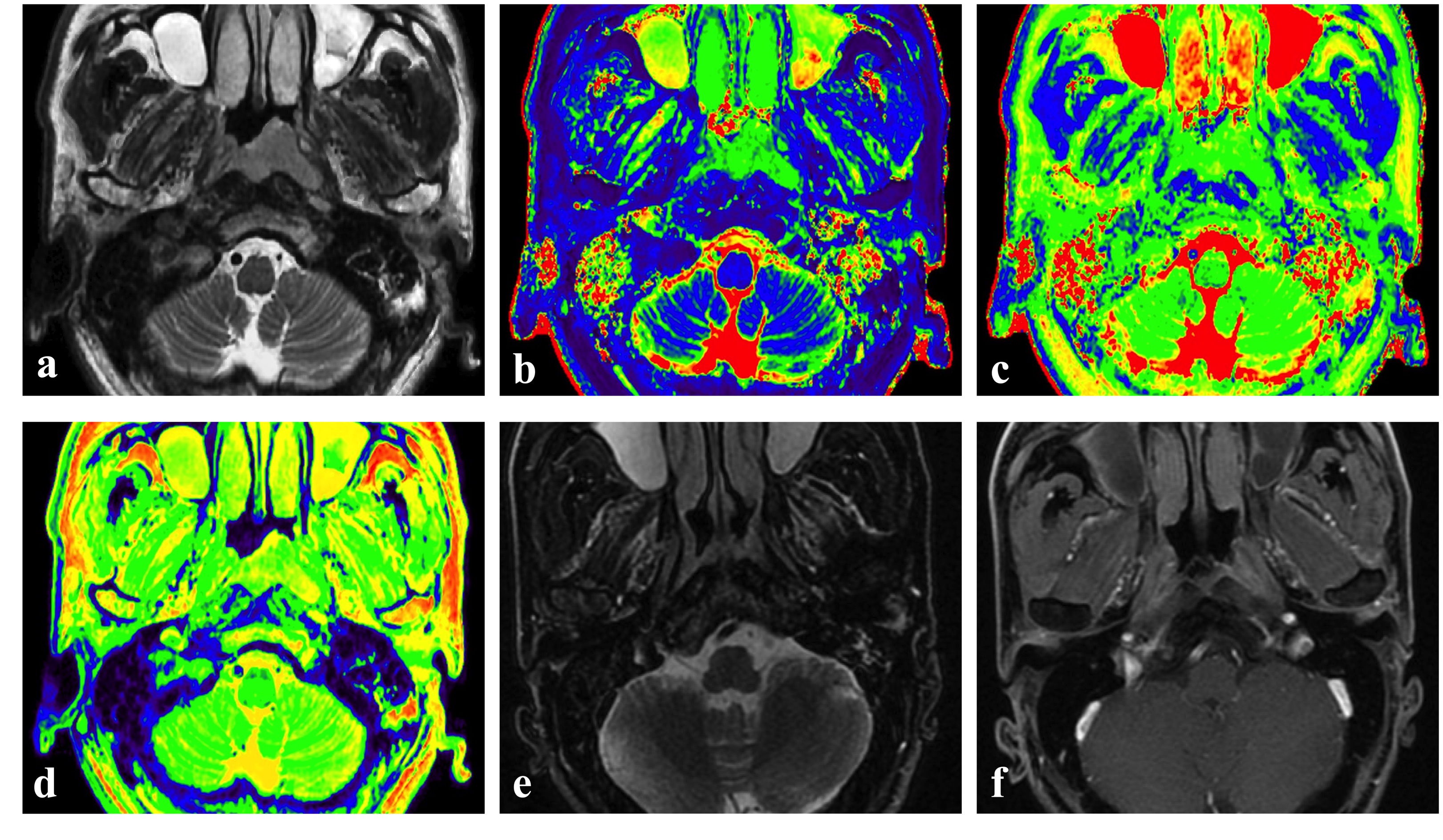
Fig.1 Representative pretreatment images of NPC patients with complete response after IC + CCRT. (a) SyT2WI shows a nasopharyngeal asymmetric mass in 56-year-old male NPC patient (b) T1 map; (c) T2 map; (d) PD map at the same level. After 12 weeks after completion of IC + CCRT treatment, T2WI with fat suppress (e) and contrast-enhanced T1WI (f) shows uniformly slightly thickened nasopharyngeal mucosa. It was pathologically confirmed to be free of tumor remnants by nasopharyngoscopy.
Table 1 Characteristics of patients
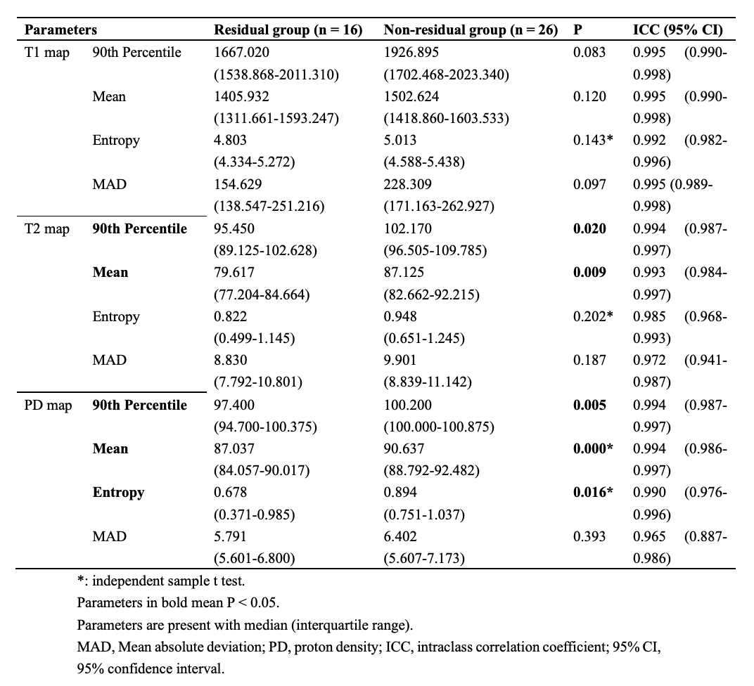
Table 2 SyMRI histogram parameters of patients with or without residue
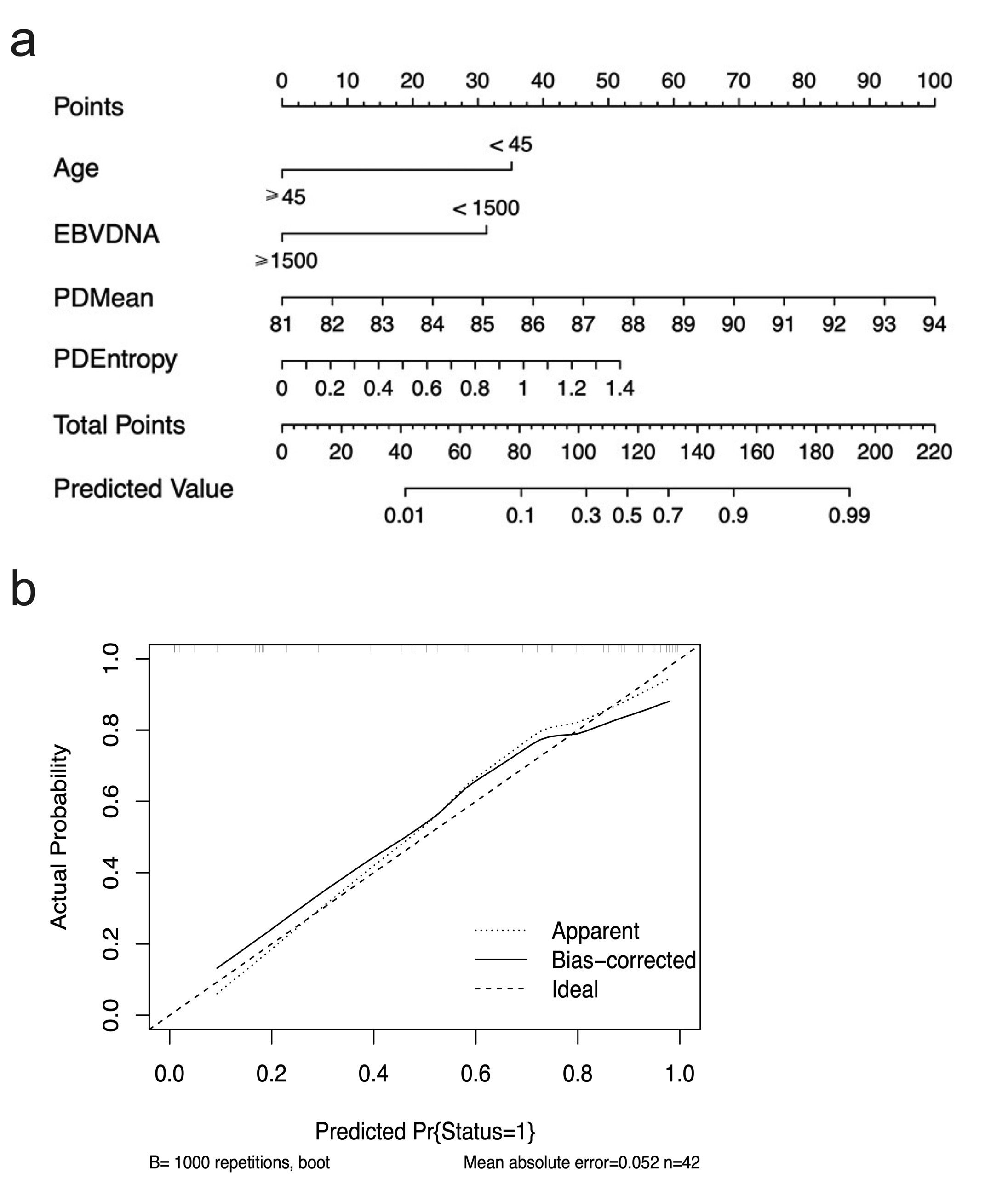
Fig.2 Nomogram and calibration curve of combined model
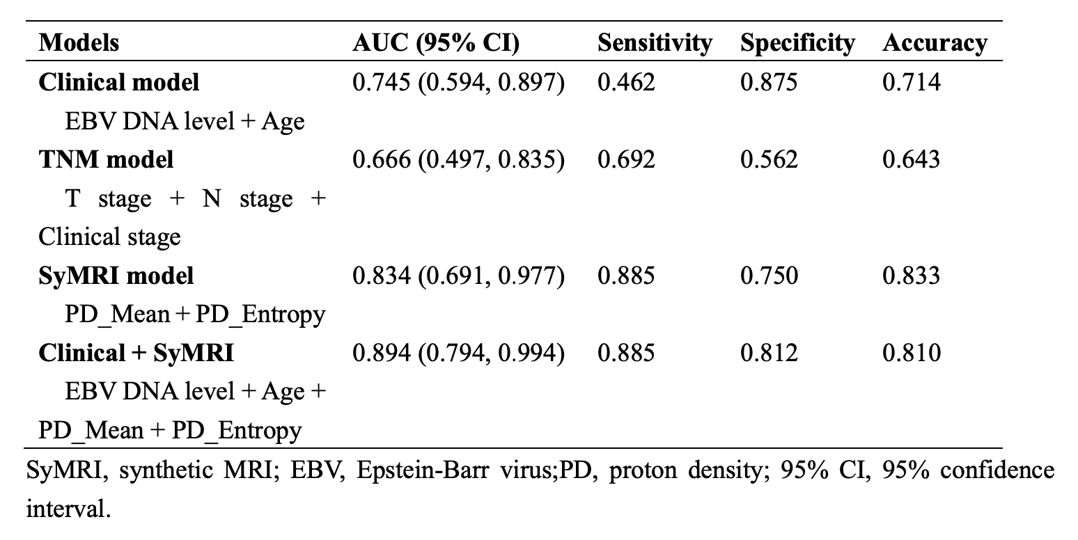
Table 3 Performance of four models in predicting treatment response
DOI: https://doi.org/10.58530/2023/5343