5340
Glymphatic system impairment in migraine: relation with chronic pain1Department of Radiology, Nanjing first hospital,Nanjing Medical University, Nanjing, China, 2Central Research Institute, United Imaging Healthcare, Shanghai, China
Synopsis
Keywords: Blood vessels, Diffusion Tensor Imaging
The glymphatic system is a recently discovered waste drainage system in the brain that involves movement of the cerebrospinal fluid along the perivascular space. In this study, we conducted a non-invasive method, diffusion tensor image analysis along the perivascular space (DTI-ALPS), to investigate the glymphatic system’s function in migraine and its association with brain atrophy and clinical symptoms. Abnormalities were identified in the glymphatic system of patients with migraine, compared with normal controls. The associations between ALPS-index and neuropsychological performance suggested the potential of ALPS-index as a biomarker for brain dysfunction.Since the introduction of the glymphatic system hypothesis by Illif et al. [1], increasing number of studies have attempted to describe the fluid dynamics in the brain parenchyma [2]. Recently, “neurofluids” has been accepted as a collective term for the fluids in which the central nervous system (CNS) is immersed, including the blood, cerebrospinal fluid, and interstitial fluid [3]. This concept helps explore and understand the fluid dynamics within the brain parenchyma. As the impairment of neurofluid dynamics is closely associated with various pathologies, the concept of ‘CNS interstitial fluidopathy’ has also been proposed to indicate the pathologies caused by the abnormal neurofluid dynamics [4]. The diffusion tensor technique is widely used for evaluating the white matter tract in the brain. This non-invasive method evaluates CNS physiology and various pathologies by measuring fractional anisotropy (FA) or apparent diffusion coefficient (ADC) [5]. Diffusion tensor image analysis along the perivascular space (DTI-ALPS) is another application of the diffusion tensor method to non-invasively evaluate the interstitial fluid dynamics using diffusion tensor image (DTI) on MRI [6]. This study aimed to evaluate the activity of glymphatic system in migraine with a diffusion-based technique called diffusion tensor image analysis along the perivascular space (DTI-ALPS) and its relationship with chronic pain.
Methods
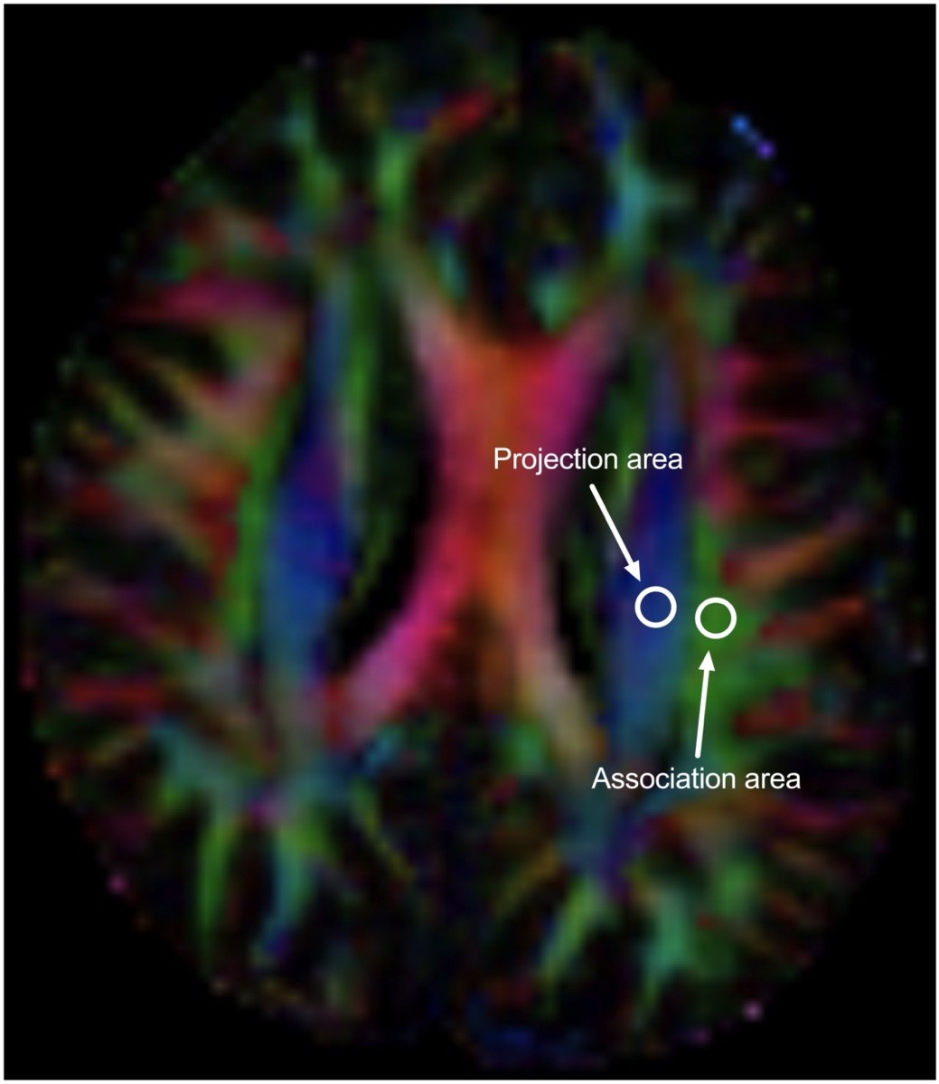
Diffusion tensor images (echo-planar imaging, 64 weighted directions and 2 b0 images, b = 1000 s/mm2, resolution 2 × 2 × 2 mm3, TE/TR = 80 ms/8300 ms) were acquired to calculate diffusivities in the x-, y-, and z- axes of the plane of the lateral ventricle body in 88 migraine patients and 47 healthy control subjects (HCs) , using a 3T MRI system (uMR 780, United Imaging Healthcare, Shanghai, China). Circular regions of interest (ROIs) with a diameter of 6 mm were placed in the projection and association areas at the level of the lateral ventricle body (Figure 1), and diffusivity of the x, y, and z directions were measured. On the ROIs, the x-, y-, and z-axis diffusivity were measured, and the ALPS index was calculated for each case, which is a ratio of the mean of the x-axis diffusivity in the projection area (Dxxproj) and the x-axis diffusivity in the association area (Dxxassoc) to the mean of the y-axis diffusivity in the projection area (Dyyproj) and the z-axis diffusivity in the association area (Dzzaccoc): ALPS index = (mean(Dxxproj, Dxxassoc))/(mean(Dyyproj, Dzzassoc)). We correlated the ALPS index with clinical characteristics and multiple rating scales.
Results
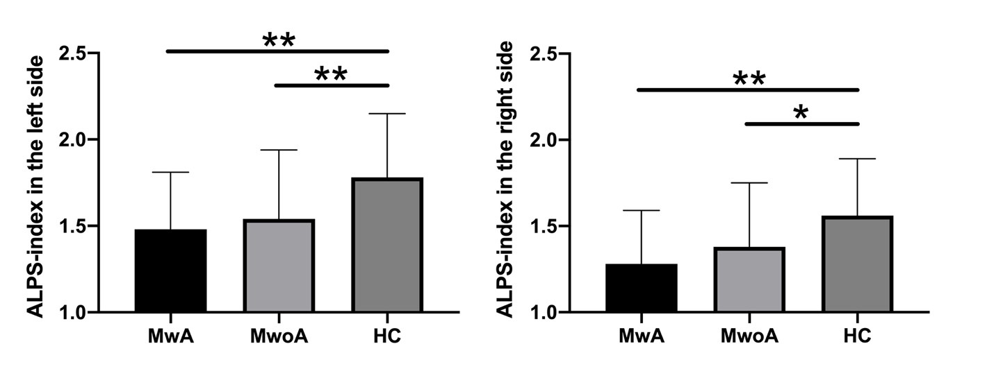
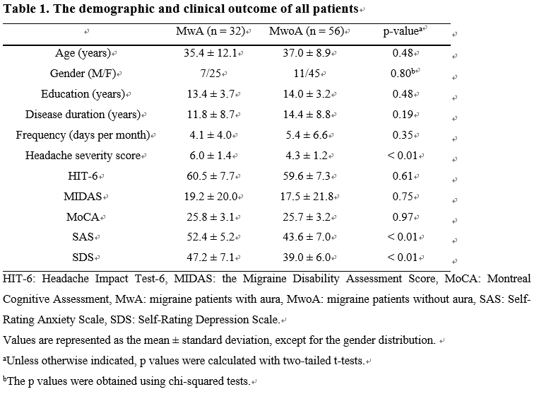
The demographic characteristics and clinical assessment of all patients were summarized in Table 1. There were no significant differences in age and gender between migraine patients and HCs, and also in age, gender, education, disease duration and frequency between patients with (MwA) and without aura (MwoA), using a chi-squared test for gender and two-tailed t-tests for continuous variables. The MwA group showed higher headache severity score, SAS and SDS scores compared to the MwoA group (all p values < 0.01). Patients showed a lower ALPS-index than HCs whether left, right or the whole brain average (all p values < 0.05, Figure 2). But there no significant difference in the ALPS-index of the left or right side between MwA and MwoA patients (Figure 2). In all participants, the left ALPS-index (1.61 ± 0.48) was significantly greater than that of right side (1.42 ± 0.35), using a paired t-test (p < 0.001). On correlation analysis, lower unilateral- and bilateral- average ALPS-index were significantly associated with higher headache severity score (all p values < 0.01).
Discussion & Conclusion
In brief, our results using the DTI-ALPS method demonstrated a significant negative correlation between the diffusivity along the perivascular space and the disease severity in migraine, indicating impaired water diffusivity related to chronic pain. Lower diffusivity along the perivascular space or lower ALPS-index seems to reflect impairment of the glymphatic system. Thus, the ALPS-index may be applied for evaluating conditions that affect the activity of the glymphatic system of individual cases in migraine. The DTI-ALPS method is used for evaluating the glymphatic system’s function or the interstitial fluid dynamics, and this method has been adopted in several studies evaluating the alteration of the glymphatic system or interstitial fluid dynamics in various pathologies [7-9]. One of the report showed that results of ALPS method were significantly related to glymphatic clearance function calculated on MRI by intrathecal administration of gadolinium-based contrast agent (GBCA) [9]. In our study, DTI-ALPS was applied to measure glymphatic function in migraine patients and HCs. Overall, our results suggested an impairment of the glymphatic system in patients compared with HCs. The lymphatic vessels collect glymphatic fluid from brain parenchyma and drain this fluid in the cervical lymph nodes contributing to the overall glymphatic system proper functioning. Thus, glymphatic system damage may promote inflammatory activation by altering lymphatic vessels-dependent CNS immune surveillance, which could elucidate the pathological mechanisms underpinning the migraine. In conclusion, abnormalities were identified in the glymphatic system of patients with migraine. This study also revealed associations between ALPS-index and neuropsychological performances, suggesting the potential of ALPS-index as a biomarker for brain dysfunction.
Acknowledgements
This research was supported by Nanjing Science and Technology Planning Project (No. 202002056).References
1. Iliff JJ, Wang M, Liao Y, Plogg BA, Peng W, Gundersen GA, et al. A paravascular pathway facilitates CSF flow through the brain parenchyma and the clearance of interstitial solutes, including amyloid beta. Sci Transl Med. 2012;4(147):147ra11.
2. Taoka T, Naganawa S. Glymphatic imaging using MRI. J Magn Reson Imaging. 2020;51(1):11-24.
3. Agarwal N, Contarino C, Toro EF. Neurofluids: a holistic approach to their physiology, interactive dynamics and clinical implications for neurological diseases. Veins Lymphat. 2019;8(3):8470.
4. Taoka T, Naganawa S. Imaging for central nervous system (CNS) interstitial fluidopathy: disorders with impaired interstitial fluid dynamics. Jpn J Radiol. 2021;39(1):1-14.
5. Kiuchi K, Morikawa M, Taoka T, Kitamura S, Nagashima T, Makinodan M, et al. White matter changes in dementia with Lewy bodies and Alzheimer’s disease: a tractography-based study. J Psychiatr Res. 2011;45(8):1095-100.
6. Taoka T, Masutani Y, Kawai H, Nakane T, Matsuoka K, Yasuno F, et al. Evaluation of glymphatic system activity with the dif- fusion MR technique: diffusion tensor image analysis along the perivascular space (DTI-ALPS) in Alzheimer’s disease cases. Jpn J Radiol. 2017;35(4):172-8.
7. Steward CE, Venkatraman VK, Lui E, Malpas CB, Ellis KA, Cyarto EV, et al. Assessment of the DTI-ALPS parameter along the perivascular space in older adults at risk of dementia. J Neu- roimaging. 2021;31:569-78.
8. Bae YJ, Choi BS, Kim JM, Choi JH, Cho SJ, Kim JH. Altered glymphatic system in idiopathic normal pressure hydrocephalus. Parkinsonism Relat Disord. 2021;82:56-60.
9. Zhang W, Zhou Y, Wang J, Gong X, Chen Z, Zhang X, et al. Glymphatic clearance function in patients with cerebral small vessel disease. Neuroimage. 2021;238:118257.
Figures