5336
Brain atrophy and cognitive impairment in patients with end-stage renal disease: A Voxel-Based Morphometry Study1the First Affiliated Hospital of Dalian Medical University, Dalian, China
Synopsis
Keywords: Nerves, Kidney
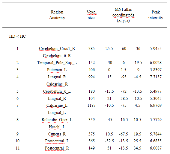
End-stage renal disease is the final stage of chronic kidney disease, often complicated by abnormal brain structure and neurocognitive function. In this study, a voxel-based morphometry (VBM) measurement was used to observe the volume change of gray matter structure in ESRD patients. Patients with ESRD were found to have atrophy in the right Cerebelum_Crus1, bilateral Cerebelum_6, left Temporal_Pole_Sup, left Putamen, bilateral Calcarine (CAL), bilateral Lingual (LING), left Rolandic_Oper, left Heschl, right Cuneus, bilateral Postcentral. Furthermore, the volumes of the right Cerebelum_Crus1, bilateral CAL, left Cerebelum_6, bilateral LING were positively correlated with overall cognitive score.Summary of Main Findings
In this study, the volume of whole brain gray matter in ESRD patients was measured and compared with healthy controls, and found that multiple brain regions atrophied and were positively correlated with cognitive function.Introduction
Chronic kidney disease (CKD) is an important factor in the morbidity and mortality of noncommunicable diseases, and the final stage is called end-stage renal disease (ESRD), during which patients require renal replacement therapy1. ESRD not only has a significant impact on brain structure, but concurrent cognitive impairment is also common, studies have shown that the decrease in executive function is the most significant2. Voxel-based morphometry (VBM) can quantitatively detect the density and volume of brain tissue at the voxel level and has been widely used to characterize subtle changes in brain structure. In this study, we aim to apply VBM analysis to compare the whole brain gray matter volume between ESRD patients and health controls (HCs) at the voxel level, and explore the relationship between gray matter volume and cognitive function.Methods
Thirty-nine ESRD patients (17 females, 22 males, mean age: 57.49 ± 10.01 years) and 51 healthy controls (HCs, 28 females, 23 males, mean age: 56.47 ± 8.62 years) were prospectively recruited and all were right-handed. Informed consent was acquired from each subject. Participants were scanned using a a 3.0 T MR scanner (Ingenia CX, Philips Healthcare, Best, the Netherlands) with a 32-channel receive-only head coil. On whole-brain, T1-weighted multishot turbo field echo (MS-TFE) was acquired: repetition time (TR)/echo time (TE) = 6.6/3.0 ms, field of view (FOV) = 256 × 256 mm, flip = 12°, 188 sagittal slices, and 1 mm3 spatial resolution. The VBM was performed on the dataset as an alternative method of detecting atrophy in the whole brain gray matter, using MATLAB R2013b (Mathworks, Natick, MA) and SPM8 (Wellcome Department of Cognitive Neurology, London). Data analyses were performed using SPM8, independent-sample t-test was used to compare the whole brain gray matter volume between the ESRD group and HCs. Age, gender, education, and estimated total intracranial volume were included as covariates. The significance levels were set at a voxel-level threshold of p < 0.001 (uncorrected) combined with a cluster-level threshold of p < 0.05 (FWE-corrected), and the voxel threshold > 100 was statistically significant. The brain regions with significant differences in gray matter volume between the two groups were taken as the regions of interest (ROI). The ROI gray matter volume of ESRD patients was extracted and further to perform partial correlation analysis with the score of the neuropsychiatric scale. The differences in clinical data and neuropsychiatric scale scores between the two groups were analyzed by SPSS 26.0 statistical software. P < 0.05 was considered statistically significant. FDR correction was applied to reduce the false-positive errors.Results
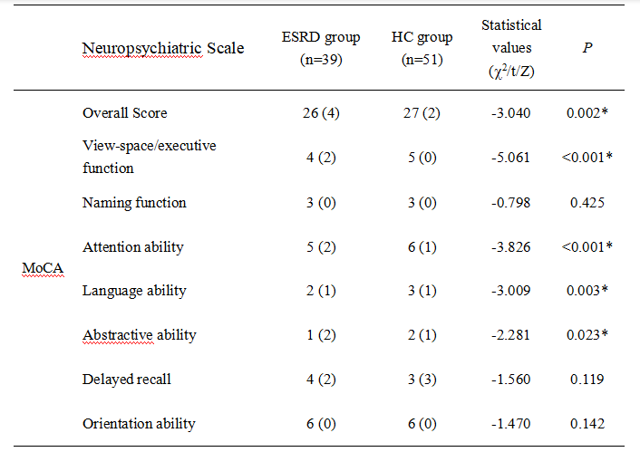
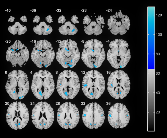
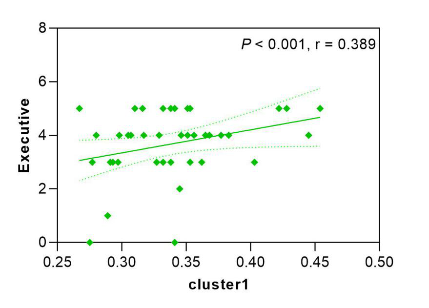
Patients with ESRD were found to have atrophy in right Cerebelum_Crus1, bilateral Cerebelum_6, left Temporal_Pole_Sup (TPOsup), left Putamen (PUT), bilateral Calcarine (CAL), bilateral Lingual (LING), left Rolandic_Oper (ROL), left Heschl (HES), right Cuneus (CUN), bilateral Postcentral (PoCG)(Table 1, Figure 1). The total MOCA scores in the ESRD group decreased compared to HCs(Table 2). The partial correlation analysis showed that after FDR correction, the volumes of right Cerebelum_Crus1(P = 0.006, r = 0.364), bilateral CAL (right: P = 0.014, r = 0.301; left: P = 0.015, r = 0.289), left Cerebelum_6 (P = 0.007, r = 0.322), and bilateral LING(P < 0.001, r = 0.419) were positively correlated with overall cognitive scores, and positively correlated with executive and attention scores (P < 0.05, FDR-corrected), the left TPOsup, bilateral CAL, left Cerebelum_6 and bilateral LINGwere were positively correlated with language ability (P < 0.05, FDR-corrected), the left Cerebelum_6, bilateral LING, left CAL and bilateral PoCG were positively correlated with abstraction ability (P < 0.05, FDR-corrected)(Figure 2).Discussion
This study investigated the volumes of the gray matter in patients with ESRD, and explored the correlation with cognition. We observed the patients with ESRD have diffuse reduction in gray matter, mainly located in the bilateral cerebellum, temporal lobe, parietal lobe, and occipital lobe. Cerebelum_Crus1, Cerebelum_6 and Cerebelum_7 are associated with cognitive functions, including language, working memory, and executive function3,4, similar results were found in this study. Furthermore, the volumes of multiple gray matter regions is associated with overall cognitive function, with predominance of executive ability, attention, language ability, and abstraction. In addition, follow-up studies could combine the functional connectivity between brain regions to explore the interaction between brain regions.Conclusion
The volume of the gray matter was reduced in the ESRD group, especially in right Cerebelum_Crus1, bilateral Cerebelum_6, left Temporal_Pole_Sup, left Putamen, bilateral Calcarine (CAL), bilateral Lingual (LING), left Rolandic_Oper, left Heschl, right Cuneus, bilateral Postcentral. The partial correlation analysis showed that the volumes of right Cerebelum_Crus1, bilateral CAL, left Cerebelum_6, bilateral LING were positively correlated with overall cognitive scores.Acknowledgements
No acknowledgement found.References
1. GBD Chronic Kidney Disease Collaboration. Global, regional, and national burden of chronic kidney disease, 1990-2017: a systematic analysis for the Global Burden of Disease Study 2017. Lancet. 2020 Feb 29;395(10225):709-733.
2. Berger I, Wu S, Masson P, et al. Cognition in chronic kidney disease: a systematic review and meta-analysis. BMC Med 2016;14:2063.
3. Buckner RL, Krienen FM, Castellanos A, et al. The organization of the human cerebellum estimated by intrinsic functional connectivity. J Neurophysiol. 2011 Nov;106(5):2322-45.
4. Sugihara I. Crus I in the Rodent Cerebellum: Its Homology to Crus I and II in the Primate Cerebellum and Its Anatomical Uniqueness Among Neighboring Lobules. Cerebellum. 2018 Feb;17(1):49-55.
Figures