5305
Changes in Cellular and Vascular Phenotype in Pediatric Ependymoma Models by Multi-Parametric MRI: Effects of Tumor Size and Radiation Treatment
Jane Manalo1, Andrea M Griesinger2, Jenna Steiner1, Angela Pierce2, Nicholas K Foreman3, and Natalie Julie Serkova4
1Radiology, University of Colorado Anschutz, Aurora, CO, United States, 2Pediatric Neurooncology, University of Colorado Anschutz, Aurora, CO, United States, 3Pediatric Hematology Oncology, The Children's Hospital of Colorado, Aurora, CO, United States, 4Radiology, University of Colorado Anschutz, Denver, CO, United States
1Radiology, University of Colorado Anschutz, Aurora, CO, United States, 2Pediatric Neurooncology, University of Colorado Anschutz, Aurora, CO, United States, 3Pediatric Hematology Oncology, The Children's Hospital of Colorado, Aurora, CO, United States, 4Radiology, University of Colorado Anschutz, Denver, CO, United States
Synopsis
Keywords: Cancer, Animals, cell size imaging
At 2022 ISMRM, we presented on simulation results of the selective size imaging using filters via diffusion times (SSIFT) in perfused irradiated cells and flank xenograft models. Here, using SSIFT and iron-oxide vessel-size imaging, we report on ependymoma (EPN) phenotypes, comparing small and large intracranial lesions: variable cell sizes fitted into SSIFT iAUC (S=14 microns small EPN, S=19 medium and S=12 large), increased vessel density index (Q=0.54 small, Q=0.62 large EPN), and low ADC (0.63x10⁻³ small, 0.58x10⁻³ mm2/s large EPN). Chemo-radiation treatment led to decreased gross tumor volumes, necrosis with decreased cell sizes and increased ADCs.INTRODUCTION
Ependymoma (EPN) is an aggressive pediatric brain tumor that contributes significantly to poor overall outcomes in children (1). Our group has previously established aggressive behaviors of EPN, including high tumor cellularity, cytological anaplasia, high mitotic index, tumor necrosis, and the presence of inflammatory cells such as M2-type myeloid cells (2). Last year at the ISMRM, we have reported on computational modeling and algorithms to derive quantitative imaging biomarkers for cell- and vessel-size imaging. The purpose of this study is to apply these technologies, including mpMRI sequences and computational post-processing, for characterizing the progression phenotype of small and advanced EPN tumors and to assess chemo-radiation treatment (CRT) response in an orthotopic mouse of patient-derived xenografts (PDX) of pediatric EPN.METHODS
All animal protocols were reviewed and approved by the local IACUC. Female severely immune deficient (SCID) mice were used for intracranial orthotopical inoculation of disaggregated tumors from pediatric EPN patients (n=8). The untreated animals (n=4) were imaged for up to 3 months, following on tumor progression. In the CRT group, animals were treated with 10 Gy radiation plus 30 mg/kg 5-fluorouracil for 5 days (n=4). All radiation treatment was perform of the animal image-guided precision XRAD irradiator, using MRI and CT guided EPN localization. For each MRI session, the animal were inserted into a Bruker 9.4 Tesla BioSpec MRI scanner with a Bruker mouse head array RF coil. Each mpMRI session consisted of high resolution T2w turboRARE (sagittal and axial) for tumor volume; echo-planar imaging (EPI) diffusion weighted sequence for tumor necrosis, edema and selective size imaging using filters via diffusion times (SSIFT); quantitative T2/T2* maps (qT2) for inflammation (before and 24hr after ferumoxytol injection) and vessel size imaging (VSI) modeling. The total acquisition time was 32 minutes based on high-resolution T2w turboRARE (3D, 12min45s), fast spin echo diffusion weighted imaging with 6 b-values (DWI axial, 1min30s), and quantitative MSME/ GRE T2/T2* maps with 8 echoes before and after ferumoxytol (iron oxide SPION) administration (qT2/T2*maps axial, 10min/ 7min30sec). Analytical methodologies included (i) ROI-based conventional volumetric analysis, apparent diffusion coefficient (ADC) values and T2 relaxation times using ParaVision NEO360 v2.0 software; in-house MATLAB simulations to calculate SSIFT incremental iAUC, fitted cell sizes, vessel size imaging (VSI) and density indices (Q) Q = DR2/(DR2*)2/3 [sec-1/3]RESULTS
High-resolution turboRARE T2w-MRI was able to detect cerebellar EPN lesions with a 48 microns in-plane resolution. The sensitivity of T2w-MRI scans was 0.2 mm for the smallest tumor detected; the median tumor volume at baseline was 2.41 mm3. Untreated mice, monitored over the course of 3 months, showed rapid progression of tumor growth from small to intermediate to large EPN (Figure 1A-C). Over the course of 3 months, untreated tumors grew an average of 30.3 mm3 (Figure 2A). During the tumor progression, a significant changes in SSIFT iAUC as well as ADC were observed, rather in an expected way. While ADC have progressively decreased (down to 0.58x10⁻³ mm2/s) with the increased tumor burden indicating restricted extracellular diffusion and high cellularity (Figure 3), the fitted tumor cells sizes have been initially increasing with the tumor growth (S=14 microns in small lesions up to 3 mm3, and S=19 in intermediate tumors up to 14 mm3), but then decreasing in large tumors (S=12 microns in 20-45 mm3 EPN). The decreased SSIFT iAUC of large tumors by the end of the study correlated with increased cellularity and cell compression of large untreated EPN (Figure 3). In addition, increased blood vessel densities (Q=0.53±0.11, Figure 3), abnormal ventricles, olfactory bulb metastases, and peritumoral edema were observed in large untreated EPN (Figure 2). The 5-day CRT with 2Gy/day and 30 mg/kg 5-FU resulted in a significant decrease in tumor volumes (Figure 1D-F, Figure 2B). The treated group also had increased ADC values (up to 1.5x10⁻³ mm2/s) and decreased SSIFT iAUC (median 3.7% versus 9.1% untreated) and fitted cell size (median 11 microns versus 21 untreated) two weeks after CRT (Figure 3). The most immediate response, seen as soon as 2 days after the CRT, was related to a decreased blood vessel density and an increased presence of inflammatory macrophages and microglial cells in irradiated EPN.DISCUSSION
It has been recently shown that SSIFT iAUC modeling can selectively detect signals arising from large cancer cells (3), and in 2022 we also shown a strong correlation between iAUC and mean cell size modelling. Orthotopically implanted PDX EPN xenografts closely mimic histological features, anatomical location and radiological features of the primary tumors. Our advanced mpMRI protocol followed by novel MATLAB algorithm analysis allows for a unique characterization of pediatric EPN as the tumors progresses. The increased tumor sizes in untreated EPN were characterized by low ADC, highly elevated blood vessel density and large tumor cell size. A significant decrease in vessel size density and an increase in inflammatory cells were seen as soon as 2 days after CRT. The late response (2 weeks post CRT) is characteristic by decreased ADC values and cell size, resulting in significantly decreased tumor volumes.Acknowledgements
The study was supported by the NIH Shared Instrumentation Grant Program (S10 OD023485 and S10 OD027023), NCI R01 CA239302, the University of Colorado Cancer Center grant (NCI P30 CA046934), and Michele Plachy-Rubin Pilot Grant ProgramReferences
1. Merchant TE. Current Clinical Challenges in Childhood Ependymoma: A Focused Review. J Clin Oncol. 2017;35: 2364-2369.
2. Pierce AM, Witt DA, Donson AM, et al. Establishment of patient-derived orthotopic xenograft model of 1q+ posterior fossa group A ependymoma. Neuro Oncol. 2019;21: 1540-1551.
3. Devan SP, Jiang X, Luo G, et al. Selective Cell Size MRI Differentiates Brain Tumors from Radiation Necrosis. Cancer Res. 2022;82: 3603-3613.
Figures
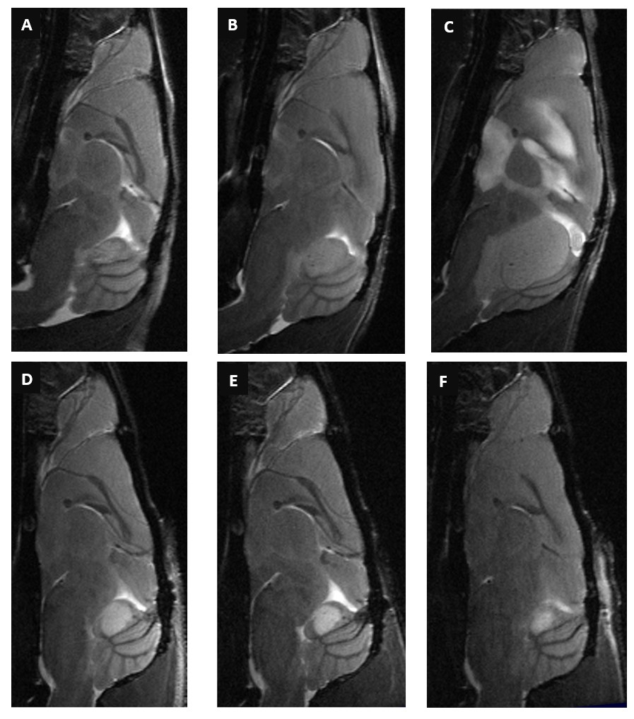
Figure 1. T2w sagittal turboRARE shows
significantly increased tumor size and appearance in an untreated animal (A-C) versus
the reduced tumor volume in the RT+5FU group (D-F).
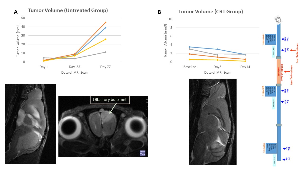
Figure 2. Changes in EPN tumor size in untreated
and CRT animals
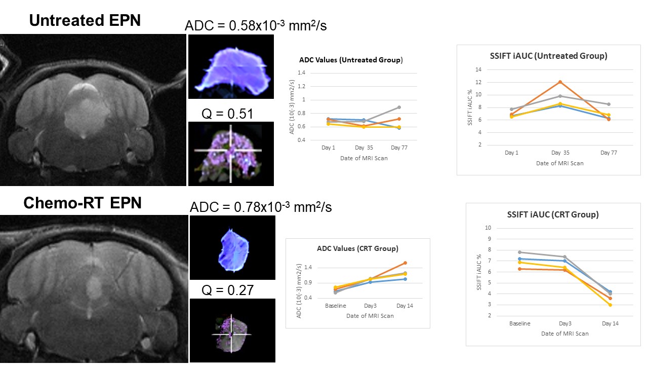
Figure 3. Changes in DWI, ADC, and SSIFT iAUC in
untreated EPN PDX mice and two weeks after CRT
DOI: https://doi.org/10.58530/2023/5305