5250
Comparing 129Xe gas MRI across major MRI vendors and sites1Pulmonary Research, Cincinnati Children's Hospital Medical Center, Cincinnati, OH, United States, 2Physics, University of Cincinnati, Cincinnati, OH, United States, 3University of Iowa, Iowa City, IA, United States, 4University of Wisconsin, Madison, WI, United States, 5University of Virginia, Charlottesville, VA, United States, 6Duke University, Durham, NC, United States, 7University of Cincinnati, Cincinnati, OH, United States
Synopsis
Keywords: Data Acquisition, Quantitative Imaging
Multi-site studies and clinical hyperpolarized 129Xe are imminent, yet image quality is challenging to quantify across MR platforms since hardware, gradient-corrections, and data-filtering are proprietary. We developed a straightforward protocol for scanning a commercial, thermally-polarized 129Xe phantom, and implemented it across three major MRI vendors, with near-identical coils. Images were acquired at 5 sites (Cincinnati, Iowa, Wisconsin, Duke, and Virginia). SNR was 18.4±1.6, with low variation in SNR between sites (13%), and vendors (10%). This confirms the ability to quantitatively compare signal and noise across sites and vendors. It is also demonstrated, here, that quality control can be implemented simply in multi-site studies.Purpose
We sought to compare 129Xe MR image quality across sites and scanner platforms with a rigorous protocol compatible for multi-center studies. MRI signal from non-renewable hyperpolarized 129Xe magnetization is challenging to objectively quantify across major MR platforms since hardware for transmit and receive coils, field gradient-corrections, and multiple levels of data filtering are all proprietary for vendors. However, for multi-site studies involving hyperpolarized 129Xe MRI, which are currently underway1, quantitative comparisons across major vendors are essential. Here, we compared 129Xe MR imaging across vendors and sites with a near-identical protocol intended to harmonize performance across sites. Building on an initial study recently published2, the approach here advances these methods by implementing harmonized scan parameters, and near-identical phantoms, chest coils, and rf-pulse characteristics.Methods
Two large high-pressure 129Xe phantoms (Phantom_1: 171.5 and Phantom_2: 174.2 psia; ~ 4 Liters each; Polarean LLC) were created by filling thick-walled high-density polyethylene cylinders with 58% naturally abundant Xe (26.4% of which is 129Xe), 5% He, and 37% O2 to reduce the intrinsic T1. The phantoms were imaged at Cincinnati Children’s Hospital Medical Center and at four collaborating imaging research centers (University of Virginia, University of Wisconsin, University of Iowa, and Duke University), spanning three major MRI scanner vendors. Near-identical whole-chest quadrature 129Xe adult-sized wrap coils were used at each site, from one manufacturer (Clinical MR Solutions), operating at approximately 34 MHz at 3T. At each site the chest coil was wrapped around an annular loader shell filled with a standard saline solution[designed to emulate the shape and loading of human lungs. The shell opening was sized to hold the high-pressure 129Xe phantoms. The protocol used for the scans was a simple, non-slice-selective, fully-sampled gradient echo sequence1, with parameters listed in Table 1. The location of each phantom in the coil (left/right) was recorded, with 3 sites imaging both phantoms simultaneously in the coil and 2 sites imaging phantoms in the left and right positions separately.Utilizing MATLAB and Python, the raw k-space data from each site were reconstructed into images via 2D Fourier transform with no additional post-processing or filtering. Images were segmented to measure the mean signal of the phantoms, avoiding edges. The standard deviation of the noise was measured using a central region of interest to avoid the edges of the field of view, which can be distorted from magnet and gradient imperfections (see Figure 2).
Results
The signal-to-noise values listed in Table 2 reveal very similar results between the MRI research sites and major vendors. SNR was 18.4±1.6, averaged across all scans. The variations in SNR between sites (13%), and vendors (10%), were both reasonably low.Discussion and Conclusion
We were able to obtain very similar quantitative results for 129Xe MRI across 5 sites and 3 major MRI vendors by following a simple, rigorous MRI protocol using near-identical phantoms and coils. This is what we expect when no hardware issues or additional sources of noise are present. There are some limitations: no resolution or distortion assessment is available with this phantom design, and scans were performed with TR and TE that are easily matched across platforms, which may not be the case in settings with more rapid scanning. In the future, however, we can perform quantitative imaging comparisons across sites in vivo with a similarly reliable algorithm for quality control. Multi-site studies using hyperpolarized 129Xe gas MRI are currently underway2. With multi-site results and clinical application of this technology ongoing, simple cross-vendor quality control is essential.Acknowledgements
CF Foundation Grant: WOODS19A0References
1. Bier EA, Nouls JC, Wang Z, et al. A thermally polarized 129 Xe phantom for quality assurance in multi‐center hyperpolarized gas MRI studies. Magnetic Resonance in Medicine. 2019;82(5):1961-1968. doi:10.1002/mrm.27836
2. Niedbalski PJ, Hall CS, Castro M, et al. Protocols for multi‐site trials using hyperpolarized 129 Xe MRI for imaging of ventilation, alveolar‐airspace size, and gas exchange: A position paper from the 129 Xe MRI clinical trials consortium. Magnetic Resonance in Medicine. 2021;86(6):2966-2986. doi:10.1002/mrm.28985
Figures
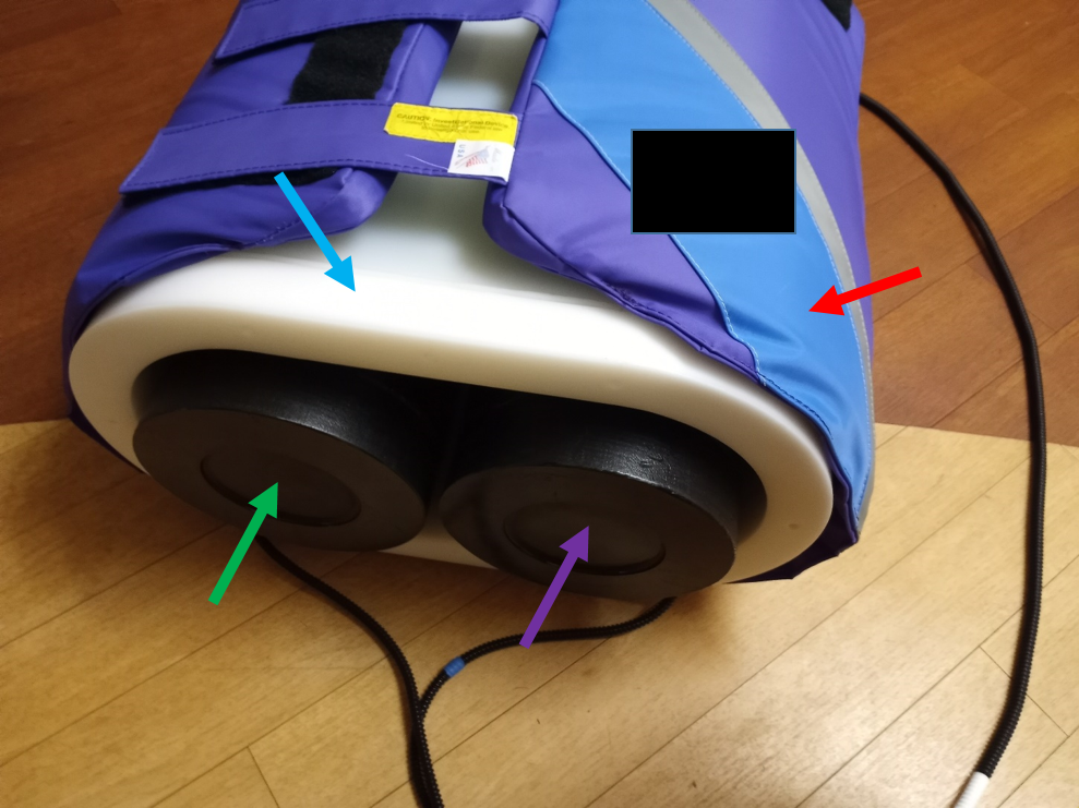
Figure 1: The Xe phantom MR imaging setup used is shown above. Red Arrow: Chest quadrature coil. Blue Arrow: Loader shell filled with saline solution. Green Arrow: Phantom_1. Purple Arrow: Phantom_2.
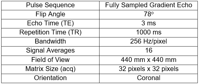
Table 1: 2D scan protocol utilized by each imaging research center to acquire the Xe - phantom images.
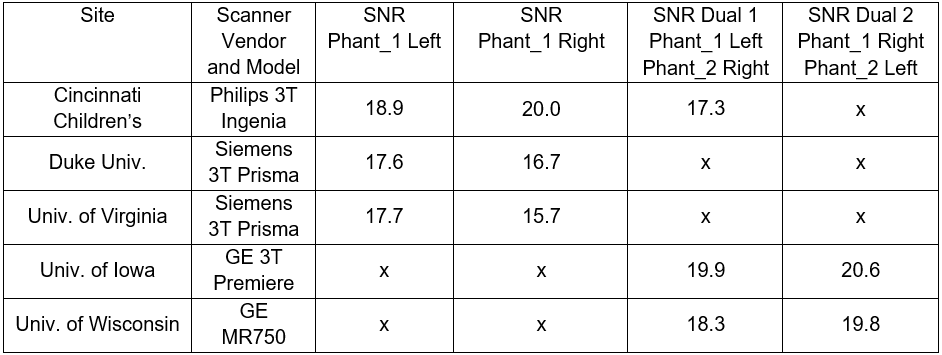
Table 2: Measured SNR values of images reconstructed offline from raw data, listing research institution and MRI vendors. The coefficient of variation (CV) amongst all SNR values is 8.46%. An analysis of variance run on the SNR values revealed a p-value of 0.09 considering these 5 research sites as distinct groups
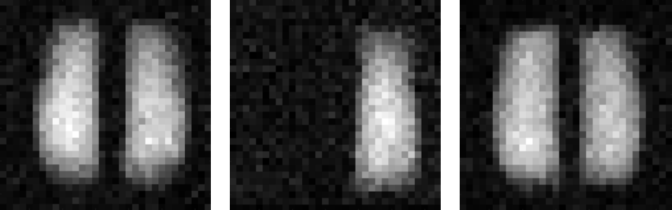
Figure 2: Left: Cincinnati (Philips), Phantom_1 on right side of image, Average SNR: 17.3. Middle: Duke (Siemens), Phantom_1 on right, Average SNR: 17.7. Right: Iowa (GE), Phantom_1 on right, Average SNR: 19.9.