5248
Determination of T1 and T2 Relaxation Times of Phosphorus-31 in a Low-Field MRI by developing a Phosphorus-31 resonant Low-Field MRI coil.1Department of Electrical Engineering and Information Technology, South Westphalia University of Applied Sciences, Luedenscheid, Germany
Synopsis
Keywords: Low-Field MRI, RF Arrays & Systems, X-nuclei, phosphorus-31
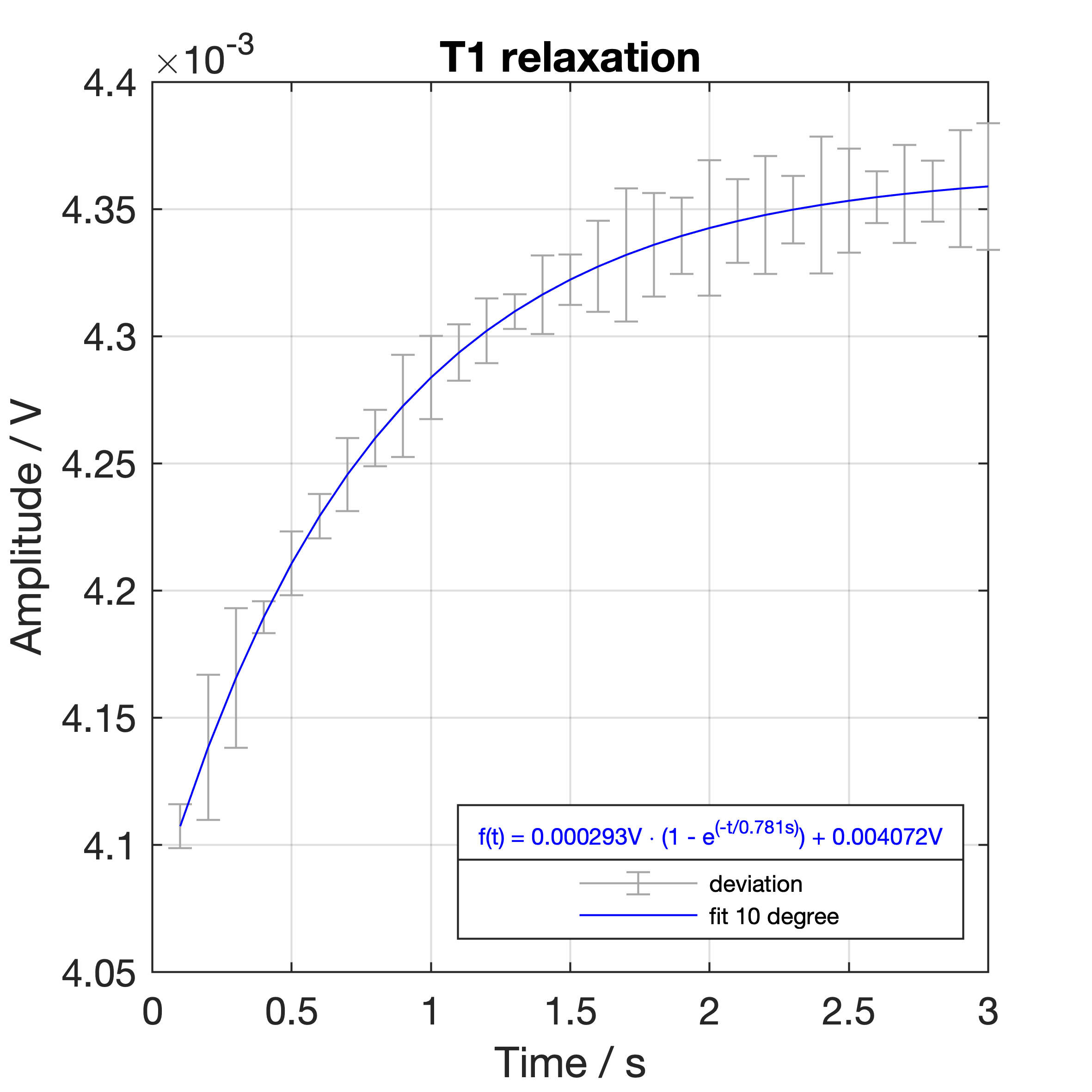
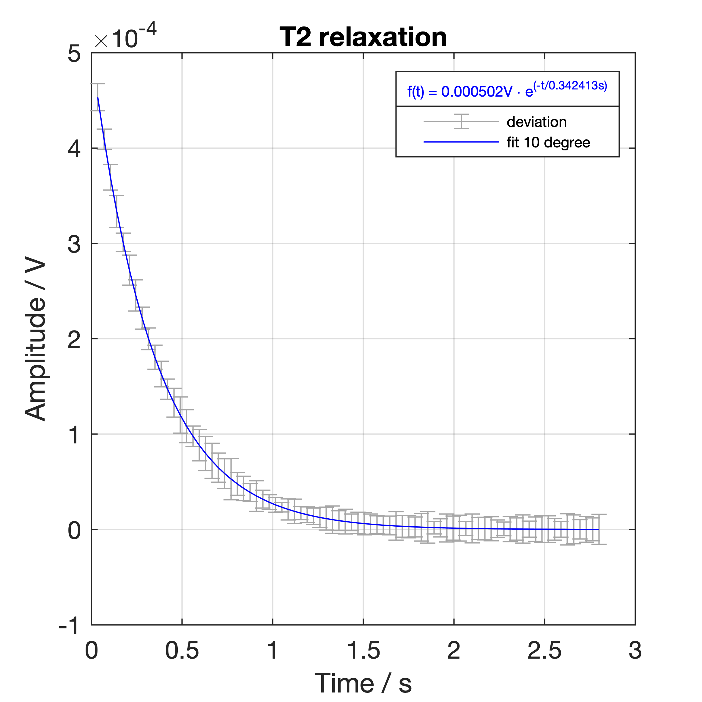
Development of a phosphorus-31 resonant NMR coil in order to estimate the T1 and T2 relaxation times. A didactic low-field 0.57 T MR-system and a 85% phosphorus acid solution as phantom are used to generate a sectional image of the phantom. In addition the spin-lattice and spin-spin relaxation signals are recorded, averaged and fitted in order to determine the T1 and T2 relaxation times. After exponential curve fitting the following relaxation times T1=0.781 s and T2=0.342 s are calculated.
Introduction
Phosphorus is present in the human body in the form of phosphates and is a major component of all human tissues and functions. It is important for energy storage and metabolism, as well as for cell communication and enzyme activation. Furthermore phosphates are relevant for preservation of bone substance and the apoptosis of chondrocytes [1]. This is the reason why phosphorus is interesting for MRI in order to get information about metabolic processes in the human body. For example, by examining phosphocreatins with MRI, ATP deficiencies and thus an abnormal metabolic response can be detected [2]. However, high-field MR-systems are expensive to purchase and operate, so low-field MRI offers a good alternative for MRI examinations of phosphorus. In addition, low-field MRI does not require as stringent safety measures as high-field MRI, due to the lower magnetic flux density. To demonstrate the feasibility of phosphorus-31 imaging in low-field MRI a transmit-receive coil for 0.57 T was developed for T1 and T2 relaxation time as well as initial imaging experiments.Materials and Methods
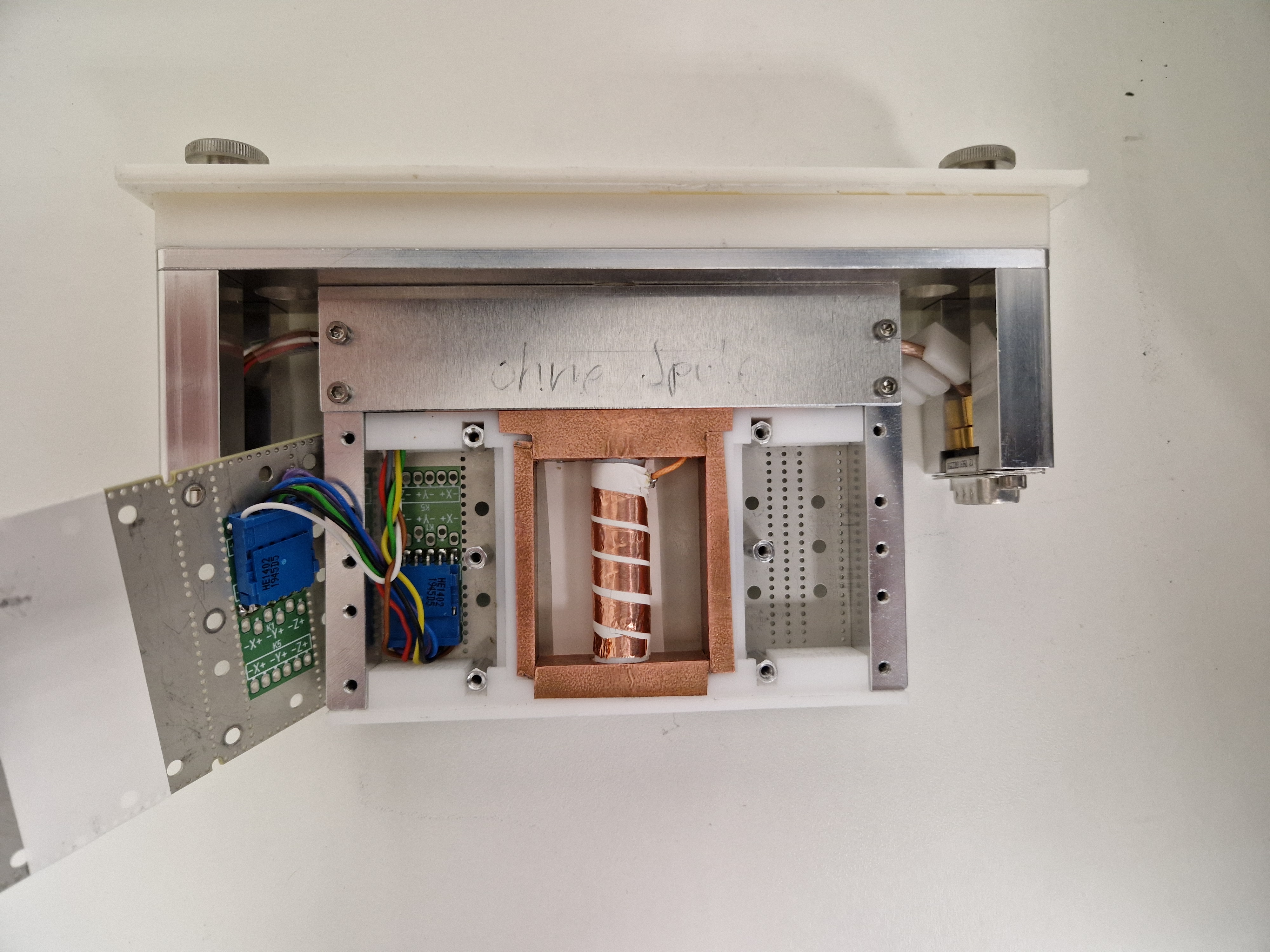
For the experimental setup, a didactic MR-system (Research MRI System, Pure Devices GmbH, Rimpar, Germany [3]) is used. The didactic MR system comprises a shielded permament magnet generating a magnetic flux density of 0.57 T, a removable sample head with integrated gradient coils, and an external control unit that outputs gradients and transmits and receives RF signals. The control unit is connected to a PC [3]. To develop a coil resonant to phosphorus-31 for the experimental setup, a coil with ten windings and an inner diameter of 11 mm was wound to a 3D-printed holder. The coil was tuned to 9.816 MHz and matched to 50 Ohm. The coil was integrated in an empty probe head and connected to the control unit. To test the setup in low field MRI a highly concentrated phosphorus solution containing 85% phosphoric acid diluted in water. The solution was filled in a standard 10 mm NMR sample tube. After frequency adjustments T1 and T2 relaxation experiments were performed and repeated 5 times respectively. An exponential function is fitted to the recorded and averaged data. For T1 a saturation recovery sequence and for T2 a multi-echo spin-echo sequence was used. In order to proof the imaging capability oft the phosphorus-31 coil a fast gradient echo sequence with following sequence parameters was used: Mtx: 64×64, TR=200ms, TE=1ms, avg 40, phase and read gradient strength = 40 μTs/m, TA=512s.Results and Discussion
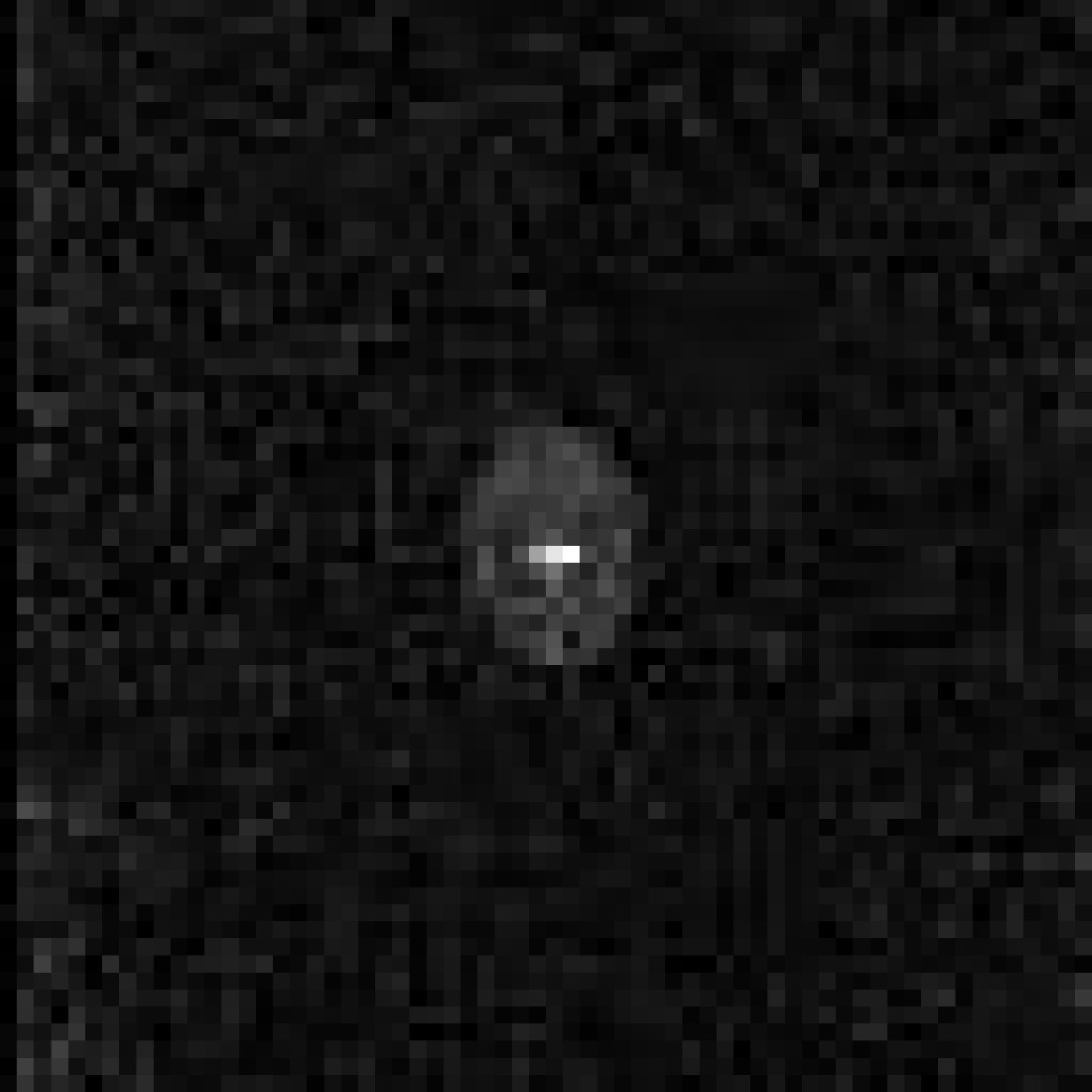
The developed NMR Coil is shown in Fig.2. Fig.1 displays the sectional image of the fast gradient echo sequence. The phosphorus phantom is clearly visible in the center. Due to low signal intensities shimming of the magnetic field is not carried out. Because of this the phantom is distorted and shows inhomogeneities. The spatial resolution of the image is approximately 6.15 mm x 6.15 mm and the signal to noise ratio is 4.187. Figures 3 and 4 show the fitted curves and deviations. T1 relaxation time is T1=0.781 s and T2 relaxation time is T2=0.342 s. In summary the detected signal shows sensitivity of the developed coil towards phosphorus. In future an improved coil design, resulting in an increased signal intensity, allows the shimming of the magnetic field yielding a better spatial resolution and an less distorted image of the phantom.Acknowledgements
No acknowledgement found.References
[1] Manghat P, Sodi R, Swaminathan R. Phosphate homeostasis and disorders. Annals of Clinical Biochemistry. 2014;51(6):631-656. doi:10.1177/0004563214521399.
[2] Buchthal SD, den Hollander JA, Merz CN, Rogers WJ, Pepine CJ, Reichek N, Sharaf BL, Reis S, Kelsey SF, Pohost GM. Abnormal myocardial phosphorus-31 nuclear magnetic resonance spectroscopy in women with chest pain but normal coronary angiograms. N Engl J Med. 2000 Mar 23;342(12):829-35. doi: 10.1056/NEJM200003233421201. PMID: 10727587.
[3] Overview - Research Lab (pure-devices.com) (access 0911.2022 19:29)
Figures