5239
Evaluation of multi-echo ICA denoising for an olfactory task experiment.1National Institute of Information and Communications Technology, Osaka, Japan, 2The University of Tokyo, Tokyo, Japan, 3Graduate School of Agricultural and Life Sciences, The University of Tokyo, Tokyo, Japan, 4International Research Center for Neurointelligence (WPI-IRCN), Institutes for Advanced Study, the University of Tokyo, Tokyo, Japan, 5Graduate School of Frontier Biosciences, Osaka University, Osaka, Japan
Synopsis
Keywords: fMRI (task based), fMRI (task based)
To remove noises from fMRI signals, ME-ICA was proposed. However, it is still unclear whether the denoising method shows good performance for an olfactory task experiment. Here, we compared the GLM and MVPA results between conventional denoising method and ME-ICA. In the 2nd level GLM, ME-ICA showed larger activation than conventional one, which is in line with the previous studies for visual or auditory experiments. On the other hand, ME-ICA did not show better MVPA performance. In conclusion, ME-ICA was a good denoising method for an olfactory experiment, but the benefit was small for MVPA.INTRODUCTION
Since fMRI signals contain many noises in addition to blood oxygenation level dependent (BOLD) signals, it is important to remove noises when investigating neural activity. When examining task-related brain activation, most previous studies have regressed out motion-related confounding factors using general liner model (GLM). Denoising methods using independent component analysis (ICA) have been proposed1, followed by derived methods such as ICA-based strategy for Automatic Removal of Motion Artifacts (ICA-AROMA)2, and FMRIB's ICA-based X-noiseifier (FIX)3. The combination of multi-echo fMRI with ICA denoising (ME-ICA)4,5 is a promising method for distinguishing BOLD signals from non-BOLD signals containing motion-related artifacts based on TE-dependence profiles. Although ME-ICA has been reported as a good denoising method for resting-state experiments6, its advantages in the detection of brain activation and in multivariate pattern analysis (MVPA) in task experiments are not well understood. Because multi-echo fMRI is advantageous to detect brain activation in areas near the nasal and ear cavities which are sensitive to magnetic susceptibility, we aim to reveal the benefits of ME-ICA denoising for the detection of brain activation and MVPA in the olfactory regions for odorant stimulation.METHODS
We measured BOLD signals for six verbally labeled odors using a 3T MRI with multi-echo gradient-echo echo-planar imaging sequence (N = 24). We conducted three types of denoising: 1) regressing out six motion-related factors in the 1st level GLM (conventional method), 2) ME-ICA, and 3) combining ME-ICA and conventional one. Among these denoising methods, we compared two metrics: 1) magnitude of brain responses for odors quantified by 2nd level GLM, 2) differences of activation patterns among odors quantified by MVPA with linear kernel support vector machine.RESULTS
When examining magnitude of brain responses evoked by odors, conventional denoising method showed significant activity in the secondary olfactory areas, such as the insula and orbitofrontal cortex (N = 24, Student’s t test, P < 0.05, cluster size corrected). On the other hand, two denoising methods using ME-ICA showed significant activity in the primary olfactory cortex, such as the piriform cortex, as well as the secondary ones (N = 24, Student’s t test, P < 0.05, cluster size corrected). Also, the approach combining ME-ICA and conventional method showed slightly larger activity than ME-ICA. On the other hand, when examining activation patterns, each denoising methods showed significant differences in the secondary olfactory areas, such as the insula and orbitofrontal cortex (N = 24, sign-permutation test, P < 0.05, cluster size corrected), and the results were not so different among them.DISCUSSION
The comparison of magnitude of brain responses suggests that ME-ICA removes noise which could not be removed by conventional method, and that conventional method also removes noise which could not be removed by only ME-ICA in the olfactory regions. The fact that ME-ICA outperformed conventional denoising method is in line with previous studies based on visual, auditory, or motor tasks7. On the other hand, denoising using ME-ICA did not showed better MVPA performance. This suggests that the signal to noise ratios in the smoothed univariate data and unsmoothed multivariate data may be different, or ME-ICA may have some smoothing effects.CONCLUSION
ME-ICA was a good denoising method for an olfactory experiment, and combining ME-ICA and conventional method also may be beneficial. On the other hand, ME-ICA did not show clear advantages for MVPA, at least for an olfactory task experiment.Acknowledgements
This work was supported by JST Mirai Program to K.T. (Grant Number JPMJMI19D1), and JSPS KAKENHI to M.O. (Grant Number JP18H04998 and JP21H05808). We appreciate the support of the IRCN Human fMRI Core, the University of Tokyo Institutes for Advanced Studies.References
1. McKeown, M. J. et al. Spatially independent activity patterns in functional MRI data during the stroop color-naming task. Proc Natl Acad Sci U S A 95, 803-810, doi:10.1073/pnas.95.3.803 (1998).
2. Pruim, R. H. R. et al. ICA-AROMA: A robust ICA-based strategy for removing motion artifacts from fMRI data. Neuroimage 112, 267-277, doi:10.1016/j.neuroimage.2015.02.064 (2015).
3. Salimi-Khorshidi, G. et al. Automatic denoising of functional MRI data: combining independent component analysis and hierarchical fusion of classifiers. Neuroimage 90, 449-468, doi:10.1016/j.neuroimage.2013.11.046 (2014).
4. Kundu, P. et al. Integrated strategy for improving functional connectivity mapping using multiecho fMRI. Proc Natl Acad Sci U S A 110, 16187-16192, doi:10.1073/pnas.1301725110 (2013).
5. Kundu, P., Inati, S. J., Evans, J. W., Luh, W. M. & Bandettini, P. A. Differentiating BOLD and non-BOLD signals in fMRI time series using multi-echo EPI. Neuroimage 60, 1759-1770, doi:10.1016/j.neuroimage.2011.12.028 (2012).
6. Kundu, P. et al. Multi-echo fMRI: A review of applications in fMRI denoising and analysis of BOLD signals. Neuroimage 154, 59-80, doi:10.1016/j.neuroimage.2017.03.033 (2017).
7. Gonzalez-Castillo, J. et al. Evaluation of multi-echo ICA denoising for task based fMRI studies: Block designs, rapid event-related designs, and cardiac-gated fMRI. Neuroimage 141, 452-468, doi:10.1016/j.neuroimage.2016.07.049 (2016).
Figures
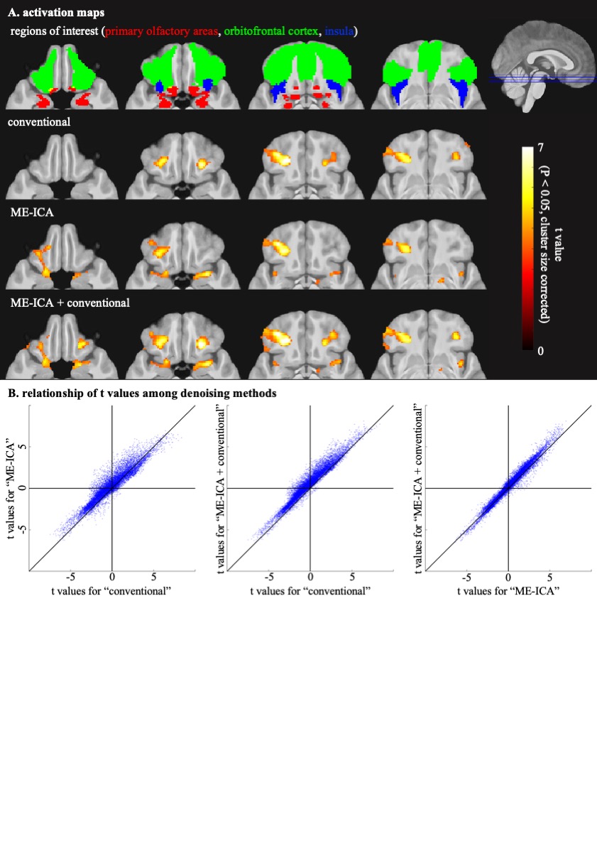
Figure 1. Brain activations evoked by odors.
A. In the top panel, regions of interest are shown, and blue lines indicate the slice locations. In the other panels, colormap indicates t values for the 2nd level GLM for each denoising method. B. Blue dots indicate t values for the 2nd level GLM for each voxel within the primary olfactory areas, orbitofrontal cortex or insula.
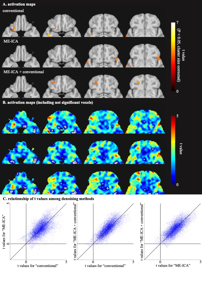
Figure 2. Differences of activation patterns evoked by odors.
A, B. Colormap indicates t values for the MVPA for each denoising method, A) for significant voxels and B) for voxels which showed positive t values. C. Blue dots indicate t values for the MVPA for each voxel within the primary olfactory areas, orbitofrontal cortex or insula.