5203
Connectome gradient dysfunction in benign childhood epilepsy with centrotemporal spikes: Initial discovery
Guiqin Chen1, Jie Hu1, Haifeng Ran1, Heng Liu1, and Tijiang Zhang1
1Department of Radiology, The Affiliated Hospital of Zunyi Medical University, Zunyi, China
1Department of Radiology, The Affiliated Hospital of Zunyi Medical University, Zunyi, China
Synopsis
Keywords: Epilepsy, Brain
Large-scale brain network abnormalities and cognitive impairment in patients with benign childhood epilepsy with centrotemporal spikes (BECTS). And cognitive function needs hierarchical interaction support between brain-collateral systems. Whether there are changes in the interaction and functional arrangement between different network systems in patients with BECTS still ambiguous. The purpose of this study is to use the method of gradient connection to investigate the changes of macro-network function hierarchy of BECTS and its potential contribution to cognitive function in BECTS children .Purpose
Large-scale brain network abnormalities and cognitive impairment in patients with benign childhood epilepsy with centrotemporal spikes (BECTS). And cognitive function needs hierarchical interaction support between brain-collateral systems. Whether there are changes in the interaction and functional arrangement between different network systems in patients with BECTS still ambiguous. The purpose of this study is to use the method of gradient connection to investigate the changes of macro-network function hierarchy of BECTS and its potential contribution to cognitive function in BECTS children.Methods
We recruited 50 BECTS patients and 69 healthy controls (HCs)). The brain network hierarchy of each group was depicted by connectome gradient analyses. We assessed the network hierarchy changes by comparing the gradient values in each network across the BECTS and HCs groups. Whole-brain voxel-level gradient values were compared across the BECTSI and HC groups to identify abnormal brain regions. Finally, we examined the relationships between altered gradient values and clinical variables. Based on the principal gradient map of the patients, the relevance vector regression (RVR) algorithm was used to predict the cognitive function score of BECTS children.Result
Compare to the controls, BECTS children extended gradient at different network-level and voxel-level. There was an apparent increase in the frontoparietal network (FPN) in the principal gradient of patients with BECTS. The dorsal attention network (DAN) and FPN exhibited a higher trend in the secondary gradient (P <0.05, FDR-corrected). Individuals with BECTS showed significantly higher functional gradient scores in the left precentral gyrus (PCG) and right angular gyrus (ANG) in the principal gradient (P <0.05, GRF-corrected). Left inferior frontal gyrus, triangular part (IFGtriang) showed an increase gradient score in the secondary gradient, and left precuneus (PCUN) and bilateral cuneus (CUN) exhibited lower trend in the secondary gradient (P <0.05, GRF-corrected). In the principal gradient, the left precentral gyrus gradient score of BECTS children was negatively correlated with the Verbal intelligence quotient(VIQ) (P=0.023,r =-0.372); in the secondary gradient, the connectome gradient alterations in FPN of BECTS children was positively correlated with the age of onset (P=0.045, r=0.297). The principal gradient map of the patients could significantly predict their VIQ (r = 0.388, P < 0.05). Most contributive features were located in the VAN (20.7%), FPN (19.9%), VIS (17.6%), and DMN (17.2%).Conclusions
These results indicate the connectome gradient dysfunction in BECTS and its linkage with age of onset and cognitive function, increasing our understanding of the functional connectome hierarchy and the pathophysiology of the cognitive impairment in BECTS, providing potential biomarkers for cognitive impairment in this disorder.Acknowledgements
None.References
None.Figures
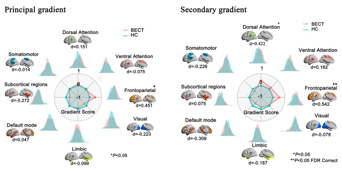
Figure 1 Network-level gradient comparisons. (A)The radar chart shows the gradient score (with respect to healthy controls) of the two groups. The FPN was increased (P < 0.05, uncorrected). (B) the FPN was significantly increased(P <0.05, false discovery rate corrected) and DAN were significantly increased ( (P < 0.05, uncorrected). Each network’s spatial location was anchored in the corresponding position, and Cohen’s d was computed.DAN dorsal attention network, FPN frontoparietal network.
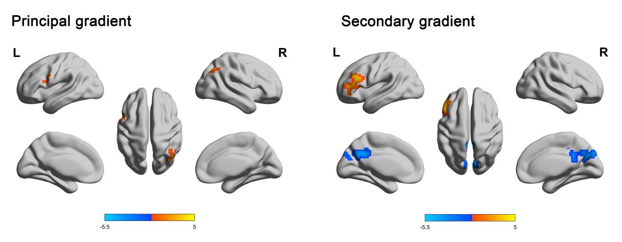
Figure 2 Voxel wise statistical comparisons between the healthy controls and patients with BECTS and distribution of the regional case-control difference in different systems. Higher/lower values in BECTS are presented as warm/cold colors. The statistical significance level was set as GRF-corrected (voxel P value < 0.001 and cluster P value<0.05).
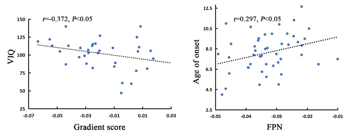
Figure 5 (A) Spearman correlation between gradient score of left PreCG and VIQ of the principal gradient in BECT patients. (B) Spearman correlation between the alterations in FPN and age of onset of the secondary gradient in BECT patients. PreCG precentral gyrus, FPN frontoparietal network, VIQ verbal intelligence quotient.Note: There were 13 patients lacking intelligence test results due to non-cooperation or inability to perform intelligence tests during patient testing, and 3 patients lacking onset age results.
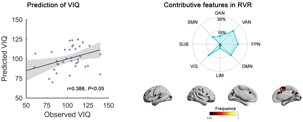
Figure 4 The
gradient topography and prediction of VIQ in patients with BECTS. (A)
Scatter plot presents the correlation between the observed VIQ and the
predicted VIQ change derived from the RVR analysis ( P < 0.05). Each dot represents the data from one patient, and
the dashes indicate the 95% prediction error bounds. (B) The absolute summed
weights in leave-one-out cross validation were mapped onto the brain surface.
Regions with higher/lower predictive power were colored in white/red. The radar
map represents the distribution of predictive power in different systems.
DOI: https://doi.org/10.58530/2023/5203