5149
Development of in vivo human brain multifrequency DTIMRE (mDTIMRE)1Biomedical Engineering, University of Illinois Chicago, Chicago, IL, United States, 2Beckman Institute, University of Illinois Urbana-Champaign, Urbana, IL, United States
Synopsis
Keywords: Elastography, Elastography
Simultaneous acquisition of diffusion tensor imaging and multifrequency magnetic resonance elastography was examined in this preliminary study on in vivo human brain. The mDTIMRE was achieved by ensuring the same diffusion encoding using experiment parameters with different mechanical frequencies, while fulfilling the timing condition of simultaneous acquisition. By applying a multifrequency dual elasto-visco inversion approach, the effective image resolution of mechanical property maps is improved and enables quantitatively assessment of tissue properties in brain sub-regions. The experiment results show a good correlation between mDTIMRE and conventional measurements.Introduction
Magnetic resonance elastography (MRE) and diffusion tensor imaging (DTI) are two non-invasive MRI techniques that use the phase and magnitude of the complex-valued MRI signal, respectively. In previous feasibility studies1,2,3, the simultaneous acquisition of MRE and DTI was fulfilled by applying 1) vibration to the in vivo human brain at a single mechanical frequency and 2) a motion encoding gradient that is sensitive to both vibration and diffusion. Monofrequency MRE can assess the regional brain stiffness, but with relatively low spatial resolution. Multi-frequency MRE (mMRE) acquires wave images at multiple mechanical frequencies and has been used to study the frequency-dependent mechanical properties of tissue using different rheological models4. Furthermore, by applying a multifrequency dual elasto-visco (MDEV) inversion approach, the effective image resolution is improved and enables quantitatively assessment of tissue mechanical properties in brain sub-regions, which can potentially change at the onset of neurodegenerative pathology5. In this study, we propose that the simultaneous acquisition of DTI and mMRE can be achieved by ensuring the same diffusion encoding using experiment parameters with different mechanical frequencies, while fulfilling the timing condition of simultaneous acquisition6,7.Methods
Three subjects were recruited and the consent forms that are approved by the Institutional Review Board, UIC, were obtained. Each subject experienced the same scan sessions repeatedly on four different days. The study was conducted on a 3T human MRI scanner (Prisma, Siemens). mDTIMRE was implemented using two mechanical frequencies and four phase offsets, each offset has three encoding directions with positive and negative polarities. The mechanical frequencies, gradient amplitudes, diffusion time and b-values were 40/50Hz, 54/78 mT/m, 37.5/30 ms and 850.7/849.6 s/mm2 respectively. The matrix size, number of slices, field of view and TE were 80x80, 48, 240x240 mm2 and 71 ms, respectively. The diffusion encoding acquired 48 diffusion encoding directions across the mDTIMRE acquisition at the two frequencies. For comparison, a 48-direction DTI and 3D-mMRE with matching experimental parameters were also acquired. The estimation of mean diffusivity (MD) and fractional anisotropy (FA) maps used the DTIFIT tool in FSL (FMRIL, Oxford). The shear stiffness (|G*|) was calculated using MDEV inversion5. All data sets were registered to the reference plane of each subject using their T2 images, and the generated affine matrices were then applied to the property maps. Six regions of interest (ROIs), white matter (WM), putamen, caudate, thalamus, hippocampus and corpus callosum, were selected after brain parcellation on MPRAGE anatomical scan in Freesurfer (Harvard University) in order to study the correlation of ROI-wise averaged values. On separate property maps, the voxel-wise correlation of WM between mDTIMRE and conventional methods from repeated scans were plotted and fitted using least-squares method. In study of reproducibility of each sequence, the voxel-wise correlation of separate property maps in WM was done on permutation of the repeated scans.Results
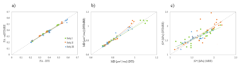
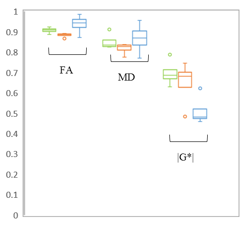
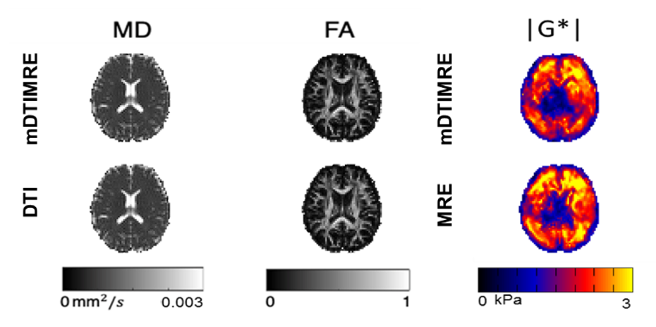
The correlation of regional averaged values between mDTIMRE and conventional measurements is plotted in Fig. 1. Therein, the Pearson’s correlation coefficients for FA, MD and |G*| values in the three subjects (I | II | III) is (1.00 | 1.00 | 0.99), (0.99 | 0.98 | 0.97) and (0.85 | 0.81 | 0.84), respectively. The boxplots in Fig. 2 show the voxel-wise correlation coefficients in WM between mDTIMRE and conventional DTI / mMRE. The individual mean values of WM on separate property maps is shown in Table 1. In the reproducibility study, the voxel-wise correlation analysis in WM was done on any two sets of the repeated scans from same sequence and the mean Pearson’s rho is displayed in Table 2. An example of acquired property maps of a central brain slice is displayed in Fig. 3.Discussion
The correlation plot displayed a linear relationship between regional average values from mDTIMRE and conventional measurements. The least-square fitting results showed a slope close to 1 in FA and MD, and a trend of decreased |G*| in mDTIMRE. The same trend of correlation on separate property maps was observed in all three subjects. An excellent voxel-wise correlation between mDTIMRE and DTI was found on the diffusive property maps, while the correlation between mDTIMRE and mMRE on the mechanical property maps was good. The repeatability of mDTIMRE was similar as for conventional methods, while the overall repeatability of diffusive property maps was higher than the mechanical property maps, as shown in Table 2. Currently, we are in the process of confirming our results using a larger sample size.Acknowledgements
This work was supported by the NIH NIBIB under grant R21EB026238, Adding MRE to DTI for free. The work represents the views of the authors and not of the NIH.References
1. Lin S, Sutton B, Magin RL, Klatt D: Development of in vivo human brain DTI-MRE. Proceedings of the 28th Annual Meeting of the ISMRM, p. 3325, virtual, 2020.
2. Lin S, Sutton BP, Magin RL, Klatt D: Development of in vivo human brain DTI-MRE: Optimization of experimental parameters. Proceedings of the 29th Annual Meeting of the ISMRM, p. 3656, virtual, 2021. 3. Lin S, Sutton BP, Magin RL, Klatt D: Development of in vivo human brain DTI-MRE: Preliminary results. Proceedings of the 30th Annual Meeting of the ISMRM, p. 3940, hybrid, 2022.
4. Klatt D, Hamhaber U, Asbach P, Braun J, Sack I. Noninvasive assessment of the rheological behavior of human organs using multifrequency MR elastography: a study of brain and liver viscoelasticity. Phys Med Biol. 2007;52(24):7281-7294.
5. Guo J, Hirsch S, Fehlner A, Papazoglou S, Scheel M, Braun J, Sack I. Towards an Elastographic Atlas of Brain Anatomy. PlosOne 2013; Vol 8(8).
6. Yin Z, Kearney SP, Magin RL, Klatt D. Concurrent 3D Acquisition of Diffusion Tensor Imaging and Magnetic Resonance Elastography Displacement Data (DTI-MRE): Theory and In Vivo Application. Magnetic Resonance in Medicine 2017; 77(1): 273-284.
7. Yin Z, Magin RL, Klatt D. Simultaneous MR elastography and diffusion acquisitions: Diffusion-MRE (dMRE). Magnetic Resonance in Medicine 2014; 71(5): 1682-1688.
Figures