5133
“ShortCardiac” - an open-source tool for assessing cardiac function quickly
Karl Ludger Radke1, Janina Hußmann1,2, Lena Röwer1,2, Dirk Voit3, Jens Frahm3, Gerald Antoch1, Dirk Klee1, Frank Pillekamp1,2, and Hans-Jörg Wittsack1
1University Düsseldorf, Department of Diagnostic and Interventional Radiology, Düsseldorf, Germany, 2Department of General Pediatrics, Neonatology and Pediatric Cardiology, University Children's Hospital Düsseldorf, D-40225 Düsseldorf, Germany, Düsseldorf, Germany, 3Biomedical NMR, Max Planck Institute for Multidisciplinary Sciences, D-37070 Göttingen, Germany, Göttingen, Germany
1University Düsseldorf, Department of Diagnostic and Interventional Radiology, Düsseldorf, Germany, 2Department of General Pediatrics, Neonatology and Pediatric Cardiology, University Children's Hospital Düsseldorf, D-40225 Düsseldorf, Germany, Düsseldorf, Germany, 3Biomedical NMR, Max Planck Institute for Multidisciplinary Sciences, D-37070 Göttingen, Germany, Göttingen, Germany
Synopsis
Keywords: Heart, Software Tools
In this work, we propose a new open-source framework for cardiac functional analysis. Our proposed framework can be used in a pipeline with the commercial software Circle cvi42 as well as the free software ITKSnap and enables a step towards a fully automated, reproducible, and quantitative assessment of MR images in clinical routine.Introduction
In recent years, numerous quantitative MR techniques have been developed as well as techniques that enable a high temporal resolution of physiological processes. However, standardization and the huge amount of acquired data remain challenging; therefore, many techniques have yet to reach clinical applications. In particular, cardiovascular imaging has a great clinical and scientific interest in acquiring data in different cardiac and respiratory phases. However, this is only possible with fast imaging techniques leading to large amounts of images. Therefore, our study aimed to develop an instrument that allows standardized and reproducible evaluation of the heart and significantly reduces the analysis time required.Methods
First, we acquired a healthy 27-year-old male example volunteer using both conventional retrospective ECG-gated MR acquisition with a slice thickness of 8 mm, a repetition time (TR) of 56.98 ms, an echo time (TE) of 1.1 ms, and a flip angle of 14°. In Addition, a real-time gradient-echo MRI sequence with pronounced radial under-sampling and balanced steady-state free precession (bSSFP) contrast, which provided a temporal resolution of 33.3 ms. All MR images were acquired in the supine position on a clinical 1.5 T MRI scanner (MAGNETOM Avanto fit, Siemens Healthineers, Erlangen, Germany) using an 18-channel body coil (Body, 18, Siemens Healthineers) centrally aligned with the heart and a 32-channel spine matrix coil (direct connect spine 32, Siemens Healthineers) installed in the MRI table. For quantitative analysis, we developed "shortCardiac," an open-source framework that is available under the GNU General Public License (GPU GPL) license (Figure 1) and can be downloaded from GitHub (https://github.com/MPR-UKD/shortCardiac). “ShortCardiac” was developed in Python 3.8 and can be used as Source Code, executable (.exe) GUI, as well as JupyterNotebook. After the MR images were segmented fully automatically using Circle, "shortCardiac" allows the angle-dependent assessment of ventricular shapes (Figure 2), centroid motion, septal rotation, and radiometric image feature extraction based on the DICOM image values (Figure 3). To validate our system, we compared the results of "shortCardiac" with the manual evaluation of an experienced radiologist using Bland-Altman (Figure 4) and intraclass correlation coefficient (ICC) analyses.Results
We observed excellent agreement between "shortCardiac" and an experienced radiologist's manual measurement (ICC 0.91-0.98) for both real-time imaging and conventional ECG-triggered MR imaging. In addition, "shortCardiac" enabled a substantial reduction in evaluation time. While the manual evaluation of a subset of 30 images with a measurement of five features per image took about an hour, "shortCardiac" needs only 12 seconds for the calculations and even determines 349 features like angle-dependent ventricular diameters, eccentricity indexes, the center of gravity movement and more.Discussion
The developed framework "shortCardiac" provides an approach for fast and standardized evaluation of cardiac MR images. Combining automatic deep-learning-based segmentation methods such as Circle and "shortCardiac" leads to a standardized assessment of cardiac MR images and enables inter-institutional comparability of future studies. In addition, computer-assisted evaluation methods allow for an accelerated evaluation, which on the one hand, enables the assessment of larger amounts of data in subsequent clinical studies and, on the other hand, is an important milestone towards improved clinical patient care.Conclusion
In our study, we successfully demonstrated that a framework such as "shortCardiac" can accelerate the analysis of cardiac MR data. Using a combination of Circle and "shortCardiac", we achieved comparable results to a radiologist while significantly shortening the evaluation time.Acknowledgements
We would like to thank the "Elterninitiative Kinderkrebsklinik e. V." for funding this research.References
No reference found.Figures
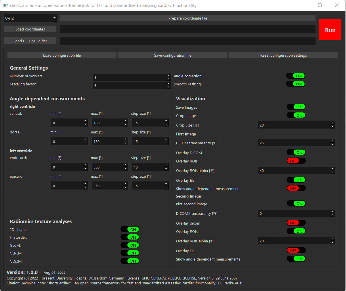
Visualization of the main window of shortCardiac to easily apply the
framework in clinical routine.
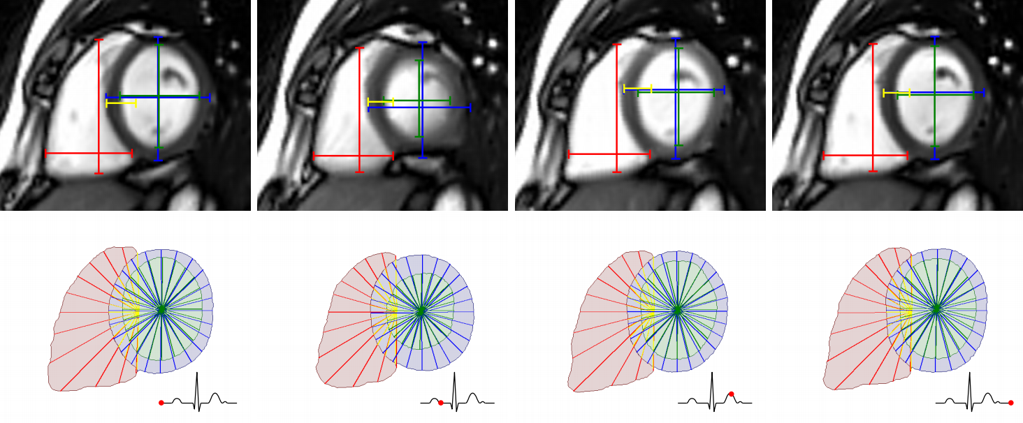
Graphical visualization of the 2D vector measurement with
"shortCardiac". Shown are four
exemplary real-time MR images representing a complete cardiac cycle.
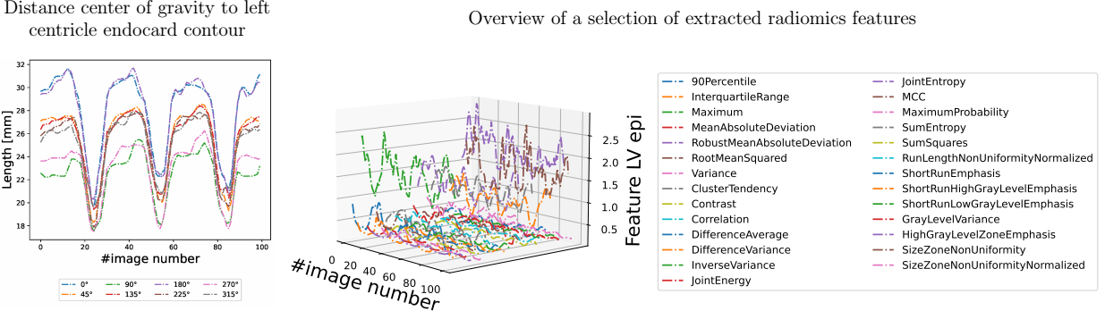
Exemplary plot of the distance changes of the endocardial contour as a
function of the angle for the left ventricle and the changes of a selection of
specific radiomics Features for the epicardium of the left ventricle as a
function of time.
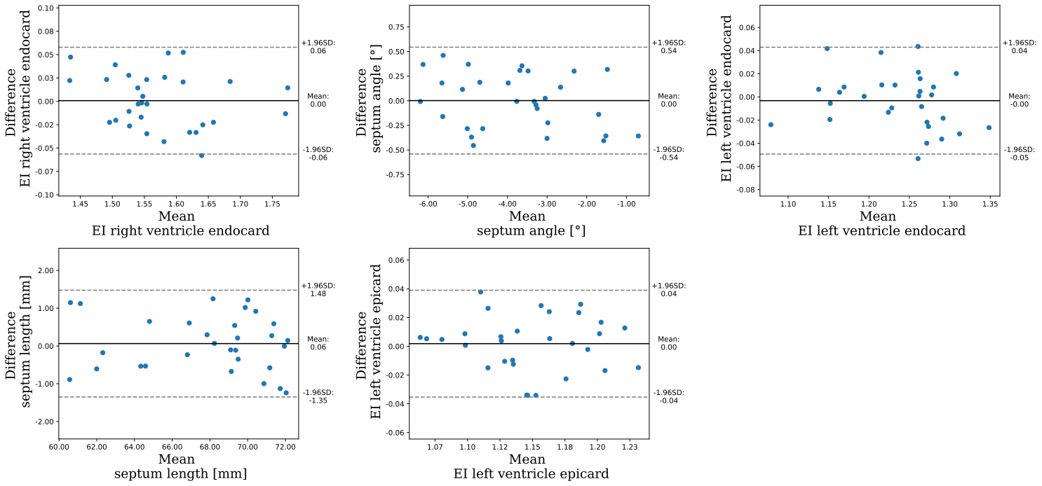
Bland-Altman diagrams to evaluate the reliability between
"shortCardiac" and the manual reference measurements of an
experienced radiologist. Shown are the values of 30 randomly selected images
from real-time MRI.
DOI: https://doi.org/10.58530/2023/5133