5129
Deep-learning-based super-resolution technique for cine cardiac magnetic resonance1Department of Diagnostic and Interventional Radiology, Hokkaido University Hospital, Sapporo, Japan, 2Department of Radiological Technology, Hokkaido University Hospital, Sapporo, Japan, 3Philips Japan, Ltd., Tokyo, Japan, 4Department of Diagnostic Imaging, Hokkaido University Graduate School of Medicine; Global Center for Biomedical Science and Engineering, Faculty of Medicine, Hokkaido University, Sapporo, Japan
Synopsis
Keywords: Myocardium, Data Processing
Cine cardiac magnetic resonance (CMR) imaging is an optimal cardiac volumetric analysis method because of its high contrast resolution. However, its spatial resolution is limited owing to prolonged scanning and breath-holding. Although compressed sensing–sensitivity encoding (Compressed SENSE; CS) and its deep-learning-based advancement (SmartSpeed AI; SSAI) can reduce the scan time, the spatial resolutions remain unchanged. Herein, we investigated the effect of a deep-learning-based super-resolution technique (SmartSpeed Precise Image; SSPI) on the cine CMR visual image quality in comparison with CS and SSAI with conventional zero-filling interpolation; resultantly, the SSPI significantly improved the visual image quality scores.Introduction
Cine cardiac magnetic resonance (CMR) based on a balanced steady-state free precession pulse sequence (bSSFP) is a widely used and essential sequence for CMR. Wall motion abnormalities, ventricular volumes, and ejection fractions of the left and right ventricles (LV and RV, respectively) can be evaluated using cine CMR1. Although cine CMR is the gold standard for volumetric analysis because of its high contrast resolution, its spatial resolution is lower than that of ultrasonography. Prolonged scanning and breath-holding times make it difficult to improve the spatial resolutions of two-dimensional bSSFP. Although compressed sensing–sensitivity encoding (Compressed SENSE; CS) is applied to accelerate image acquisition, a higher acceleration factor results in poorer image quality. A deep-learning (DL) (Adaptive-CS-Net)-based image reconstruction technique (SmartSpeed AI; SSAI) has the potential to improve the image quality2. However, because SSAI cannot affect spatial resolution, its impact on image quality is limited. The conventional zero-filling interpolation (ZIP) technique is widely used to improve “apparent” spatial resolution; however, it cannot improve the actual spatial resolution. Recently, a DL-based super-resolution technique (SSPI) has been developed to improve actual spatial resolution. We hypothesized that this DL-based super-resolution technique would reduce the scan time while improving the image quality. This study aimed to compare the image quality of cine CMR images reconstructed using CS with ZIP (CS-ZIP), SSAI with ZIP (SSAI-ZIP), and SSAI with SSPI (SSAI-SSPI).Methods
This prospective study was approved by the institutional review board. Written informed consent was obtained from all the participants. Six healthy volunteers (all men; median age, 36 years (range, 32–57 years)) were scanned using a 3.0T-MRI scanner (Ingenia Elition X, Philips Healthcare, Best, the Netherlands) with a dS Torso coil. The bSSFP images with the left ventricular short-axis plane were acquired with two different acceleration factors (acceleration factors of 2 and 4) and reconstructed using three different reconstruction methods (CS-ZIP, SSAI-ZIP, and SSAI-SSPI). Thus, six types of images were reconstructed. The acquisition parameters were as follows: TR/TE (ms) = 3.8/1.79, Flip angle (degree) = 70, Slice thickness (mm) = 10, Field of view (mm) = 300×300, acquisition matrix (mm) = 2.0×2.0, reconstruction matrix (mm) = 0.94×0.94, number of phases per cardiac cycle (phases) = 20.SSAI is a convolutional neural network called Adaptive-CS-Net2 that allows the reconstruction of images acquired with CS-based variable density undersampling patterns3. SSPI is a DL-based reconstruction technique combined with Adaptive-CS-Net and Precise Image Net, which is a DL-model applied to remove ringing artifacts and to replace the traditional zero-filling strategy to increase the matrix size and thus the image sharpness; these types of networks are known as SuperResolution networks4,5.
The acquisition time per slice for each acquisition method was measured. The visual image quality scores using whole cardiac phase images were assessed by two independent radiologists (seven and two years of experience in cardiovascular imaging) using a 3-point scale score (3=excellent, 2=moderate, 1=poor) with each of four categories: the sharpness between the LV or RV free wall and the ventricular cavity, the sharpness between LV or RV trabeculae and the ventricular cavity, the overall image quality considering image noise, and any artifact. All variables were presented as medians (ranges). The summed scores of the two observers were compared using Wilcoxon’s signed-rank test with Bonferroni correction because of the multiple comparison test problems. Inter-observer reproducibility was assessed using kappa-statistics (0.0–0.20, poor; 0.21–0.40, fair; 0.41–0.60, moderate; 0.61–0.80, good; 0.81–1.0, excellent). A p-value < 0.00033 was considered statistically significant based on a fifteen-times statistical test from six types of images.
Results
The heart rate was 60 (52–65) per minute. The acquisition time was 6.0 (5.3–6.3) seconds with an acceleration factor of 2 and 3.9 (3.3–4.0) seconds with an acceleration factor of 4. SSPI significantly improved the visual image quality scores regarding the sharpness between the myocardial wall and cavity (Fig. 1), sharpness between the trabeculae and cavity (Fig. 2), and overall quality (Fig. 3) in comparison with conventional ZIP. A representative case is shown in Fig. 4. The scores for any artifacts were not significant among the six types of images. Although inter-observer reproducibility of the scores of the overall quality was poor (kappa=0.18), the others were moderate or good (kappa = 0.54 and 0.58 for the sharpness of the LV and RV myocardia, respectively; 0.70 and 0.50 for the sharpness of the LV and RV trabeculae, respectively; 0.48 for artifacts).Discussion
The novel DL-based reconstruction method, SSPI, provides better image quality than that of the conventional ZIP. Moreover, the image quality of a doubled acceleration factor with SSPI is better than that of the initial acceleration factor with ZIP. Furthermore, SSPI neither removed nor worsened the artifacts. SSPI with a higher acceleration factor may improve the image quality of cine CMR, while reducing the imaging time.Conclusion
The novel DL-based super-resolution method (SSPI) could improve the image quality of cine CMR in comparison with conventional ZIP.Acknowledgements
None.References
- Kramer et al. Standardized cardiovascular magnetic resonance imaging (CMR) protocols: 2020 update. Journal of Cardiovascular Magnetic Resonance (2020) 22:17
- Pezzotti N, de Weerdt E, Yousefi S, et al. Adaptive-CS-Net: FastMRI with Adaptive Intelligence. arxiv. 2019;(NeurIPS).
- Hans Peeters PhD, Hayley Chung PhD, Giuseppe Valvano PhD, Deniz Yakisikli MSc., Jeroen van Gemert PhD, Elwin de Weerdt PhD and Kim van de Ven PhD. Philips SmartSpeed. No compromise Image quality and speed at your fingertips. https://www.philips.com/c-dam/b2bhc/master/landing-pages/mri-innovations/philips-smart-speed-brochure-lr.pdf
- Chao Dong, Chen Change Loy, Kaiming He, Xiaoou Tang. Super-Resolution Using Deep Convolutional Networks. 2014 arXiv:1501.00092.
- Y. Li, B. Sixou, F.Peyrin. A review of the deep learning methods for medical images super resolution problems. IRBM, Volume 42, Issue 2, April 2021, Pages 120-133.
Figures
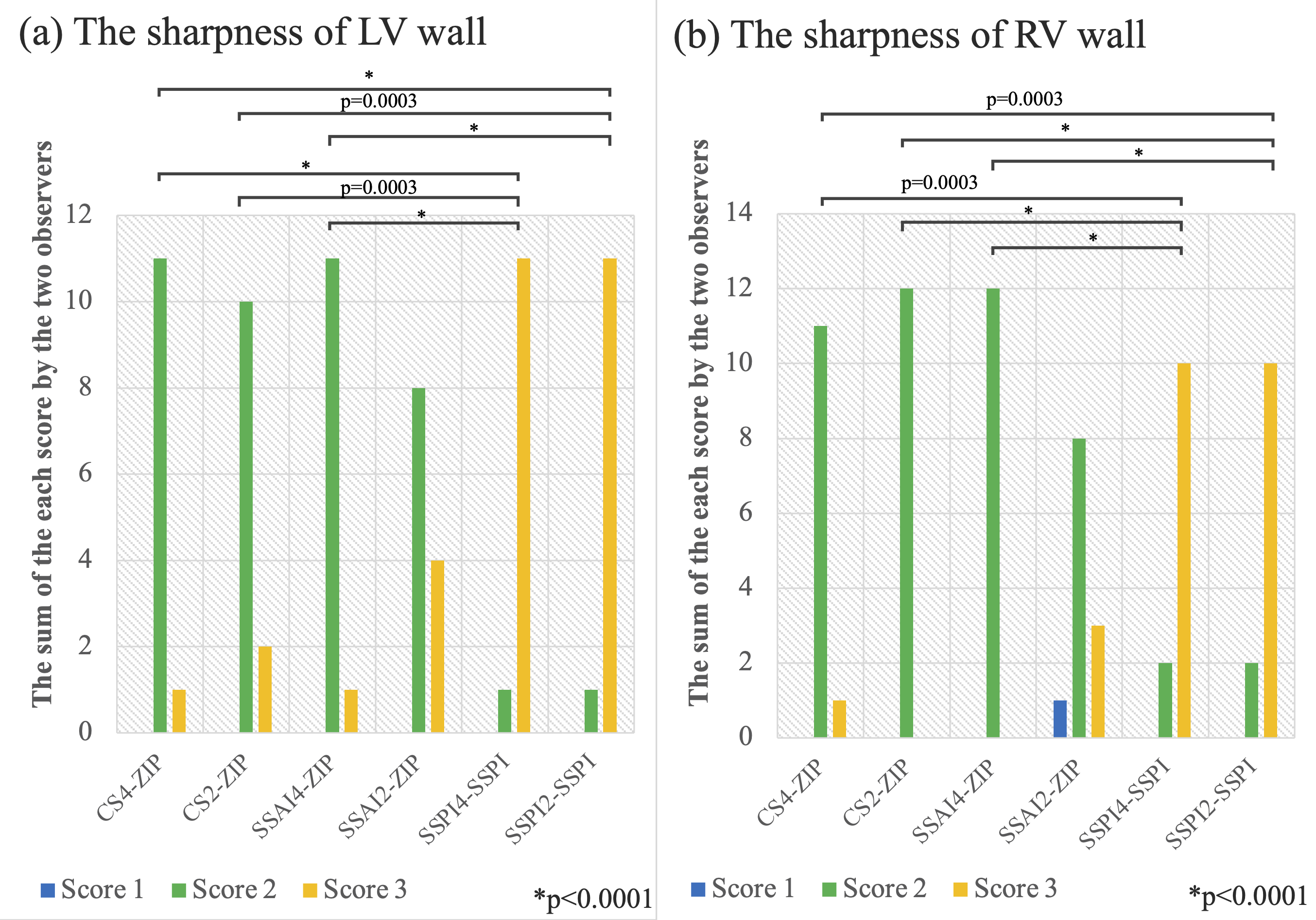
Figure 1. The image quality score of the sharpness between the myocardial wall of LV (a) or RV (b) and its cavity. SmartSpeed Precise Image significantly improved the sharpness with acceleration factors of both 2 and 4 in comparison with that of conventional ZIP.
CS: compressed sensing – sensitivity encoding, LV: left ventricle, RV: right ventricle, SSAI: SmartSpeed AI, SSPI: SmartSpeed Precise Image, ZIP: zero-filling interpolation
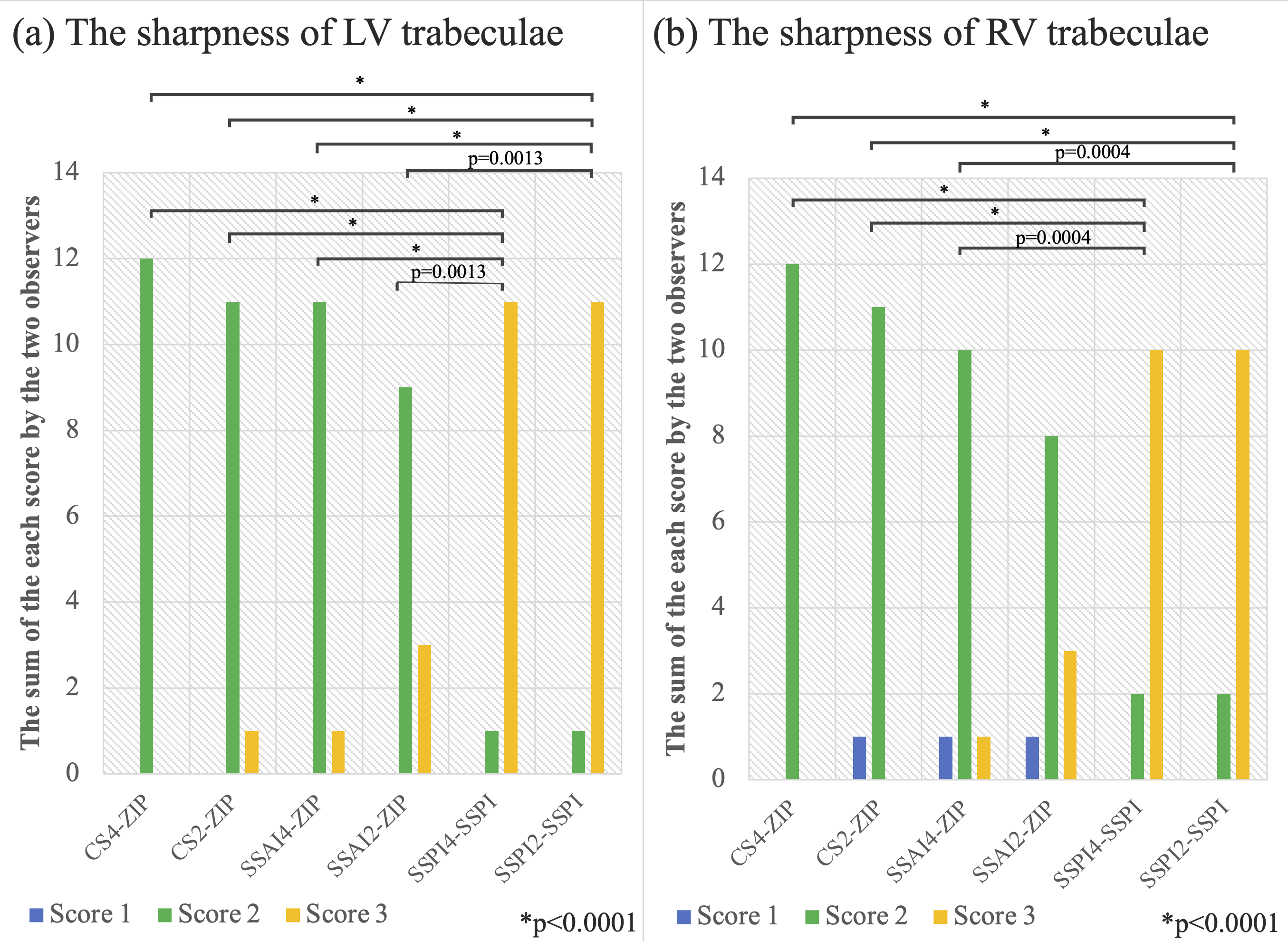
Figure 2. The image quality score of the sharpness between the LV (a) or RV (b) trabeculae and its cavity. SmartSpeed Precise Image significantly improved the sharpness with acceleration factors of both 2 and 4 in comparison with that of conventional ZIP.
CS: compressed sensing – sensitivity encoding, LV: left ventricle, RV: right ventricle, SSAI: SmartSpeed AI, SSPI: SmartSpeed Precise Image, ZIP: zero-filling interpolation
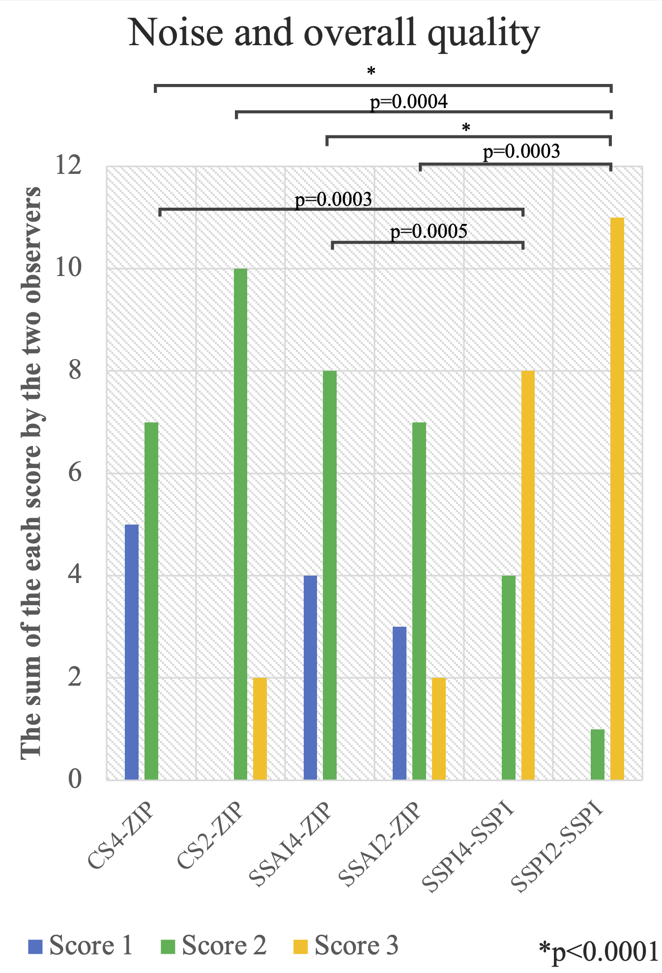
Figure 3. The image quality score of the overall quality. SmartSpeed Precise Image significantly improved overall image quality and reduced noise in comparison with those in conventional ZIP.
CS, compressed sensing – sensitivity encoding; LV, left ventricle; RV, right ventricle; SSAI, SmartSpeed AI; SSPI, SmartSpeed Precise Image; ZIP, zero-filling interpolation
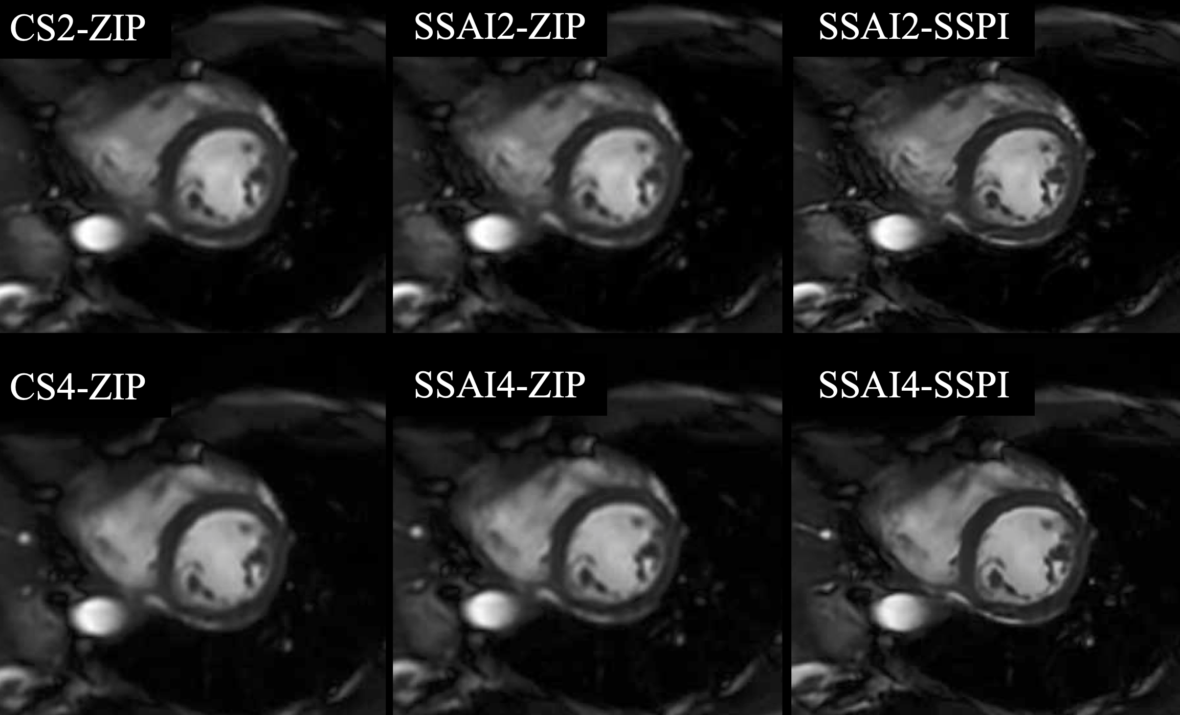
Figure 4. A representative case of the effect of SmartSpeed Precise Image for the sharpness. Left ventricular short-axis image at end-diastolic phase are shown. The myocardial wall and trabeculae are clearly depicted with SSPI with acceleration factors of both 2 and 4.
CS, compressed sensing – sensitivity encoding; SSAI, SmartSpeed AI; SSPI, SmartSpeed Precise Image; ZIP, zero-filling interpolation