5120
Assessing the clinical value of PETRA sequence for detection of subsolid pulmonary nodules:comparison with CT
Hui Feng1, Li Yang1, Chen Zhang2, Hui Liu1, Ning Zhang1, and Gaofeng Shi1
1The Fourth Hospital of Hebei Medical University, Shijiazhuang, China, 2Siemens Healthineers, Beijing, China
1The Fourth Hospital of Hebei Medical University, Shijiazhuang, China, 2Siemens Healthineers, Beijing, China
Synopsis
Keywords: Visualization, Lung
The PETRA technique had high sensitivity for the detection of subsolid pulmonary nodules and can accurately assess their diameter and morphologic characteristics.Introduction
Pulmonary nodules are the main imaging manifestations of lung cancer, and subsolid nodules, especially ground-glass nodules, are common manifestations in early stage lung cancer. According to the Fleischener Association, the risk of malignancy is lower for nodules less than 6 mm in diameter, while nodules more than 6 mm in diameter require frequent follow-up. Low-dose computed tomography (LDCT) remains the primary screening and follow-up method for pulmonary nodules in high-risk populations, however, repetitive use of CT increases the risk of developing cancer due to ionizing radiation exposure. Magnetic resonance imaging (MRI) is a non-ionizing technique, that is particularly useful for the assessment of lung diseases, including pneumonia and cystic fibrosis, in children or patients who require frequent follow-up examinations [1]. Ultra short echo time technique have been shown to be crucial in improving the quality of lung proton MRI [2]. The Pointwise Encoding Time Reduction with Radial Acquisition (PETRA) sequence is a noise-free prototype hybrid approach to ultrashort-echo-time three-dimensional imaging [3]. This technique has several advantages over other conventional sequences, including three-dimensional isotropic imaging with submillimeter spatial resolution.Therefore, this study aimed to evaluate the detection ability of subsolid nodules using the PETRA sequence on a 3.0T MRI scanner and compared with CT .Objective
This study aimed to evaluate the visibility of subsolid pulmonary nodules using the PETRA sequence[1] on 3T MRI in comparison with that obtained using LDCT.Methods
A total of 82 patients from a lung cancer screening program (36 males and 46 females; age ranges 52–85) were enrolled for this prospective study. The exclusion criteria included patients who had contraindications to MRI, including pacemakers, metal implants, and severe claustrophobia, and patients with severe emphysema and/or fibrosis. Written informed consent was obtained from each participant. All patients were scanned by PETRA sequence on 3.0T MRI (Magnetom Skyra, Siemens Healthcare) and CT (Definition Flash, Siemens Healthcare, Forchheim, Germany). The PETRA sequence parameters were as follows: TR = 3 ms; TE = 0.07 ms; matrix = 256 × 256mm2; FOV = 400 × 400mm2; slice thickness = 1.6 mm; acquisition plane = axial; bandwidth=260;total scan time=3min25s. The appropriate detection rate was calculated for subsolid pulmonary nodules of less than 3 cm in diameter. The mean diameter of each detected nodule was determined. The sensitivity of detection for the pure ground glass nodules and partial solid nodules was determined based on the location, size, type of nodules and morphologic characteristics. The agreement of nodule characteristics between CT and MRI were assessed by intraclass correlation coefficient (ICC) and Kappa test.Results
The CT scanner detected 185 subsolid pulmonary nodules, including 97 partial solid nodules(PSNs) and 88 pure groud glass nodules(pGGNs) with a mean nodule diameter of 8.9 mm(Table 1). For the PSNs, PETRA technique identified 18 nodules among 23 nodules of less than 6 mm in diameter and 72 of 74 more than 6 mm in diameter(Fig1). For 36 pGGNs less than 6mm, the PETRA detected 17 nodules, and 48 nodules of 52 more than 6 mm in diameter(Fig2). The detection rate of PETRA sequences was high (83.7%).The detection rates of pGGNs and PSNs were 74%, and 92.7%, respectively. For nodules more than 6 mm in diameter, the sensitivity of PETRA sequences was 95%. For nodules with a CT value greater than 600 HU, the sensitivity of PETRA sequence reached 94.7%, which was higher for nodules located in the upper lung fields than those in the middle and lower lung fields.Strong agreement was found between the CT and PETRA results (correlation coefficients = 0.956).Conclusion
The PETRA technique had high sensitivity for the detection of subsolid pulmonary nodules and can accurately assess their diameter and morphologic characteristics. It may be an effective alternative to CT as a tool for screening and follow up pulmonary nodules.Keywords
Lung nodule; MRI; CT; PETRA;Subsolid pulmonary noduleAcknowledgements
We are particularly grateful to all the people who have given us help on our article.References
1.Meier-Schroers M, Homsi R, Schild HH, Thomas D. Lung cancer screening with MRI: characterization of nodules with different non-enhanced MRI sequence. Acta Radiol. 2019;60(2):168-176.
2. Bae K, Jeon KN, Hwang MJ, Kim HC. Comparison of lung imaging using three-dimensional ultrashort echo time and zero time sequence: preliminary study. Eur Radiol.2019;29(5):2253-2262.
3. Nozawa K, Niwa T, Aida N. Imaging of cystic lung lesions in infants using pointwise encoding time reduction with radial acquisition (PETRA). Magn Reson Med Sci. 2019;18(4):299-300.
Figures
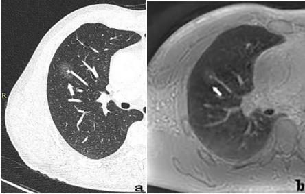
Fig1. A 65-year-old man with 24.1mm PSN(arrow)in the right middle lobe. The nodule is demonstrated on CT image(a) and PETRA sequence(b).

Fig2. A 50-year-old woman with 8.5mm GGN(arrow)in the right upper lobe. The nodule is demonstrated on CT image(a) and PETRA sequence(b).
DOI: https://doi.org/10.58530/2023/5120