5119
Multi-Echo FLORET UTE MRI1Cincinnati Children’s Hospital Medical Center, Cincinnati, OH, United States, 2Philips Healthcare, Rochester, MN, United States, 3Mayo Clinic, Rochester, MN, United States, 4University of Cincinnati, Cincinnati, OH, United States, 5University of Cincinnati Medical Center, Cincinnati, OH, United States
Synopsis
Keywords: Data Acquisition, Data Acquisition
FLORET UTE MRI has been used for many applications including lung, brain, musculoskeletal, and multinuclear imaging. The appeal of the pulse sequence comes from its high SNR and sampling efficiencies. With recent improvements minimizing artifacts, high-quality FLORET images are obtained routinely. However, acquisition of multiple echoes has not been investigated. Here, we determined the feasibility and optimal pulse-sequence design of multi-echo FLORET. Collecting echoes independently, but interleaved, produces the highest quality images, especially for ≥3 echoes. Furthermore, additional echoes provide improvements to image quality and add clinically useful contrast like T2*.
Introduction
Ultrashort echo time (UTE) MRI with multiple echoes improves image quality and provides additional information, especially for short T2* species[1]. Qualitatively, different echo times (TE) have different tissue contrast that can be harnesses to generate quantitative maps of T2*, magnetic susceptibility, B0, fat-fraction, and proton density. Rather than acquiring each TE separately, most multi-echo implementations acquire subsequent echoes from a single excitation after rewinding to the center of k-space[2]. However, this limits the minimum ∆TE to the length of the encoding and rewinder and makes it subject to progressive trajectory errors that accumulate across echoes.Fermat looped, orthogonally encoded trajectories (FLORET) is an efficient 3D UTE spiral implementation which has shown benefits for musculoskeletal, lung, and multi-nuclear imaging[3-8]. However, acquisition of valuable multi-echo images has yet to be demonstrated for FLORET. In this study, we implemented and investigated multi-echo imaging with FLORET.
Methods
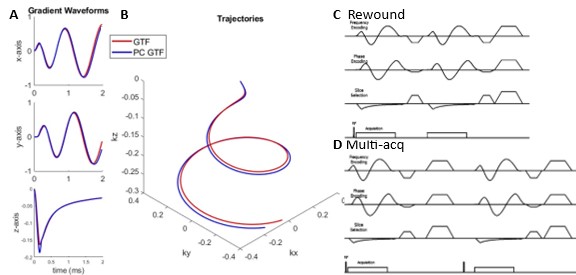
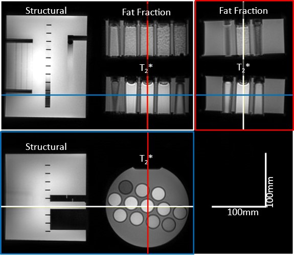
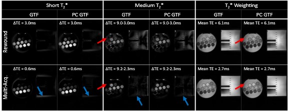
FLORET trajectories were designed as previously described[3, 7, 9]. Trajectory calculations included a gradient transform function (GTF) inspired gradient frequency limitation (1kHz), an optional pre-compensation (PC)(Figure 1A,B). RF-spoiled acquisitions were collected in two different ways for 5 echoes: 1) collected following rewinding back to the center of k-space (rewound, Figure 1C) or 2) collected as a separate interleave (multi-acquisition, Figure 1D). MRI was acquired for structural and quantitative phantoms (Figure 2). All images were fully sampled with the following parameters: Philips Ingenia Elition, 3.0T; FOV = 350mm isotropic; resolution = 2mm isotropic; readout = 2.0ms; FLORET hubs = 3; FLORET alpha = 36°; FLORET interleaves: 34; max gradient strength = 65mT/m; max slew = 220T/m/s, with TE and TR dependent on method. For rewound echoes, TEs(shortest) = 0.10, 3.1, 6.1, 9.1, and 12.1ms, and TR = 16ms; for multi-acquisition echoes, TE = 0.10, 0.58, 1.15, 2.3, and 9.2ms and TR = 13ms. Scan times ranged from 2 minutes to 9 minutes. A high-resolution, single-echo image was collected for reference with the same parameters except for 1mm isotropic voxels, TE = 0.10ms, TR = 5.4ms, no PC, and a scan duration of 6 minutes (Figure 2).Images were reconstructed using the Philips Recon2.0 platform as previously described[7]. Image post-processing, parameters maps, and image combinations were performed in MATLAB2021b.
Results
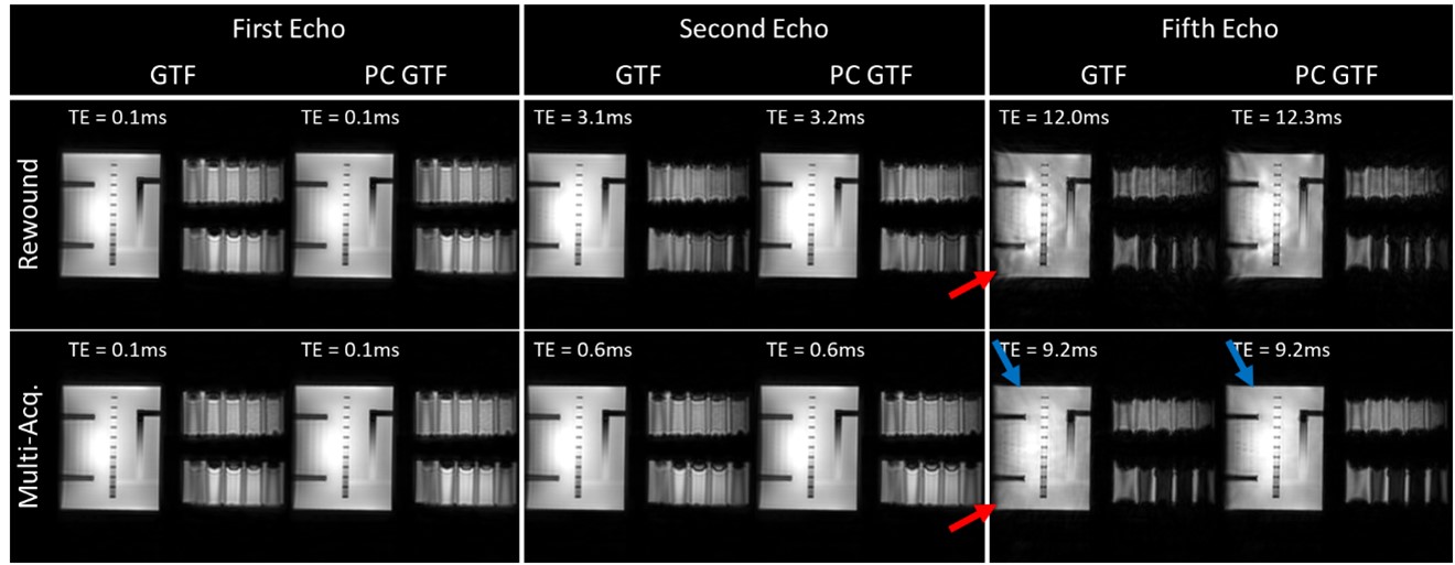
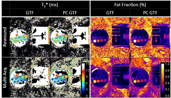
Both echo-acquisition methods, and gradient-waveforms calculations produced high-quality UTE images (Figure 3). For the second echo, the rewound method shows more signal decay in the quantitative phantoms, which is expected with the longer TE. Without PC, the 5th-echo images contained artifacts for both echo acquisition methods, with the artifacts being more intense with the rewound method. Adding PC to the gradient-waveform calculation slightly improved the image quality for the multi-acquisition method but did not provide any clear benefit for the rewound method. As expected, the acquisitions with images of higher quality produced higher-quality parameter maps (Figure 4) and image combinations (Figure 5), e.g., in the T2* maps in Figure 4, the rewound method acquisitions have artifacts and poor differentiation of the phantom’s vials. Quantitatively, the rewound method reduced the average percent error in T2* by 200%. For fat-fraction, the average percent error increased 20% with the rewound method, while the standard deviation of the maps reduced by 60%. No noticeable changes were observed for gradient PC or for other measures of percent error and standard deviation.Discussion
The high-quality single-echo images are consistent with a past study showing that frequency-limiting of FLORET waveforms produced higher quality images[7]. For additional echoes, two likely factors lead to reduced image quality: trajectory imperfections and off-resonance artifacts. As with many UTE and spiral sequences, FLORET uses large gradient magnitudes and slew rates, which make the k-space trajectories prone to inaccuracies from gradient imperfections. These imperfections progressively accumulate over echoes, resulting in the artifacts using the rewound method. For longer TE, B0 imperfections and susceptibility produce artifacts as seen in both the multi-acquisition and rewound methods. Thus, the rewound method should be limited to only a few echoes.In practice, the multi-acquisition method is less time efficient. However, the concomitant higher image quality also enables greater control of TEs and does not suffer from the same potential bias due to diffusion-weighting as the rewound method[10, 11].
T2* maps, B0 maps, water-only images, fat-only images, and fat-fraction maps can all be obtained using multi-echo FLORET. Each of these can provide clinically relevant contrast or be harnessed to improve image quality (such as off-resonance corrections). For example, T2-weighted lung images can assess disease severity and differentiate infection from mucus plugs in cystic fibrosis and idiopathic pulmonary fibrosis[12-14]. In the brain, multi-echo UTE can quantify myelin, which plays an important role in inflammatory and neurodegenerative disorders[15, 16]. Lastly, for musculoskeletal applications, image subtraction provides excellent visualization of short-T2* components, including tendons and ligaments[1, 17]. Future work will investigate these capabilities in-vivo.
Conclusion
Multi-echo FLORET was successfully demonstrated using two methods for echo acquisition. Due to gradient imperfections, additional echoes should be acquired individually in an interleaved fashion—particularly for high echo numbers. Water-fat separation, T2* maps, and fat-fraction maps are also feasible using multi-echo FLORET. Overall, multi-echo FLORET provides additional information, which is beneficial for clinical assessment in many applications, efficiently and at high spatial resolution.Acknowledgements
The authors thank the following sources for research funding and support: NIH R01 HL131012, NIH R01 HL146689, NIH R01HL143011, NIH 2R01HL126771, NIH R00HL138255, and Philips Healthcare.
References
1. Weiger, M. and K.P. Pruessmann, Short-T2 MRI: Principles and recent advances. Prog Nucl Magn Reson Spectrosc, 2019. 114-115: p. 237-270.
2. Reeder, S.B., et al., Quantification of hepatic steatosis with MRI: The effects of accurate fat spectral modeling. Journal of Magnetic Resonance Imaging, 2009. 29(6): p. 1332-1339.
3. Pipe, J.G., et al., A new design and rationale for 3D orthogonally oversampled k-space trajectories. Magn Reson Med, 2011. 66(5): p. 1303-11.
4. Robison, R.K., A.G. Anderson, 3rd, and J.G. Pipe, Three-dimensional ultrashort echo-time imaging using a FLORET trajectory. Magn Reson Med, 2017. 78(3): p. 1038-1049.
5. Willmering, M.M., et al., Implementation of the FLORET UTE sequence for lung imaging. Magn Reson Med, 2019. 82(3): p. 1091-1100.
6. Willmering, M.M., et al., Improved pulmonary (129) Xe ventilation imaging via 3D-spiral UTE MRI. Magn Reson Med, 2020. 84(1): p. 312-320.
7. Krishnamoorthy, G., et al. High-quality Lung imaging with FLORET UTE and Fibonacci interleaved trajectory ordering. in ISMRM Annual Meeting. 2022. London: ISMRM.
8. Bdaiwi, A.S., et al., Diffusion weighted hyperpolarized 129Xe MRI of the lung with 2D and 3D (FLORET) spiral. Magnetic Resonance in Medicine, 2022: p. 1-15.
9. Pipe, J.G. and D.D. Borup, Generating spiral gradient waveforms with a compact frequency spectrum. Magn Reson Med, 2022. 87(2): p. 791-799.
10. Chaudhari, A.S., et al., Imaging and T2 relaxometry of short‐T2 connective tissues in the knee using ultrashort echo‐time double‐echo steady‐state (UTEDESS). Magnetic Resonance in Medicine, 2017. 78(6): p. 2136-2148.
11. Hargreaves, B., Rapid gradient‐echo imaging. Journal of Magnetic Resonance Imaging, 2012. 36(6): p. 1300-1313.
12. Benlala, I., et al., Quantification of MRI T2 Interstitial Lung Disease Signal‐Intensity Volume in Idiopathic Pulmonary Fibrosis: A Pilot Study. Journal of Magnetic Resonance Imaging, 2021. 53(5): p. 1500-1507.
13. Dournes, G., et al., The Clinical Use of Lung MRI in Cystic Fibrosis. Chest, 2021. 159(6): p. 2205-2217.
14. Benlala, I., et al., Quantification of MRI T2-weighted High Signal Volume in Cystic Fibrosis: A Pilot Study. Radiology, 2020. 294(1): p. 186-196.
15. Jang, H., et al., Improved volumetric myelin imaging in human brain using 3D dual echo inversion recovery-prepared UTE with complex echo subtraction. Magn Reson Med, 2020. 83(4): p. 1168-1177.
16. Shen, X., et al., Ultra-short T2 components imaging of the whole brain using 3D dual-echo UTE MRI with rosette k-space pattern. Magn Reson Med, 2022.
17. Jang, H., et al., Ultrashort echo time Cones double echo steady state (UTE‐Cones‐DESS) for rapid morphological imaging of short T2 tissues. Magnetic Resonance in Medicine, 2021. 86(2): p. 881-892.
Figures