5115
Ultrashort Echo Time Magnetization Transfer Imaging of Knee Cartilage After Long-Distance Running1Department of Radiology,Fifth Affiliated Hospital, Sun Yat-Sen University, Zhuhai, China, 2MR Research, GE Healthcare, Beijing, China, 3University of California, San Diego, Department of Radiology, San Diego, CA, United States
Synopsis
Keywords: Cartilage, Cartilage
To assess the detection of changes in the knee cartilage of amateur marathon runners before and after long-distance running. We recruited 23 amateur marathon runners prospectively. MRI scans using UTE-MT and UTE-T2* sequences. UTE-MTR and UTE-T2* were measured for knee cartilage. The UTE-MTR values in lateral tibial plateau, central medial femoral condyle, and medial tibial plateau showed a significant decrease at 2 days post-race compared to the other two time points (P < 0.05). But no significant UTE-T2* changes were found for any cartilage subregions. UTE-MTR is a promising method for the detection of dynamic changes in knee cartilage.INTRODUCTION
Long-distance running is related to injury of the knee cartilage, which can eventually develop into osteoarthritis. Non-invasive MR monitoring of early changes in the cartilage allows for timely intervention of osteoarthritis progression and can prolong the life of exercise. This study aims to assess the detection of changes in the knee cartilage of amateur marathon runners before and after long-distance running using a 3D ultrashort echo time MRI sequence with magnetization transfer preparation (UTE-MT).METHODS
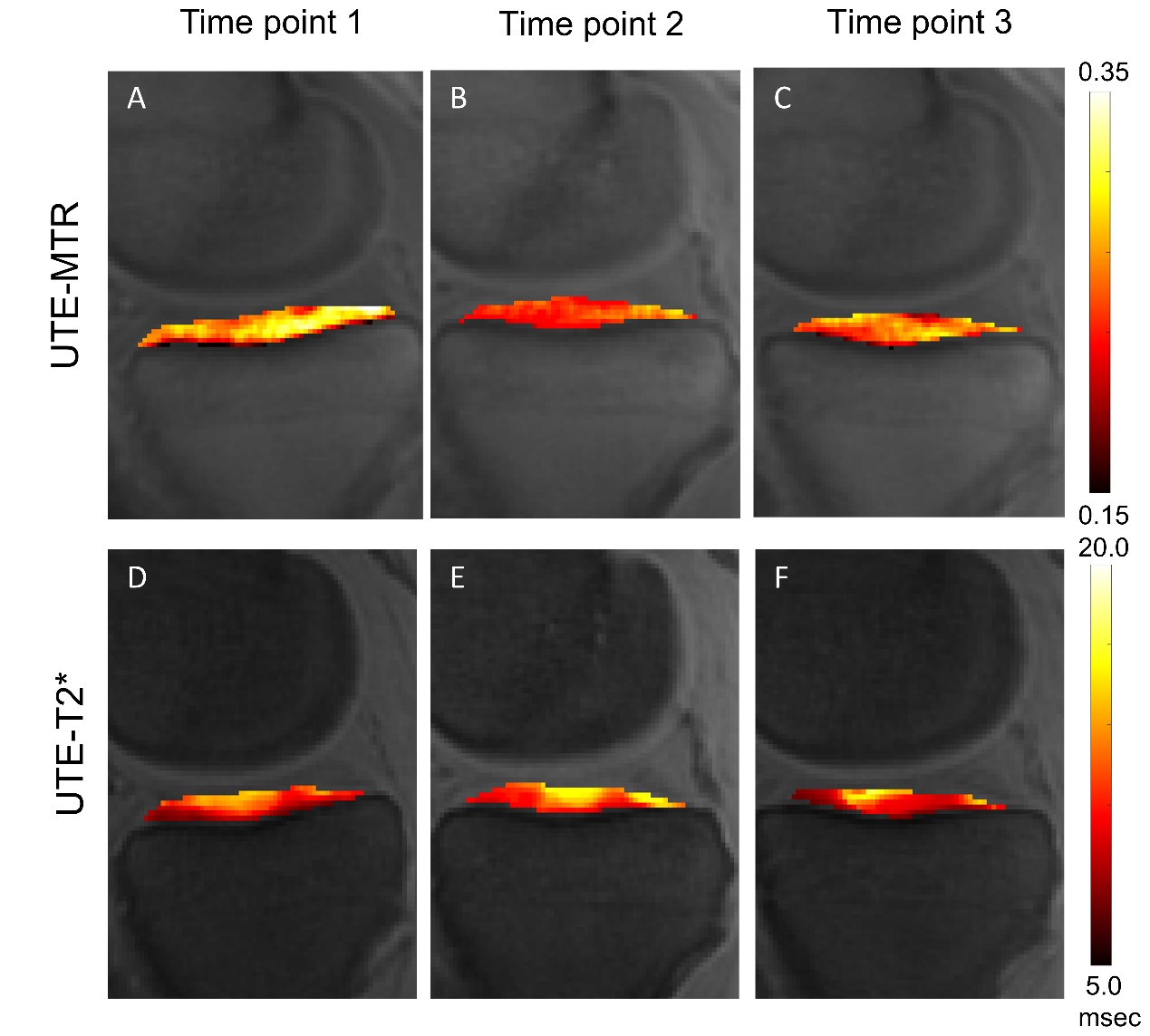
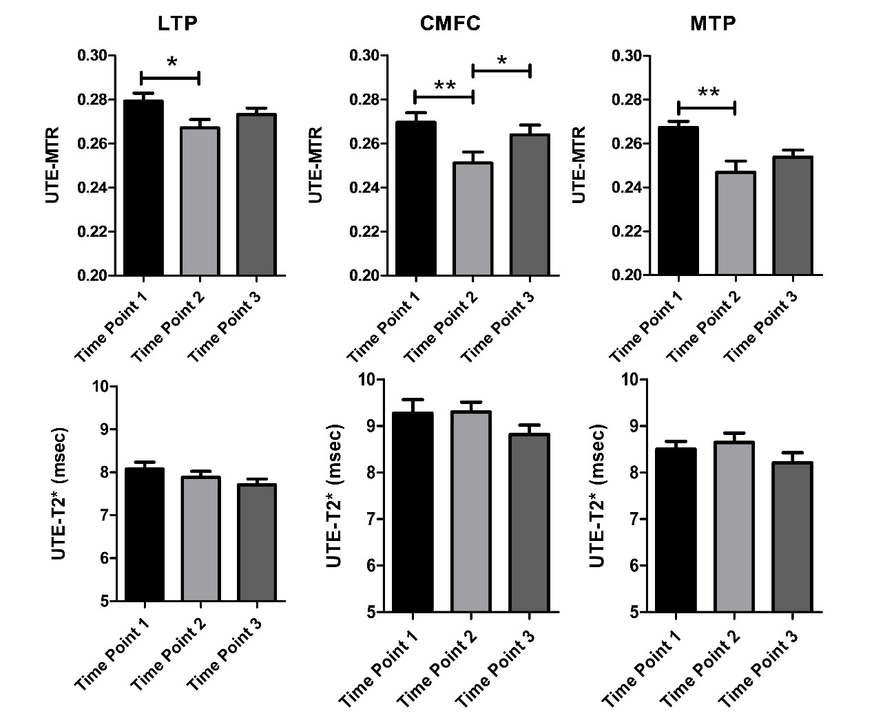
We recruited 23 amateur marathon runners (46 knees) in this prospective cohort study. MRI scans using ultrashort echo time magnetization transfer (UTE-MT) and UTE-T2* sequences were performed 3 times (pre-race, 2 days post-race, and 4 weeks post-race). UTE-MT ratio (UTE-MTR) and UTE-T2* were measured for knee cartilage (eight subregions) before and after running. The sequence reproducibility and inter-rater reliability were also investigated.RESULTS
Both the UTE-MTR and UTE-T2* measurements showed good reproducibility and inter-rater reliability. For most subregions of cartilage, the UTE-MTR values decreased 2 days post-race and increased after 4 weeks of rest. Conversely, the UTE-T2* values increased 2 days post-race and decreased after 4 weeks. The UTE-MTR values in lateral tibial plateau, central medial femoral condyle, and medial tibial plateau showed a significant decrease at 2 days post-race compared to the other two time points (P < 0.05). By comparison, no significant UTE-T2* changes were found for any cartilage subregions.DISCUSSION
The UTE-MT technique characterizes the tissue system with a two-pool model which includes the water and macromolecular proton pools1, 2. The magnetizations in these two pools are constantly being exchanged. Even though the UTE sequence is unable to detect the signal from macromolecular protons3, the UTE-MT technique provides indirect information about collagen content and integrity by utilizing the exchange effect. Li et al. using UTE-MT suggests that the MRI analysis (bound proton fraction and k) showed a statistically significant increase in correlated with the increase of glycosaminoglycan4. A recent study has found a strong correlation between UTE-MTR values and histological degeneration grades (i.e., Mankin scores, r = - 0.678, P < 0.001) for human knee cartilage samples5. This demonstrates that UTE-MT technique is also promising for the evaluation of tissue changes in knee osteoarthritis. To the best of our knowledge, this is the first UTE-MT study that evaluates changes in the knee cartilage after long-distance running.CONCLUSION
UTE-MTR is a promising method for the detection of dynamic changes in knee cartilage after long-distance running.Acknowledgements
This article is supported by the National Natural Science Found (No. 82101995); the National Natural Science Found(82172053).References
[1] Zhang, X., Y.J. Ma, Z. Wei, et al (2021) Macromolecular fraction (MMF) from 3D ultrashort echo time cones magnetization transfer (3D UTE-Cones-MT) imaging predicts meniscal degeneration and knee osteoarthritis. Osteoarthritis Cartilage 29(8):1173-1180.
[2] Ma, Y.J., E.Y. Chang, M. CarlJ. Du (2018) Quantitative magnetization transfer ultrashort echo time imaging using a time-efficient 3D multispoke Cones sequence. Magn Reson Med 79(2):692-700.
[3] Ma, Y.J., E.Y. Chang, G.M. BydderJ. Du (2016) Can ultrashort-TE (UTE) MRI sequences on a 3-T clinical scanner detect signal directly from collagen protons: freeze-dry and D2 O exchange studies of cortical bone and Achilles tendon specimens. NMR Biomed 29(7):912-7.
[4] Li, W., L. Hong, L. HuR.L. Magin (2010) Magnetization transfer imaging provides a quantitative measure of chondrogenic differentiation and tissue development. Tissue Eng Part C Methods 16(6):1407-15.
[5] Yang, J., H. Shao, Y. Ma, et al (2020) Quantitative ultrashort echo time magnetization transfer (UTE-MT) for diagnosis of early cartilage degeneration: comparison with UTE-T2* and T2 mapping. Quant Imaging Med Surg 10(1):171-183.
Figures