5106
Slice-selective Zero Echo Time imaging of ultra-short 𝑇2 tissues1Tesoro Imaging S.L., Valencia, Spain, 2Institute for Molecular Imaging and Instrumentation, Spanish National Research Council & Universitat Politècnica de València, Valencia, Spain
Synopsis
Keywords: New Trajectories & Spatial Encoding Methods, New Signal Preparation Schemes
Here we provide an MRI sequence which allows slice selection and 2D-imaging of hard tissues with T2 as short as 275 μs within clinically acceptable scan times even at fields as low as 260 mT. Our proposed sequence combines slice selection through spin-locking, which suffers a much more benign decay (T1ρ>>T2), and the fastest imaging sequence (ZTE), providing a new and robust tool for slice selection of the shortest-lived tissue signals in the body.
Introduction
This research is driven by the need to shorten acquisition times with Zero Echo Time (ZTE1) sequences, whose implicit 3D nature introduces high temporal cost as a main constraint. Our purpose is to expand the capabilities of ZTE with a slice selection method suitable for the shortest-lived tissues in the body. Here we present a new sequence that we have tested in ex vivo phantoms showing promising potential to be applied to in-vivo clinics.Methods
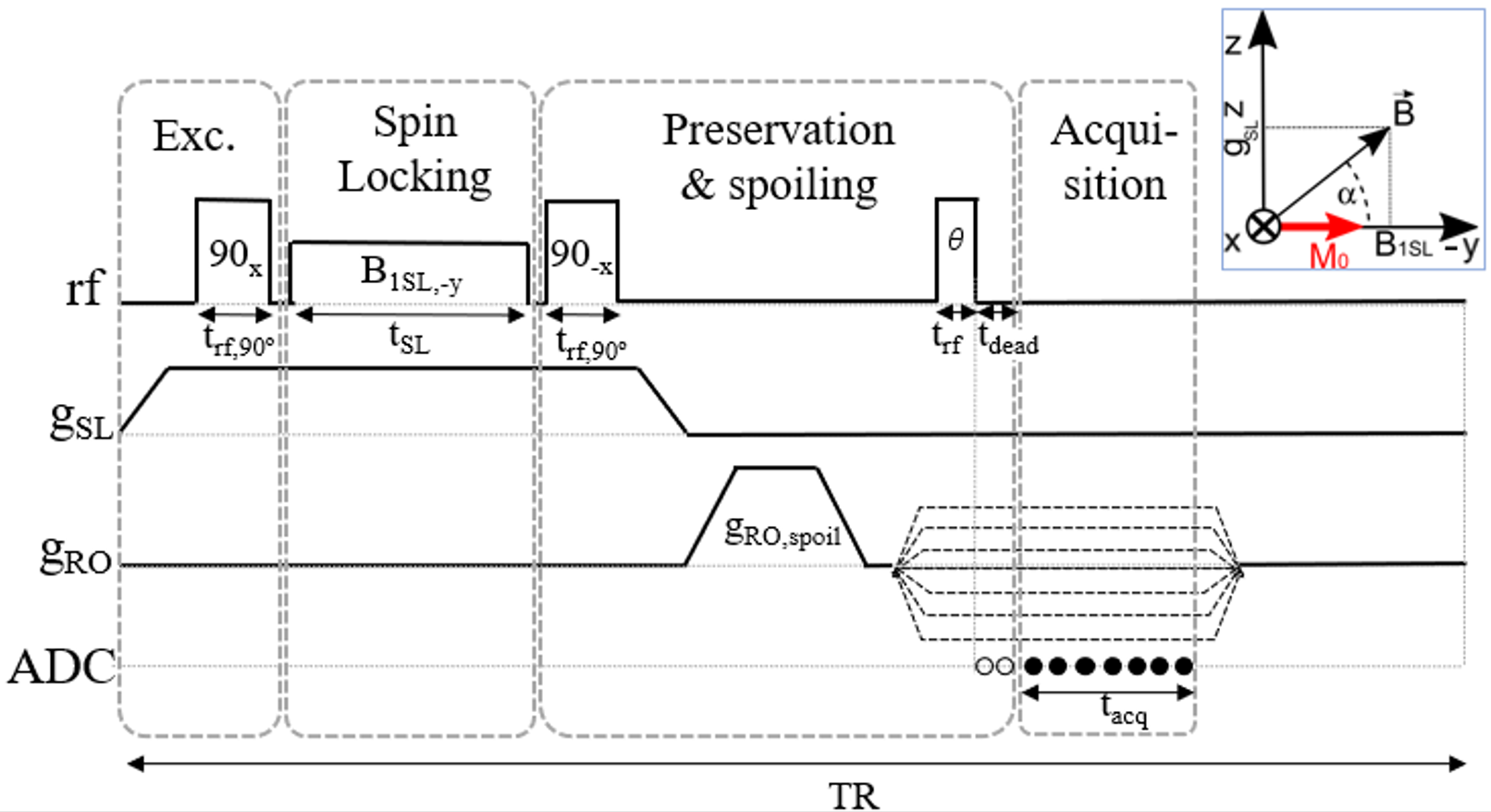
We present PreSLoP (Preserved Spin-Locked PETRA2, Fig.1), a 2D slice selective imaging sequence suitable for ultra-short 𝑇2 samples. Here, slice selection is performed by means of a spin-locking (SL) pulse of amplitude B1 in the presence of magnetic gradient of strength gSL. In-slice signal decay during SL occurs at a lower rate than conventional way (𝑇1𝜌 ≫ T2*), leaving a slice FWHM given by Δ=B1/2gSL. The SL time required for the slice to be selected can be roughly estimated as tSL=7π/2γB1, even though longer spin-locking durations can be used as tunable degree of contrast. After SL, a -90º pulse protects the in-slice magnetization from T2* decay while the slice-selection and imaging gradients are switched off/on respectively. Finally, a reexcitation pulse is applied and a FID is radially acquired in k-Space. Missing central k-Space points are then recovered in a point-wise fashion, as in PETRA. All experiments are performed in a 0.26 T scanner3.Results
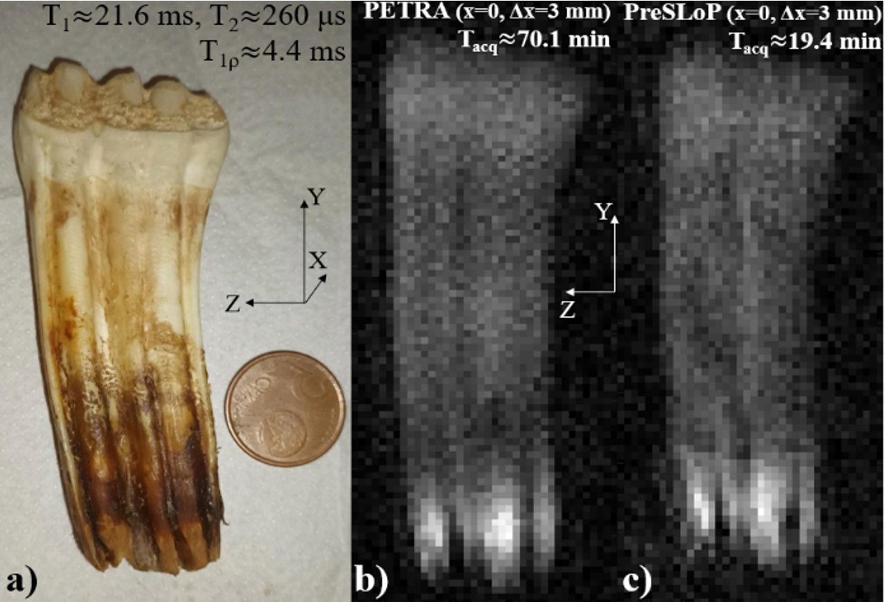
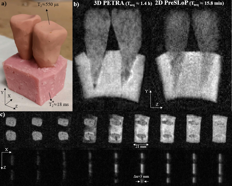
Fig.2 shows a slice of a horse tooth (left, 𝑇2* ≈ 260 us) acquired with 3D PETRA (middle) and the same slice encoded with PreSLoP (right). As compared to standard 3D ZTE sequences, PreSLoP achieves the same signal-to-noise ratio (SNR) in 3 times shorter scan times. This is due to the filling scheme of the finite gap at the center of 𝑘-space unavoidable with ZTE sequences.In Fig.3 we study the performance of PreSLoP for slice selection of an ultrashort T2* sample in the presence of softer matter (Fig.3a), containing two clay tooth molds (𝑇2* ≈ 550 µs) embedded in a piece of ham (𝑇2* ≈ 18 ms). In Fig.3b we compare the middle slice of 3D-PETRA (left) and PreSLoP (right) selecting same slice with equivalent thickness. Finally, in Fig.3c we assess the quality of the selected slice with PreSLoP (bottom row), using as reference the full phantom acquired with PETRA (top row).
Conclusions
The proposed sequence is capable of slice-selective imaging of ultra-short 𝑇2* biological tissues within clinically acceptable scan times even in our low-field MRI scanner. The nature of spin-locking enables not only image slices of isolated hard tissues, but rather any hard tissue surrounded by soft matter, whose contrast could be driven with spin locking pulse duration. For all that, these protocols may find application in clinical diagnosis of injuries in bones and bucodental exploration, among others.Acknowledgements
This work was supported by the Ministerio de Ciencia e Innovación of Spain through research grant PID2019-111436RB-C21. Action co-financed by the European Union through the Programa Operativo del Fondo Europeo de Desarrollo Regional (FEDER) of the Comunitat Valenciana (IDIFEDER/2018/022 and IDIFEDER/2021/004). JB acknowledges support from the Innodocto program of the Agencia Valenciana de la Innovación (INNTA3/2021/17).
References
1. Weiger M, Pruessmann K. Short-T2 MRI: Principles and recent advances. Prog Nucl Magn Reson Spectrosc. 2019 Oct-Dec.
2. Borreguero et al. Slice-selective Zero Echo Time imaging of ultra-short T2 tissues based on spin-locking. arXiv:2201.06305 [physics.med-ph]
3. Algarín et al. Simultaneous imaging of hard and soft biological tissues in a low-field dental MRI scanner. Scientific Reports 10, 21470 (2020)
Figures