5089
Repeatability, Distortion and SNR comparison of diffusion-weighted imaging techniques on an MR-Linac
Prashant Prabhakaran Nair1, Robin J.M. Navest2, Rosie Goodburn1, Bastien Lecoeur1, Uwe Oelfke1, and Andreas Wetscherek1
1Radiotherapy and Imaging, The Institute of Cancer Research, London, United Kingdom, 2Netherlands Cancer Institute, Amsterdam, Netherlands
1Radiotherapy and Imaging, The Institute of Cancer Research, London, United Kingdom, 2Netherlands Cancer Institute, Amsterdam, Netherlands
Synopsis
Keywords: Quantitative Imaging, Precision & Accuracy, MR-Linac
DW-acquisitions with high ADC accuracy, precision and low geometric distortion are necessary for using ADC maps for treatment planning and response assessment in MR-guided radiotherapy on MR-Linacs. We measured SNR, ADC bias, repeatability coefficient and marker distances on the NIST phantom for DW-EPI, DW-TSE and SPLICE protocols. Two volunteers were scanned in the head and neck region. The investigated TSE-based protocols showed ADC bias and DW-EPI demonstrated better repeatability. Spectral fat-suppression techniques led to higher SNR than using STIR. SPLICE DWI could be an alternative to diffusion EPI with reduced distortion and comparable SNR, if low-high profile order is used.Introduction
The apparent diffusion coefficient (ADC) is a potential quantitative biomarker for assessing response to radiotherapy and for aiding dynamic treatment planning1. Diffusion-weighted (DW) imaging acquired with an echo-planar imaging (EPI) readout suffers from geometric distortion related to field inhomogeneity and eddy currents, limiting its utility for radiotherapy treatment planning. We hypothesise that turbo spin-echo (TSE) sequences with phase error correction could reduce distortions and yield more accurate ADC measurements on an MR-Linac. We compare ADC accuracy (bias), precision (repeatability coefficient – RC), and geometric distortions between DW imaging acquired with the standard EPI readout and with various TSE-based techniques: Alsop’s method2 (TSE), SPLICE3 with linear profile order (SPLICE) and SPLICE with low-high profile order (SPLICE_LH). An additional objective is to compare spectral fat suppression techniques with short TI inversion recovery (STIR) in terms of signal-to-noise ratio (SNR).Methods
We scanned a diffusion phantom4 (Qalibre MD Inc., Boulder, CO) filled with ice-water with a DW-EPI consensus protocol5 and with two DW-TSE sequences, SPLICE and TSE on a 1.5T Unity MR-Linac (Elekta AB, Stockholm). Phantom temperatures were measured before and after the experiment. ADC maps were calculated online using b-values 0, 150, 500 s/mm2.ROIs were drawn manually, and mean ADCs were calculated using MATLAB R2022a (The MathWorks, Natick, MA) and compared (paired t-tests). ADC biases for the individual vials were evaluated. RC were calculated from two repetitions following literature6. SNR was measured on b-500 images as the coefficient of variation (CV) within the central vial across five different slices. The CVs were then averaged. Geometric distortion was assessed on b-500 images by measuring distances between phantom markers A, B and C as shown in Fig.1. Measurements were compared against 3D T2 images (2 sided paired t-test). A p-value of 0.05 was considered statistically significant.
Two volunteers were scanned in the head & neck region with different combinations of readouts and fat suppression techniques: EPI-STIR, TSE-STIR, SPLICE-STIR, EPI-SPAIR and EPI-SPIR. CVs were calculated over ROIs at selected slices as a surrogate for SNR. A repeated scan was performed for the volunteers, which included a SPLICE_LH acquisition. The scan parameters for the volunteer sequences are provided in Fig. 2.
Results
The mean ADC values of DW-EPI and DW-TSE were not statistically different from the reference values (Fig. 3a). For the EPI sequence, average distances (in cm) measured for AB (11.95) were not significantly different, but BC (5.74) and AC (10.15) were significantly different from 3D T2w (12.00, 5.94, 10.4, p-values < 0.01). The average distances measured on SPLICE (11.92, 5.97, 10.34) and TSE (11.9, 5.95, 10.33) images were not significantly different from each other, but mean AC distances differed from the 3D T2 sequence (p = 0.01 and 0.02, respectively) (Fig. 3b,c,d). The average distances measured on the SPLICE-LH (11.92, 5.94, 10.4) were not significantly different from the T2 image (Fig 3b,c,d). The ADC biases are displayed in Fig. 4. The EPI sequence had a better RC of 0.044 (10-3mm2/s) compared to the SPLICE - 0.067 (10-3mm2/s) and TSE – 0.075 (10-3mm2/s) (Fig. 4d). The mean SNRs of EPI (CV: 3.37) and SPLICE (CV: 3.28) were not significantly different; however, both had higher SNR than TSE (CV: 5.28). The SPLICE-LH acquisition (CV: 2.82) had significantly higher SNR compared to the other acquisitions (Fig 4d). The [pre, post] phantom temperatures (°C) for the repeated measurements were [0.38,0.52] and [0.24,0.62], respectively.For the volunteer scans, the DW TSE-STIR images (Fig. 5a) had significantly lower ADC compared to the DW EPI-STIR image (Fig. 5b). DW EPI SPAIR and SPIR had better SNR than the STIR scan (Fig. 5c). Further, DW EPI-STIR and the SPLICE_LH images (Fig. 5d) had comparable SNR (Fig 5e).
Discussion
For use of DW MRI for radiotherapy treatment adaptation in MR-guided radiotherapy, not only accurate ADC measurements, but also geometric accuracy is required. As hypothesized, geometric distortion was reduced in DW-TSE sequences with phase error-correction. However, ADC values measured with the SPLICE and TSE were less precise compared to the EPI sequence. The RC measures indicate that there is 95% confidence when there is an increase/ decrease in ADC > 0.044 (10-3 mm2/s) that the effect is of a physiological origin for the EPI sequence, whereas for the SPLICE acquisition this confidence is achieved only when the ADC changes > 0.067 (10-3 mm2/s). SNR was higher for SPLICE_LH compared to SPLICE linear and TSE for both volunteer and phantom images. There is some blurring of the thinner structures with SPLICE_LH (Fig. 5) which needs further study. TSE gave a lower ADC accuracy on the NIST phantom (Fig. 3), while that of the EPI, SPLICE_LH and SPLICE were comparable.Conclusion
Using TSE-based DW imaging techniques on an MR-Linac reduces geometric distortion compared to DWI, but overall we found higher variation in ADC values measured with TSE-based techniques. EPI SPAIR and SPIR fat suppression provided better SNR than STIR. Of the investigated TSE-based sequences only SPLICE with low-high profile order could match the ADC accuracy and SNR of EPI, but fine structures appeared blurred. Future work will establish repeatability for SPLICE_LH to help adopt the ADC as a reliable measure for treatment response.Acknowledgements
This work was supported by Cancer Research UK programme grant (C33589/A28284)
The Institute of Cancer Research and The Royal Marsden NHS Foundation Trust are members of the Elekta MR-Linac Research Consortium.
We acknowledge research support from Elekta and Dave Higgins (Philips MR) for providing the SPLICE diffusion sequence as a research tool.
We acknowledge Bjoern Eiben and Joan Chick for discussions.
References
1. van Houdt PJ, Saeed H, et al. Integration of quantitative imaging biomarkers in clinical trials for MR-guided radiotherapy: Conceptual guidance for multicentre studies from the MR-Linac Consortium Imaging Biomarker Working Group. Eur J Cancer 2021;153:64–71. https://doi.org/10.1016/j.ejca.2021.04.041.2. Alsop DC. Phase insensitive preparation of single-shot RARE: application to diffusion imaging in humans. Magn Reson Med 1997;38:527– 533.
3. Schick F. SPLICE: Sub-second diffusion-sensitive MR imaging using a modified fast spin-echo acquisition mode. Magn Reson Med 1997;38:638–44.
4. Keenan KE, Ainslie M, et al. Quantitative magnetic resonance imaging phantoms: A review and the need for a system phantom. Magn Reson Med 20018;79(1):48-61.
5. Kooreman ES, van Houdt PJ, Nowee ME, van Pelt VWJ, Tijssen RHN, Paulson ES, et al. Feasibility and accuracy of quantitative imaging on a 1.5 T MR-linear accelerator. Radiother Oncol 2019;133:156–62. https://doi.org/10.1016/j.radonc.2019.01.011.
6. Shukla-Dave A, Obuchowski NA, Chenevert TL, Jambawalikar S, Schwartz LH, Malyarenko D, et al. Quantitative imaging biomarkers alliance (QIBA) recommendations for improved precision of DWI and DCE-MRI derived biomarkers in multicenter oncology trials. J Magn Reson Imaging 2019;49:e101–21. https://doi.org/10.1002/jmri.26518.
Figures
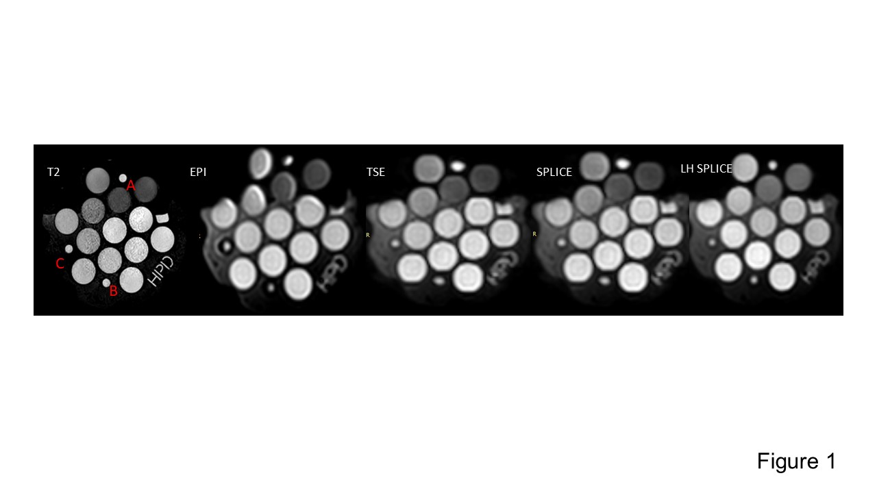
Snapshot featuring an example slice from the 3D T2, EPI, TSE, SPLICE and SPLICE_LH images of the NIST phantom. Markers for measuring geometric distortion in the phantom are indicated by red letters.
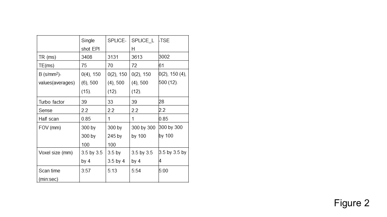
Acquisition parameters for the volunteer scans.
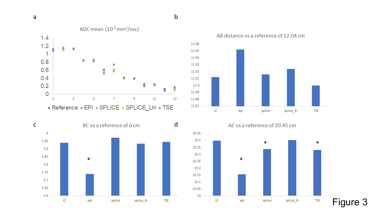
ADC mean measurements (a) and distances between markers (b,c,d) for the NIST phantom. ‘*’ indicates significant difference from the values of the T2 sequence. The differences from the reference values indicate the severity of the distortion.
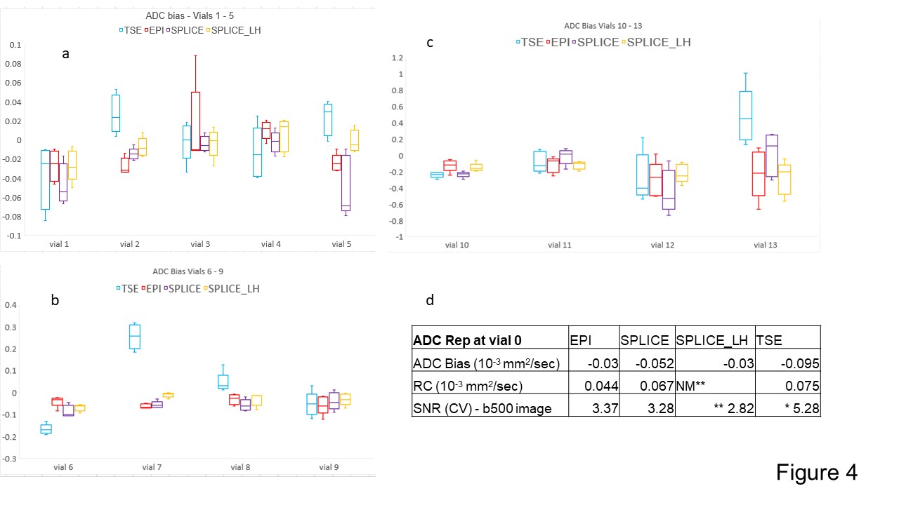
Box plots of ADC bias measured from the various vials for the sequences (a,b,c). (d) shows the ADC bias and the repeatability coefficient (RC) for the central vial. The SPLICE_LH was only acquired once, hence no repeatability measures. ‘**’ indicates that the SNR of the SPLICE_LH was significantly higher than the EPI and SPLICE. ‘*’ at the TSE indicates that the value was significantly lower than the others.
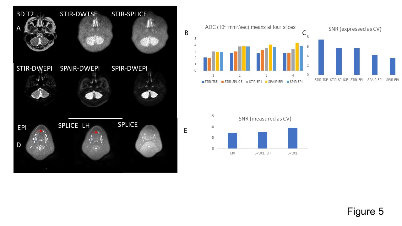
(a) Head and neck scan from a non-patient volunteer. SPIR/SPAIR fat suppression schemes had higher SNR compared to STIR fat suppression. SPLICE/TSE sequences had comparatively lower SNR than EPI-based acquisitions. (b) ADC and (c) SNR measures for (a). (d) SPLICE_LH and EPI had comparable SNRs. Minor blurring of thin structures was observed for SPLICE_LH (red arrow). The SPLICE sequence had lower SNR noticeable g-factor noise. ROIs are blue markers. (e) SNR comparison for (d).
DOI: https://doi.org/10.58530/2023/5089