5086
Hydrogel-based quantitative multi-parametric MRI phantom with controlled T2, diffusion and kurtosis
Scott D. Swanson1, Ted Lynch2, Shigeto Ono2, and Dariya I. Malyarenko1
1Department of Radiology, University of Michigan, Ann Arbor, MI, United States, 2CIRS/Mirion Inc., Norfolk, VA, United States
1Department of Radiology, University of Michigan, Ann Arbor, MI, United States, 2CIRS/Mirion Inc., Norfolk, VA, United States
Synopsis
Keywords: Quantitative Imaging, Phantoms, Diffusion, Kurtosis, Transverse Relaxation
A set of materials with tunable T2, diffusion, and kurtosis were assembled to create quantitative biomimetic MRI phantoms. T2 is controlled with variable agarose concentration, mono-exponential diffusion by polyvinylpyrrolidone, and kurtosis by addition of lamellar vesicles. The phantoms are mechanically stabilized by polyacrylamide gels to allow biomimetic morphologies. These nanostructured systems provide an ideal platform for moldable multiparametric MRI phantoms that are useful for pulse sequence design and protocol standardization for multi-site multi-vendor imaging trials, as well as for refinement of emerging AI analysis methods.Introduction
The ideal biomimetic MR phantom would faithfully reproduce all MR parametric and spatial features of tissue. In reality, MR phantoms typically mimic selective tissue properties such as T1 and T2 (1), or diffusion (1,2), or spatial structures (3). The goal of this work is to create multiparametric phantoms with controllable T2, diffusion and kurtosis properties achieved in the same spatial compartment that can be fabricated into tissue-like 3D structures. To accomplish this, we use a combination of polyvinylpyrrolidone (PVP), agarose, lamellar vesicles and polyacrylamide gels (PAG). This work represents a first step towards a quantitative, multiparametric MR phantom which can be molded into biomimetic shapes.Materials and Methods
Multiparametric phantom materials: Phantoms with monoexponential diffusion decay were fabricated by adding agarose and PVP to water in specific concentrations. Multiexponential diffusion decay was created by addition of lamellar vesicles composed of Cetearyl alcohol, Stearylamidoproply dimethylamine and Behentrimonium chloride (4,5), abbreviated "CSB". An acrylamide/bis-acrylamide mixture, 5 or 10%, was added and cross-linked to create mechanical stability. Multi-layered phantoms were assembled in 50 ml centrifuge tubes from select materials mimicking relaxation and diffusion parameters of normal and cancerous prostate tissue (6-8), placed in water in a 1 L Nalgene jar, and studied over a month period to test for compartmental stability of relaxation and diffusion parameters.Phantom DWI and T2 measurements: Diffusion weighted images (DWI) were acquired on a 3T MRI scanner using b-values of 0, 100, 200, 500, 800,1500, 2000, 2500 s/mm2, TR/TE (5000/82 ms), and 2.4x2.4x4 mm3 voxels. The transverse relaxation, T2, measurements were performed using spin-echo sequence with 8 echo-times (TE) from 15 to 155 ms for 1.5x1.5x3 mm3 voxels. T2 and ADC (b=0, 1500 s/mm2) maps, were generated on the scanner. Apparent diffusion, Da, and kurtosis, Ka, parameters for isotropic diffusion kurtosis (DK) were estimated from linear least squares fits of voxel log-signal dependence on DWI b-value, according to the kurtosis model: -Dab + Ka(Dab)2/6. The maximum b-value constraint bmax < 3/DaKa was implemented for DK model by iterative fitting, using b >100 s/mm2 (2). Data analysis utilized lscov function and the Imaging Processing toolbox from MATLAB R2019b (Mathworks, Natick MA). The quantitative parameter values were measured by manually placing 15mm diameter ROI on the middle slice of material vials.
Results and Discussion
For agarose-PVP materials, PVP concentration determines ADC, while agarose concentration determines T2, nearly independently (Figure 1a,b). A sample of 0.5% agarose and 15% PVP generates ADC and T2 values similar to those of normal prostate peripheral zone (nPZ, fast diffusion and long T2)(4). Gleason Score 7 (GS7) parameters, with lower ADC and shorter T2 values (6), are well mimicked with 1% agarose and 30% PVP. Use of agarose for T2 control (vs. solutions of paramagnetic ions) provides stable, compartmental T2 in multi-layered phantom (Figure 1c,d). The agarose-PVP materials supplied Gaussian diffusion media with Ka~0 (map not shown).In 5% PAG hydrogels with vesicle concentrations between 0.2-0.8 (w/w)% in water, transverse relaxation is largely controlled by agarose concentration (Figure 2a), achieving tissue-like T2 values (6,7) between 260ms and 65 ms. At the higher vesicle concentrations, > 0.4 (w/w)%, kurtosis increases (0.7-1.2) and diffusion decreases (1.4 - 1.0 ) μm2/ms for agarose concentrations studied here (0.3% - 2.0%, Figure 2b). Increasing agarose at fixed vesicle concentration leads to a minor decrease in Da but notable increase in Ka (Figure 2b, dashed). Ka also increases with PAG addition, compared to vesicle-free solutions (4,5). 10% PAG slightly decreased Da and preserved T2 compared to 5% PAG. Uniform cross-linking of PAG in samples with large volumes and higher PAG concentrations was challenging, leading to variable changes in T2 properties. 5% PAG was sufficient to provide mechanical structure to the castings. The measured relaxation and diffusion parameters in multi-layered phantom (Figure 2c-e) compartments were stable over one month and provided close mimics for normal and cancerous prostate tissue (5,7,8).
Conclusion
The developed materials allow control over quantitative relaxation, diffusion and kurtosis parameters in a single spatial compartment for construction of multi-compartment multi-parametric MRI phantom. Hydrogel improves stability of vesicle-based tissue-mimics and allows shaping into biomimetic morphologies.Acknowledgements
Funding support from National Institutes of Health Grants: 75N91021C00036, U01CA211205, R01CA190299, and University of Michigan Basic Radiologic Sciences pilot project fund.References
- Keenan KE, Ainslie M, Barker AJ, et al. Quantitative magnetic resonance imaging phantoms: a review and the need for a system phantom. Magn Reson Med. 2018; 79(1): 48- 61
- Malyarenko DI, Swanson SD, Konar AS, LoCastro E, Paudyal R, Liu MZ, Jambawalikar SR, Schwartz LH, Shukla-Dave A, Chenevert TL. Multicenter Repeatability Study of a Novel Quantitative Diffusion Kurtosis Imaging Phantom. Tomography. 2019;5:36-43
- Rai R, Holloway LC, Brink C, Field M, Christiansen RL, Sun Y, Barton MB, Liney GP. Multicenter evaluation of MRI-based radiomic features: A phantom study. Med Phys. 2020 Jul;47(7):3054-3063
- Malyarenko DI, Chenevert TL, Ono S, Lynch T, Swanson SD: Temperature and Concentration Dependence of Diffusion Kurtosis Parameters in a Quantitative Phantom, ISMRM 30th annual conference, London, UK, DP 2429, 2022
- Swanson SD , Chenevert TL , Shankar PR , Lynch T, and Malyarenko DI: Biomiimetic phantoms of impeded diffusion in prostate cancer using lipid nanoparticles, International Society for Magnetic Resonance in Medicine 29th Annual Conference, virtual due to COVID, Proceedings OP 697, 2021.
- Simpkin CJ, Morgan VA, Giles SL, Riches SF, Parker C, deSouza NM. Relationship between T2 relaxation and apparent diffusion coefficient in malignant and non-malignant prostate regions and the effect of peripheral zone fractional volume. Br J Radiol. 2013; 86(1024):20120469.
- Chatterjee A, Bourne RM, Wang S, Devaraj A, Gallan AJ, Antic T, Karczmar GS, Oto A. Diagnosis of Prostate Cancer with Noninvasive Estimation of Prostate Tissue Composition by Using Hybrid Multidimensional MR Imaging: A Feasibility Study. Radiology. 2018.
- Hectors SJ, Semaan S, Song C, Lewis S, et al. Advanced Diffusion-weighted Imaging Modeling for Prostate Cancer Characterization: Correlation with Quantitative Histopathologic Tumor Tissue Composition-A Hypothesis-generating Study. Radiology 2018;286(3):918-28.
Figures
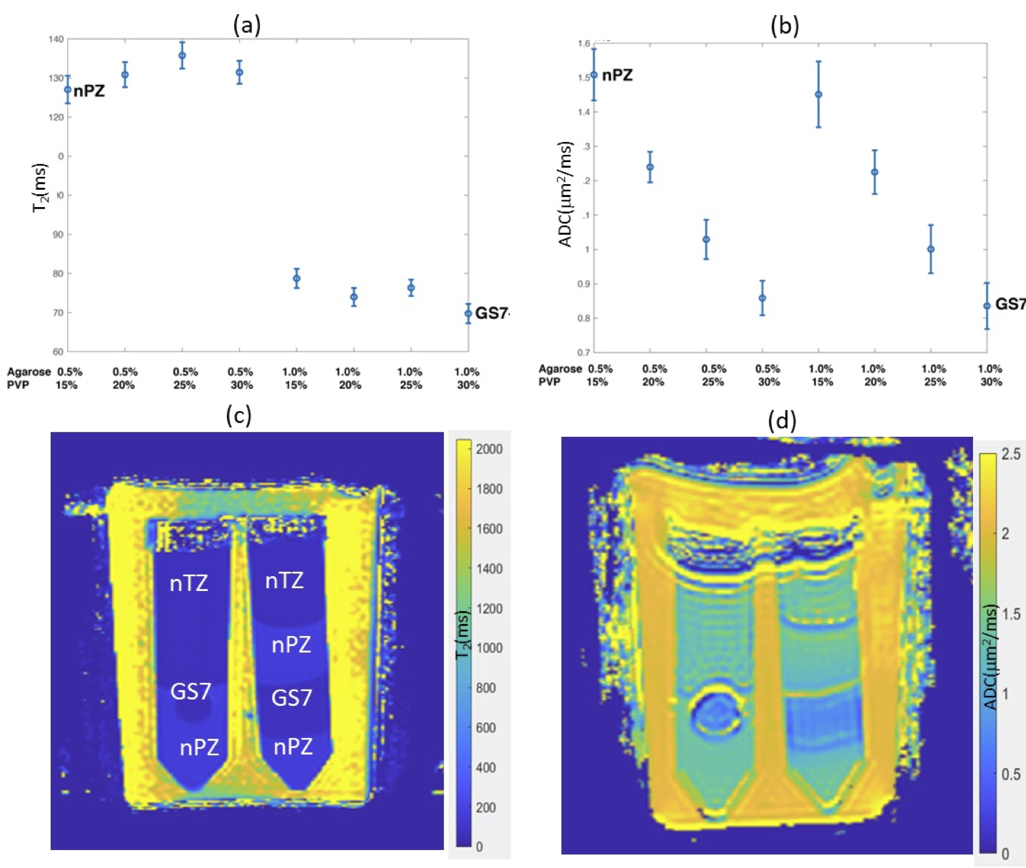
Figure 1: Measured T2 (a) and ADC (b) values of agarose-PVP materials as a function of component concentrations (error bars are 95% CI). T2 (c) and ADC (d) maps for layered agarose-PVP phantoms. Prostate tissue compartments with close quantitative parameters are labeled on T2 map: “nTZ”- normal transition zone; “nPZ” – normal peripheral zone; “GS7” – Gleason score 7.
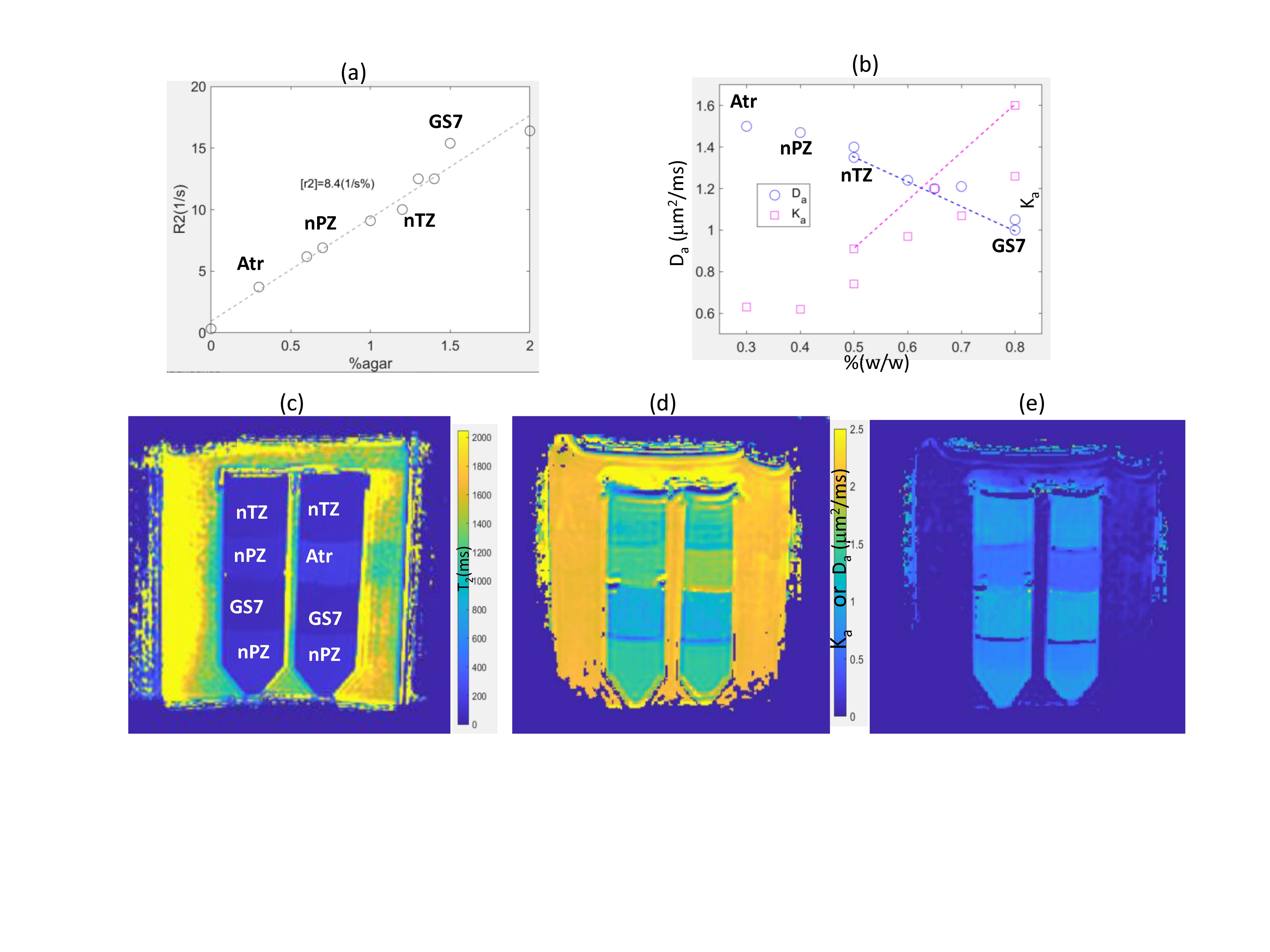
Figure 2: Results for agarose/acrylamide composite hydrogels at 3T. Top plots show R2 (1/T2) (95%CI: ±0.2s-1) and DK (95%CI [Da, Ka]: ± [5,10]%) parameter dependence on agarose (a) and vesicles (b) concentrations. All samples contained 5% PAG. The dashed lines on Da (μm2/ms), Ka plots connect values with higher agar concentration. Bottom maps show quantitative T2, Da and Ka parametric maps for the layered phantoms made of materials closely mimicking prostate tissue properties (labeled on T2 maps: “nTZ”,”nPZ”,”GS7” and “Atr” - atrophy).
DOI: https://doi.org/10.58530/2023/5086