5058
Myelination differences between cerebrum and cerebellum at early-stage schizophrenia detected by macromolecular proton fraction MRI.
Ekaterina Krupina1, Andrei Valerievich Manzhurtsev2,3,4, Maxim Vadimovich Ublinskiy2,3, Olga Vasilievna Bozhko3, Galina Mamedova5, Vadim Ushakov1,5,6, Natalia Zakharova5, Vasiliy Leonidovich Yarnykh7,8, Denis Andreyuk5, Maxim Borisovich Shlyapnikov5, Georgiy Kostiuk5, and Tolib Abdullaevich Akhadov3
1National Research Nuclear University MEPhI, Moscow, Russian Federation, 2Emanuel Institute of Biochemical Physics of the Russian Academy of Sciences, Moscow, Russian Federation, 3Clinical and Research Institute of Emergency Pediatric Surgery and Trauma, Moscow, Russian Federation, 4Moscow State University, Moscow, Russian Federation, 5Psychiatric Clinical Hospital 1 named N.A. Alekseev., Moscow, Russian Federation, 6Institute for Advanced Brain Studies, Moscow State University, Moscow, Russian Federation, 7Radiology, University of Washington, Seattle, WA, United States, 8Laboratory of Neurobiology, Research Institute of Biology and Biophysics, Tomsk State University, Tomsk, Russian Federation
1National Research Nuclear University MEPhI, Moscow, Russian Federation, 2Emanuel Institute of Biochemical Physics of the Russian Academy of Sciences, Moscow, Russian Federation, 3Clinical and Research Institute of Emergency Pediatric Surgery and Trauma, Moscow, Russian Federation, 4Moscow State University, Moscow, Russian Federation, 5Psychiatric Clinical Hospital 1 named N.A. Alekseev., Moscow, Russian Federation, 6Institute for Advanced Brain Studies, Moscow State University, Moscow, Russian Federation, 7Radiology, University of Washington, Seattle, WA, United States, 8Laboratory of Neurobiology, Research Institute of Biology and Biophysics, Tomsk State University, Tomsk, Russian Federation
Synopsis
Keywords: Psychiatric Disorders, Nervous system, myelin, schizophrenia
The purpose of this study is to identify quantitative alterations of the myelin content using the macromolecular proton fraction (MPF) method. Forty-five subjects, 22 controls and 23 schizophrenia patients participated in the study. A significant decrease in myelination in schizophrenia was observed in the left and right cerebral cortex and in the left and right cerebral white matter. No myelination alterations in the entire cerebellum (not separated into regions) were detected. The differences found in the regional and global myelination at an early stage of schizophrenia may provide additional information for understanding the biological mechanisms of the disease.Introduction.
According to literature data, there is evidence that brain myelination is impaired in schizophrenia [1]. The purpose of this study is to identify quantitative alterations of the myelin content using the macromolecular proton fraction (MPF) method [2] in various brain structures in patients with schizophrenia at an early stage, as well as to evaluate the differences in myelination of these structures.Materials and methods
Forty-five subjects, 22 controls (10m+12f, 31.6±9.7 y.o.) and 23 schizophrenia patients (F20.0, 11m+12f, 31.5±5.1 y.o.) participated in the study. Philips Achieva dStream 3T MRI scanner, standard head coil were used. The magnetization transfer (TR=20 ms, TE=4.60 ms, FA=10°), T1-weighted (TR=20 ms, TE=4.60 ms, FA=20°) and PD-weighted (TR=20 ms, TE=4.60 ms, FA=4°) were acquired.The MPF maps were reconstructed using special software in C++ (available at https://www.macromolecularmri.org/). Non-brain structures were removed from the MPF card using the bet2 function in the MRIcro program. Further, using the FSL software, MPF maps were co-registered to the standard MNI152 1 mm atlas. The quantitative myelin values were determined as the average values over the regions of interest. These areas were the left and right cerebral cortex and cerebral white matter, and the entire cerebellum. Also, myelination of all cerebral cortex and cerebellum regions (defined using Harvard Oxford Cortical Atlas and the Cerebellar Atlas in MNI 152 FLIRT) was acquired. The normality of data distribution was assessed using the Shapiro-Wilk test for each group of subjects. Depending on the result, the Student's t-test or the Mann-Whitney criterion were used to search for the between-group differences.
Results
All data sets were normally distributed. A significant decrease in myelination in schizophrenia was observed in the left and right cerebral cortex (by 3%, p=0.03 and by 3.2%, p=0.02, respectively) and in the left and right cerebral white matter (by 3%, p=0.03 and by 3.3%, p=0.02, respectively). At the same time, no myelination alterations in the entire cerebellum (not separated into regions) were detected. A separate analysis of the cerebral regions of interest revealed a significant myelination decrease in schizophrenia in Superior Temporal Gyrus (anterior division), Heschl's Gyrus, Postcentral Gyrus, Lateral Occipital Cortex (superior division), Precuneous Cortex, Frontal Pole, Paracingulate Gyrus, Cerebellum left crus 2 and Cerebellum Paramedian Lobule. In the cerebellum, a significant decrease in myelination in schizophrenia was found only in Cerebellum left crus 2 and Cerebellum Paramedian Lobule, the remaining X zones were not significantly different from the norm. The results are shown in table 1.The regions with significantly different myelination between the schizophrenia and normal groups are shown in Fig. 1 and Fig. 2
Discussion
The decrease in cerebral myelination in general is consistent with the results of other studies on the measurement of myelin in schizophrenia. At the same time, when analyzing the cerebellum as a whole structure, we showed that its myelination in schizophrenia at an early stage is not impaired. Analysis of various cerebral regions demonstrated which areas are mostly characterized by a decrease in the amount of myelin. A number of these zones, in particular, the Frontal pole and the Paracingulate Gyrus, are involved in cognitive processes [3]. At the same time, areas with significantly lower myelin content in schizophrenia are also present in the cerebellum. The cognitive role of the cerebellum is critically tied to its distributed connections throughout the brain. The revealed differences may indirectly indicate impairment of these connections with other brain structures. To sum up, the differences found in the regional and global myelination at an early stage of schizophrenia may provide additional information for understanding the biological mechanisms of the development of this disease.Acknowledgements
Grant RSF 20-15-00299 (partially)References
1. doi: 10.5498/wjp.v12.i2.264
2. doi: 10.3389/fnins.2022.819912
3. doi:10.3389/fnsys.2014.00163
Figures
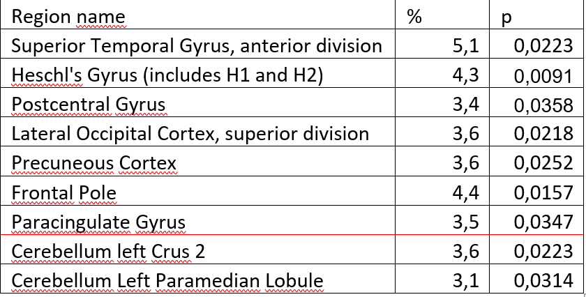
Table 1. Regions with statistically significant differences between the norm and schizophrenia.
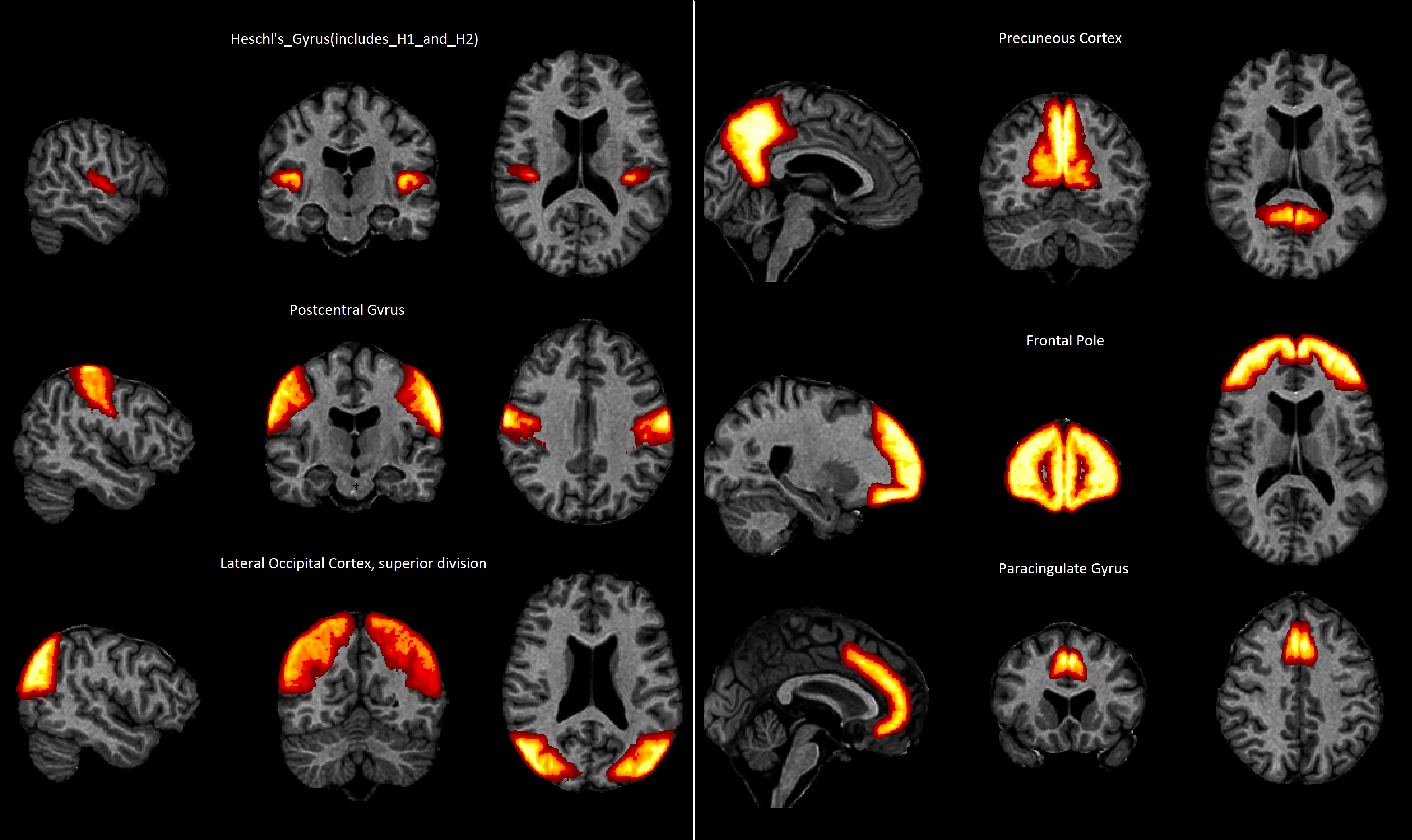
Fig.1 The cerebral regions with significantly different myelination between the schizophrenia
and normal groups

Fig 2. The regions of cerebellum with significantly different myelination between the schizophrenia
and normal groups
DOI: https://doi.org/10.58530/2023/5058