5055
Alteration in white matter tracts in early onset schizophrenia Using Diffusion tensor imaging1Dept. of NMR & MRI Facility, All India Institute of Medical Sciences, New Delhi, India, 2Department of psychiatry, All India Institute of Medical Sciences, New Delhi, India
Synopsis
Keywords: Psychiatric Disorders, Diffusion Tensor Imaging, Early-onset Schizophrenia
The study investigated white matter tracts disruption in early-onset Schizophrenia (EOS). Our finding revealed decreased white matter tracts fractional anisotropy (FA) and increased radial, axial and mean diffusivity in EOS as compared to controls, suggesting thinner packing of axon and fiber bundle. Disrupted white matter fibers may be associated with impaired neurobehavior in EOS.
Materials and Methods: Twenty-one (N=21) EOS patients (mean age 17.41±2.1 years; 8F/13M) who met ICD-10 (code F20) criteria were recruited from the psychiatric clinics. Twenty (N=20) healthy subjects (age=17.9±0.99, 6F/14M) with no history of neurological and psychiatric disorder were recruited from local community. The study was approved by the institutional ethics committee.
Data Acquisition: DTI data were acquired on a 3T MRI (Ingenia 3.0 T, M/s Philips Health Care, Netherlands) using 32-channel head coil, with parameters: b-values=0, 1000 s/mm2, 64 diffusion directions, slice thickness=3 mm with no inter-slice gap, number of slices=50, TR/TE = 6642/86 ms. Data was processed using MRtrix6 and FSL7.
Results: EOS showed decrease fractional anisotropy (FA) and increase radial, axial and mean diffusivity in corticospinal tracts (CST), Anterior thalamic radiation (ATR), Inferior fronto-occipital fasciculus (IFO), Superior longitudinal fasciculus (SLF), Inferior longitudinal fasciculus (ILF), Superior longitudinal fasciculus (SLFT), Forceps major (FM) and Uncinate fasciculus (UF) (Figure 1, Table 1).
Discussion: Atrophy in the white matter regions including CST, ATR, IFOF, decreased FA, increased MD and RD in EOS maybe suggestive of thinner packing of axon and fiber bundle2-4. Increased AD suggests impairment in axonal integrity3. Altered connection IFOF fiber suggest impaired visuospatial processing. CST fiber connection may be attributed to impaired motor function, and ATR fibers connection to impaired emotional and social withdrawal behavior in EOS.
Conclusion: Our findings suggest that EOS demonstrated widespread reduction of FA, MD, RD, AD in major white matter pathways and such abnormal white matter structure may be linked to cognitive and behavioural impairment.
Acknowledgements
MK acknowledges the funding from National DBT-RA Program Biotechnology and Life Sciences (DBT/2020/January/72), Ministry Science and Technology, Government of India.
References
1. Marder SR, Cannon TD. Schizophrenia. N Engl J Med. 2019;381(18):1753-1761.
2. Epstein KA, Cullen KR, et al. White matter abnormalities and cognitive impairment in early-onset schizophrenia-spectrum disorders. Journal of the American Academy of Child & Adolescent Psychiatry. 2014;53(3):362-72.
3. Henze R, Brunner R, et al. The Optic Radiation and the Cerebellar Peduncles in Adolescents with First‐Admission Schizophrenia—A Diffusion Tensor Imaging Study. Journal of Neuroimaging. 2014; 24(2):111-6.
4. Gawłowska M, Rabe-Jabłońska J, et al. DTI [Internal capsule integrity and its sex-related structural differences in early-onset schizophrenia - diffusion tensor imaging study]. Psychiatr Pol. 2015;49(2):349-61.
5. Gawłowska-Sawosz M, Pawełczyk A, et al. Evaluation of white matter structure changes, as assessed in tractography, and cognitive dysfunctions in patients with early onset schizophrenia and their first-degree relatives. Psychiatr Pol [Internet]. 2017; 51(4):735-50.
6. Smith SM, Jenkinson M, et al. Tract-based spatial statistics: voxelwise analysis of multi-subject diffusion data. Neuroimage. 2006;31(4):1487-505.
7. Cordero-Grande L, Christiaens D, et al. Complex diffusion-weighted image estimation via matrix recovery under general noise models. Neuroimage. 2019; 200: 391-404.
Figures
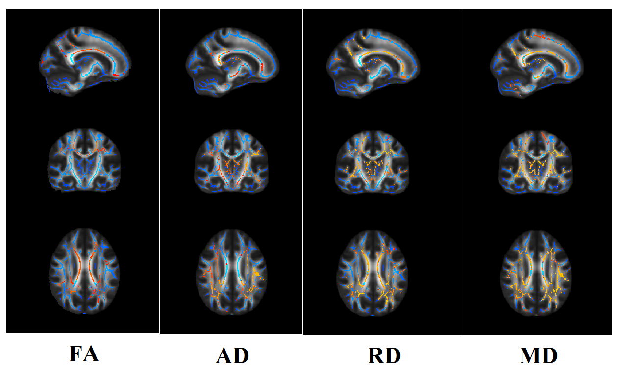
Figure 1: Fractional anisotropy (FA), radial diffusivity (RD) and axial diffusivity (AD) and mean diffusivity (MD) maps overlaid on mean FA skeleton (Green) and differences (Red-Yellow Color) in early onset schizophrenia (EOS) with respect to healthy controls.
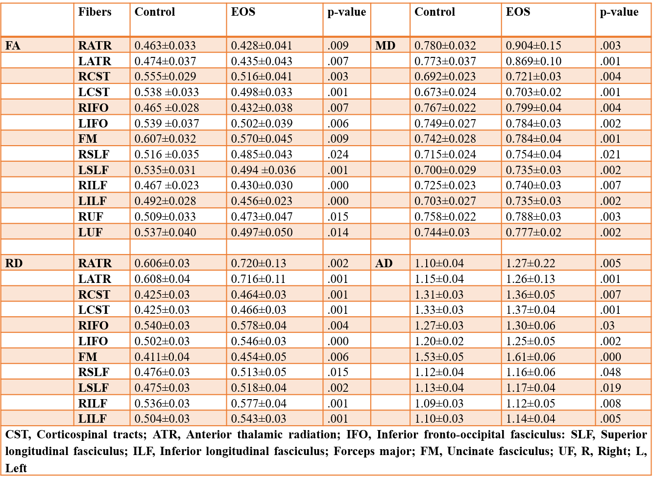
Table 1. Fractional anisotropy (FA), radial diffusivity (RD) and axial diffusivity (AD) and mean diffusivity (MD) values from the white matter tracts of the brain in early onset schizophrenia (EOS) as compared to controls.