5054
Functional and structural brain alterations in autism spectrum disorder: A multimodal meta-analysis of neuroimaging studies1Medical Imaging Center, First Affiliated Hospital of Jinan University, Guangzhou, China, 2MR Research, GE Healthcare, Beijing, China
Synopsis
Keywords: Psychiatric Disorders, Brain, fMRI(resting state); Grey matter volume
Numerous neuroimaging studies conducted have shown functional and structural brain alterations in patients with ASD. We conducted a whole-brain voxel-wise meta-analysis to summarize previous inconsistent results by using Seed-based d Mapping software. In the present multimodal meta-analysis, functional and structural alterations in specific regions were found, which provide new insights into the quest for a neuroimaging-based marker for ASD.Background
Autism spectrum disorder (ASD) is a neurodevelopmental disorder characterized by deficits in social interaction and communication, repetitive behaviors, and restricted interests1,2. Numerous resting-state functional imaging and voxel-based morphometry (VBM) studies conducted have shown functional and structural brain alterations in patients with ASD, but results have been inconsistent3-6. An updated meta-analysis is urgently required to summarize these inconsistent results.Methods
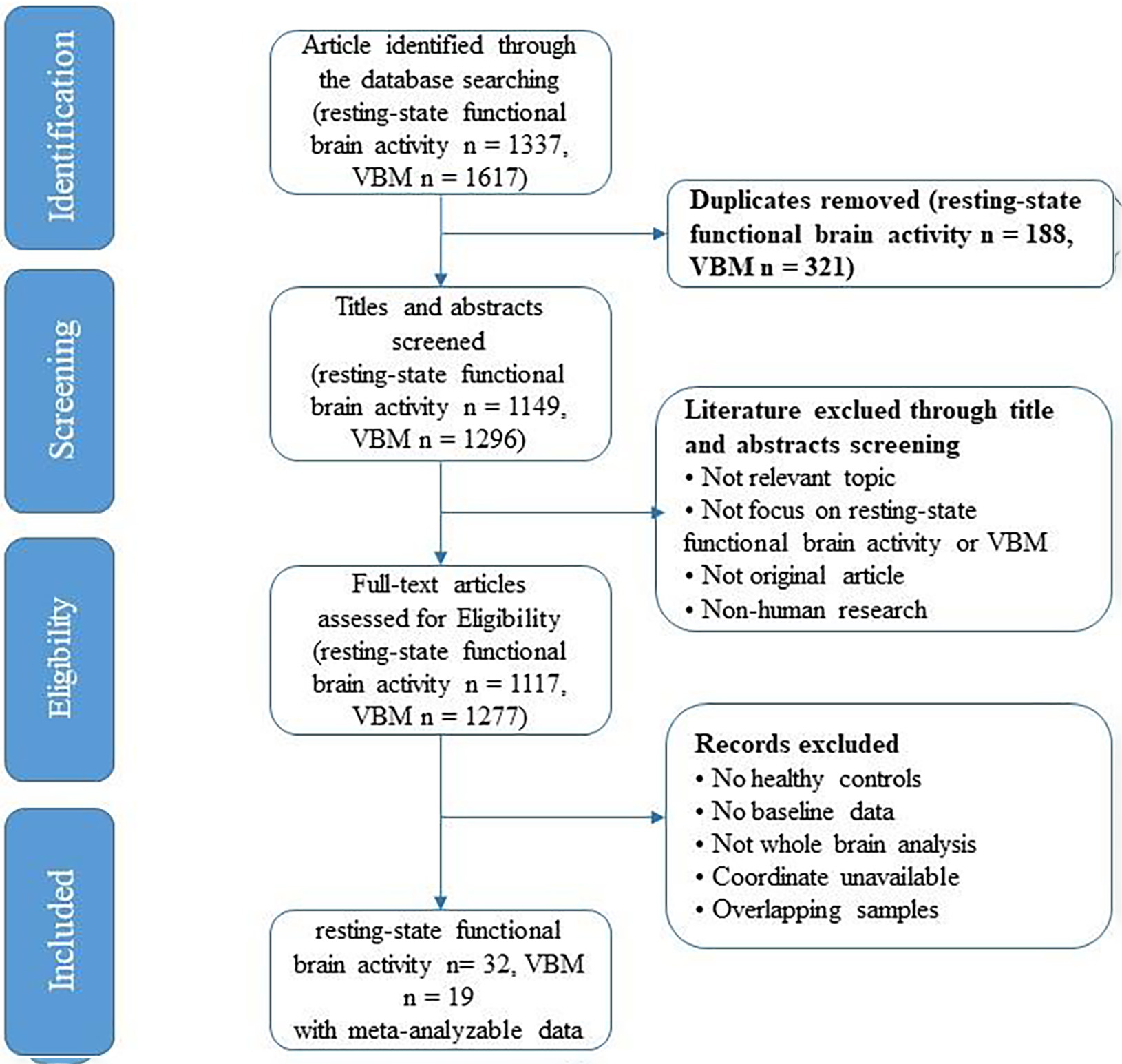
Relevant studies published before September 2022 were searched and screened, we conducted a whole-brain voxel-wise meta-analysis to compare intrinsic functional activity and gray matter volume (GMV) differences between patients with ASD and typically developed (TD) individuals by using Seed-based d Mapping software.Results
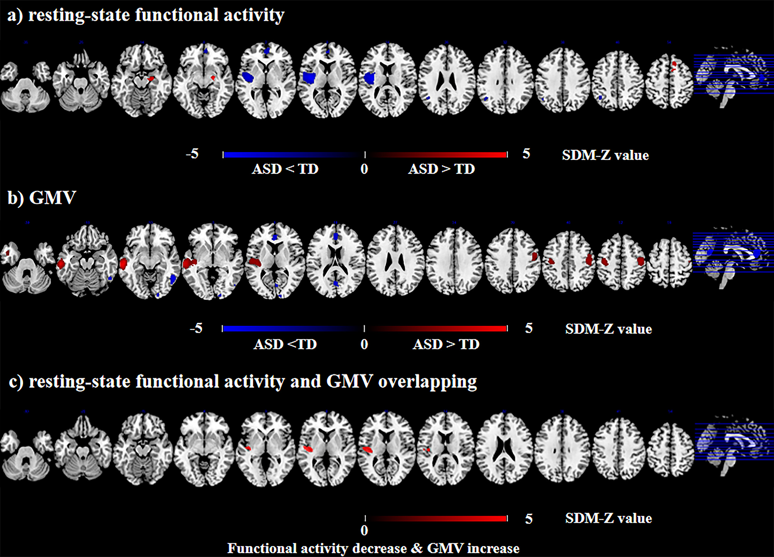
A total of 32 functional imaging studies (975 ASD, 837 TD) and 19 VBM studies (563 ASD, 588 TD) were included. Overall, compared to TD, ASD patients displayed resting-state functional increase in the right hippocampus, right supplementary motor area, left precentral gyrus, and left precuneus, as well as decrease in the left insula (extending to left superior temporal gyrus and left putamen), bilateral medial prefrontal cortex/ anterior cingulate cortex (mPFC/ACC) and left angular gyrus. For VBM meta-analysis, ASD patients displayed increased GMV in left middle temporal gyrus (extending to left superior temporal gyrus and left insula), and bilateral pre- and post-central gyrus, and decreased GMV in bilateral mPFC/ACC, right inferior temporal gyrus, and right precuneus. Further, ASD patients displayed decreased resting-state functional activity and increased VBM in the left insula (extending to left superior temporal gyrus) after overlapping the functional and structural differences.Conclusions
The present multimodal meta-analysis suggested that ASD patients showed the same and different brain region abnormalities in structural and functional MRI, which would provide valuable insights into underlying pathophysiology of ASD.Acknowledgements
The study was supported by grants from the National Natural Science Foundation of China (81671670, 81971597 and 82102003); National Key Research and Development Program of China (2020YFC2005700); Project in Basic Research and Applied Basic Research in General Colleges and Universities of Guangdong, China (2018KZDXM009); Key-Area Research and Development Program of Guangdong Province (2020B1111100001); Medical Science and Technology Research Foundation of Guangdong Province (A2021109). The funding organizations played no further role in study design, data collection, analysis and interpretation and paper writing.References
1. American Psychiatric Association. Diagnostic and Statistical Manual of Mental Disorders. 5th ed. American Psychiatric Association; 2013.
2. Hennessy A, Seguin D, Correa S, et al. Anxiety in children and youth with autism spectrum disorder and the association with amygdala subnuclei structure. Autism. 2022 Oct 22:13623613221127512.
3. Berto S, Treacher AH, Caglayan E, et al. Association between resting-state functional brain connectivity and gene expression is altered in autism spectrum disorder. Nat Commun. 2022 Jun 9;13(1):3328.
4. Jao Keehn RJ, Nair S, Pueschel EB, et al. Atypical Local and Distal Patterns of Occipito-frontal Functional Connectivity are Related to Symptom Severity in Autism. Cereb Cortex. 2019 Jul 22;29(8):3319-3330.
5. Carlisi CO, Norman LJ, Lukito SS, et al. Comparative Multimodal Meta-analysis of Structural and Functional Brain Abnormalities in Autism Spectrum Disorder and Obsessive-Compulsive Disorder. Biol Psychiatry. 2017 Jul 15;82(2):83-102.
6. Kojima M, Yassin W, Owada K, et al. Neuroanatomical Correlates of Advanced Paternal and Maternal Age at Birth in Autism Spectrum Disorder. Cereb Cortex. 2019 Jun 1;29(6):2524-2532.
Figures