5053
A study of AFQ-based method in adolescents with first-episode treatment-naive major depressive disorder1Changzhi Medcal College, Changzhi, China, 2Zigong Fourth People‘s Hospital, Zigong, China, 3Heping Hospital Affiliated to Changzhi Medical College, Changzhi, China, 4Centre for Artificial Intelligence Driven Drug Discovery, Faculty of Applied Sciences, Macao Polytechnic University, Macao, China, 5Clinical Science,Philips Healthcare, Chengdu, China, 6Zigong Fourth People‘s Hospital, ZIgong, China
Synopsis
Keywords: Psychiatric Disorders, Diffusion Tensor Imaging
The study is to explore the changes of white matter fiber tracts in adolescents with first-episode treatment-naive depression using a DTI-based AFQ method. Adolescent depression can cause damage to multiple white matter fiber tracts. The severity of depression is positively correlated with white matter fiber damage. The depression prediction model established has a certain accuracy in the diagnosis of adolescent depression. White matter fiber tract abnormalities may be the pathophysiological mechanism of depression and can be used as a potential biomarker for the diagnosis of depression.Introduction
Depression major depressive disorder (MDD) is a common and complex psychiatric disorder. Compared with adult depression, adolescent depression has atypical symptoms and higher incidence, recurrence and disability rates1,2. At present, the diagnosis of depression in adolescents mainly depends on the experience of clinicians and depression scale screening. However, the scale method has a certain degree of subjectivity, so it is of great significance to explore the objective indicators of depression for the prevention and treatment of adolescent depression. With the development of neuroimaging, magnetic resonance imaging (MRI) technology has recently provided an important research method for clinical diagnosis, drug response evaluation and recurrence prediction of depression because of its non-invasive, simple and high repeatability. In neuroimaging research, diffusion tensor imaging (DTI) technology is used to reflect the integrity and orientation of white matter fiber tracts, and then reflect the changes of brain structure3. AFQ is a new automatic analysis method proposed by Yeatman JD in 2012 to obtain the direction and distribution of nerve fiber bundles from DTI tensor information4. However, current studies mainly focus on adults5,6, using traditional artificial Regions of interest or Tract-based spatial statistics analysis to analyze DTI data7-10, and there is no study to explore brain structural changes in adolescent depression. In this study, we used Automating fiber-tract quantification (AFQ) to investigate the white matter fiber changes in adolescents with depression, and to further lay the foundation for revealing the pathogenesis of adolescent depression and the objective diagnosis of adolescent depression.Methods
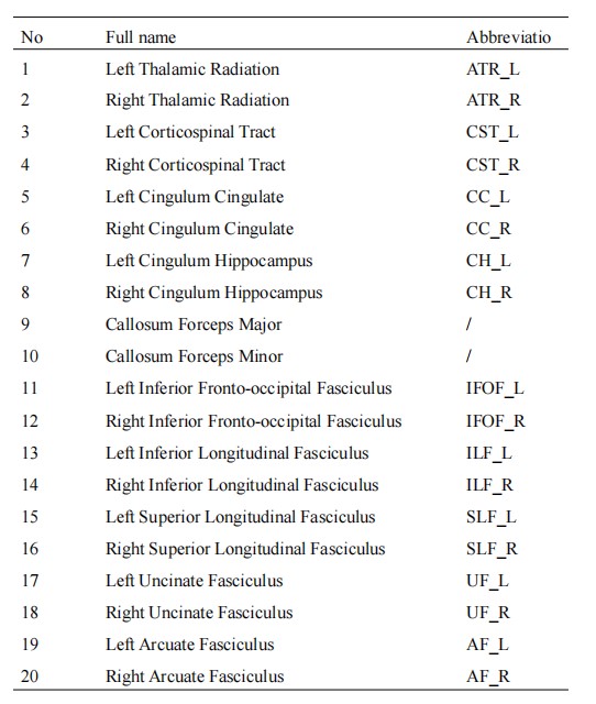
This study was approved by the institutional ethics committee. 24 first-episode untreated adolescent depression patients(mean age,15.2[SD,1.5]years;18 woman[75%]) and 29 sex- and age-matched healthy volunteers (mean age,15.8[SD,2.2]years;18 woman[62%]) ere collected. DTI was performed on a clinical Siemens Skyra 3.0T system with 32 channel coil using the scan parameters listed as follows: TR/TE=12000/77ms, field of view=224mm2×224mm2, reconstructed voxel size diffusion 30 directions, b=0/1000. 3D T1-weighted imaging was also conducted for reference. Twenty major fiber bundles in the brain were extracted using the AFQ method based on MATLAB software package (https://github.com/jyeatman/AFQ) for whole brain (Table 1) , and the 20 fiber bundles were subdivided into 100 segments. The Fractional anisotropy(FA),Radial diffusivity(RD),Mean diffusion (MD) and Axial diffusivity(AD) values were calculated for the 20 fiber bundles and the related 100 subdivided segments. Two independent sample T-Test was used to compare the differences of the 20 fiber bundles and the 100 segments between adolescents with depression and the healthy controls.The HAMD-17 depression scale was developed by Hamilton in 1960 to assess the severity of depression. Kendall rank correlation analysis was performed to analyze the correlations between the fiber bundle diffusion tensor imaging metrics and HAMD-17 scores. A random forest model was constructed to differentiate the patients with depression by using diffusion tensor imaging metrics correlated with HAMD-17 scores, and the ROC curve of the model was plotted.
Results
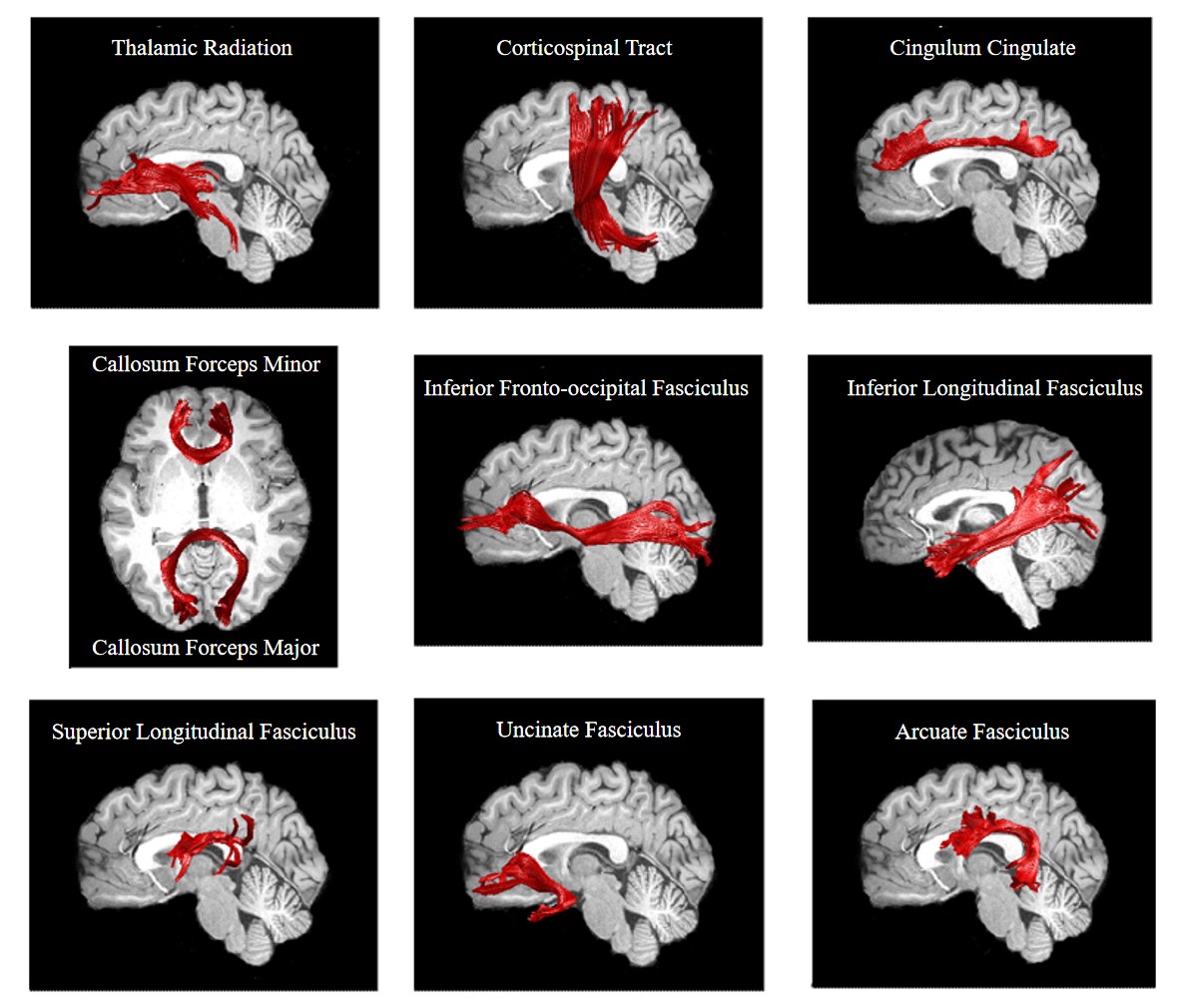
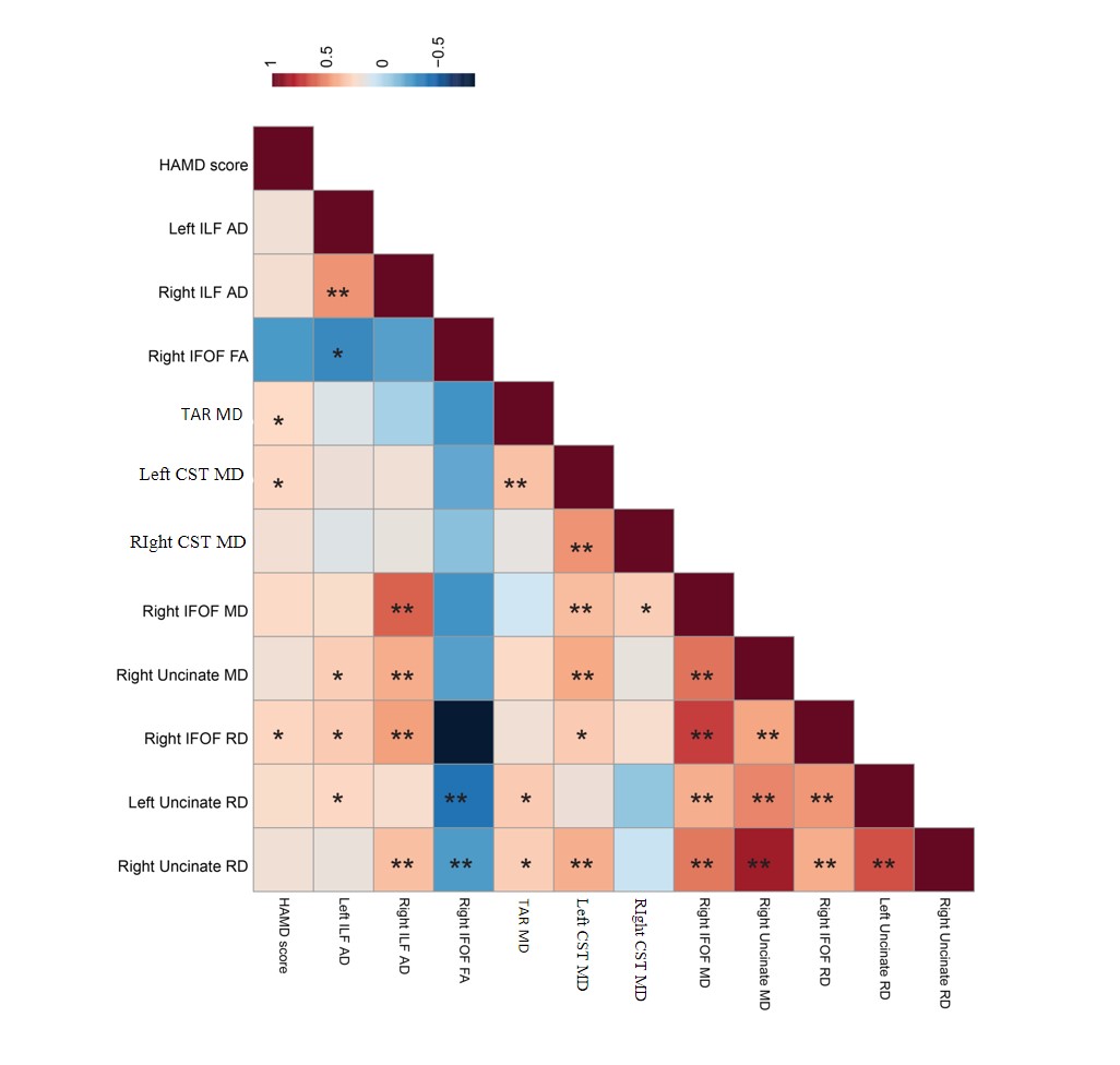
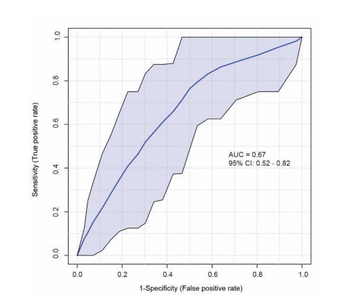
Because some of the subjects had fewer fibers in the Left Cingulum Hippocampus and Right Cingulum Hippocampus, which were difficult to extract, they were not included in the statistical analysis. In the MDD and HC groups, 18 fiber bundles were included analyses(Figure 1).Compared with the healthy-control group, MDD group had significantly lower FA values in t IFOF_R and AF_R, significantly higher MD values in ATR_L, CST_L,CST_R, IFOF_R and UF_R, significantly higher AD values in ILF_L and ILF_R, significantly higher RD values in IFOF_R, UF_L,UF_R, and AF_R (P<0.05). The 100 segments of the 20- fiber bundle in the MDD and HC groups has a small segmental difference in diffusion tensor imaging metrics (P<0.01). The MD value of ATR_L, CST_L and the RD value of IFOF_R were positively correlated with the HAMD-17 scale score(r = 0.20、0.20、0.20,P = 0.04、0.04、0.04(Figure 2). Tensor indices associated with HAMD-17 scores were the MD value of ATR_L, the MD values of CST_L, and the RD value of IFOF_R. The machine learning random forest model (n = 500 trees) was used to rank the diffusion tensor indexes of abnormal fiber tracts according to the feature selection of Mean Decease Accuracy. After screening, the MD value of ATR_L combined with the RD value of IFOF_R had the best diagnostic effect for depression. The area under the ROC curve of the model was 0.67, the sensitivity was 66.7%, and the specificity was 65.5%(Figure 3).Conclusions
The study suggests white matter fiber tract abnormalities may be a pathophysiological mechanism of depression and may serve as potential biological markers for the diagnosis of depression in adolescents.Acknowledgements
No acknowledgement found.References
1. Yu M, Linn KA, Shinohara RT, Oathes DJ, Cook PA, Duprat R, et al. Childhood trauma history is linked to abnormal brain connectivity in major depression. Proc Natl Acad Sci U S A 2019; 116(17):8582-8590.6. Gunnell D, Kidger J, Elvidge H. Adolescent mental health in crisis. Bmj 2018; 361:k2608.
2. Cullen KR, Klimes-Dougan B, Muetzel R, Mueller BA, Camchong J, Houri A, et al. Altered white matter microstructure in adolescents with major depression: a preliminary study. J Am Acad Child Adolesc Psychiatry 2010; 49(2):173-183.e171.
3. Yeatman JD, Dougherty RF, Myall NJ, Wandell BA, Feldman HM. Tract profiles of white matter properties: automating fiber-tract quantification. PLoS One 2012; 7(11):e49790.
4. LeWinn KZ, Connolly CG, Wu J, Drahos M, Hoeft F, Ho TC, et al. White matter correlates of adolescent depression: structural evidence for frontolimbic disconnectivity. J Am Acad Child Adolesc Psychiatry 2014; 53(8):899-909, 909.e891-897.
5. Wu F, Tu Z, Sun J, Geng H, Zhou Y, Jiang X, et al. Abnormal Functional and Structural Connectivity of Amygdala-Prefrontal Circuit in First-Episode Adolescent Depression: A Combined fMRI and DTI Study. Front Psychiatry 2019; 10:983.
6. Jiang W, Gong G, Wu F, Kong L, Chen K, Cui W, et al. The papez circuit in first-episode, treatment-naive adults with major depressive disorder: combined atlas-based tract-specific quantification analysis and voxel-based analysis. PLoS One 2015; 10(5):e0126673.
7. Heij GJ, Penninx B, van Velzen LS, van Tol MJ, van der Wee NJA, Veltman DJ, et al. White matter architecture in major depression with anxious distress symptoms. Prog Neuropsychopharmacol Biol Psychiatry 2019; 94:109664.
8. Sengul Y, Otcu H, Ustun I, Sengul HS, Cersonsky T, Alkan A, et al. Neuroimaging depression and anxiety in essential tremor: A diffusion tensor imaging study. Clin Imaging 2019; 58:96-104.
9. Chen G, Guo Y, Zhu H, Kuang W, Bi F, Ai H, et al. Intrinsic disruption of white matter microarchitecture in first-episode, drug-naive major depressive disorder: A voxel-based meta-analysis of diffusion tensor imaging. Prog Neuropsychopharmacol Biol Psychiatry 2017; 76:179-187.
10. Deng F, Wang Y, Huang H, Niu M, Zhong S, Zhao L, et al. Abnormal segments of right uncinate fasciculus and left anterior thalamic radiation in major and bipolar depression. Prog Neuropsychopharmacol Biol Psychiatry 2018; 81:340-349.
Figures