5023
SNR-Enhanced T2 and Diffusion-Weighted 3D Dual-Echo Steady-State MRI Using Random Matrix Theory-Based Denoising Reconstruction
Zhaohuan Zhang1, Shu-Fu Shih1, and Holden H. Wu1
1Department of Radiology, University of California, Los Angeles, Los Angeles, CA, United States
1Department of Radiology, University of California, Los Angeles, Los Angeles, CA, United States
Synopsis
Keywords: Diffusion/other diffusion imaging techniques, Diffusion/other diffusion imaging techniques
Three-dimensional (3D) dual-echo steady-state (DESS) MRI can produce multi-contrast (MC)images with T2 and diffusion weighting (DW) for simultaneous T2 and D mapping in knee and prostate. A primary challenge of MC DESS is the low signal-to-noise ratio (SNR) of DW DESS leads to less reliable diffusion mapping. In this work, we investigated a new random matrix theory-based denoising reconstruction to improve MC T2W/DW DESS by taking advantages of the inherent redundancy in noise statistics across multiple coil channel and contrasts dimensions. Our in-vivo knee and prostate experiments showed promising improvements in SNR and quality of T2 and D mapping.Introduction
Quantitative multiparametric MRI of T2 and diffusivity (D) provides MR parameters reflecting tissue chemical composition and microstructure1-4, which can facilitate the quantitative characterization of tissue physiologic states for disease diagnosis and monitoring1. To optimize the diagnostic performance, quantitative T2 and diffusion maps should be combined with spatially matched position and resolution. However, the widely used single-shot spin-echo echo-planar imaging (ss-SE-EPI) based diffusion-weighted MRI (DWI) sequence has lower spatial resolution and suffers from geometric distortion compared to T2-weighted (T2W) MRI sequences5.Three-dimensional (3D) dual-echo steady-state (DESS) MRI can produce FID and ECHO images with T1/T2-weighted, T2W, and/or DW contrasts that are inherently matched spatially and have shown promise for rapid 3D T2 mapping of the entire knee and prostate 6-8. Multi-contrast (MC) T2W and DW DESS is also being explored for in vivo T2 and D mapping9-15. One primary challenge for in vivo MC T2W/DW-DESS is the low signal-to-noise ratio (SNR) of the DW DESS ECHO signals due to additional diffusion signal decay on top of T2 decay, which degrades the reliability of D measurements15-16 and needs be addressed before clinical translation.
Recently, random matrix theory (RMT) based denoising reconstruction17-18 was developed to substantially reduce thermal noise in MRI raw signals by exploiting the redundancy of noise statistics across multiple coil channels and imaging contrasts. Promising SNR gains using RMT denoising reconstruction was first demonstrated in ss-SE-EPI diffusion MRI and other multi-contrast sequences17. Since MC T2W/DW-DESS can include up to 4 contrasts (FID, ECHO; DW-FID, DW-ECHO), it can potentially take advantage of coil-level RMT-based reconstruction to improve its SNR for robust diffusion imaging and measurements.
Therefore, the purpose of this study was to investigate a new RMT-based reconstruction method to denoise 3D MC T2W/DW-DESS MRI and evaluate it for in vivo knee and prostate T2 and D mapping.
Methods
MC-DESS: MC T2W/DW-DESS consisted of two acquisitions with identical imaging parameters except for the moment of the spoiler gradient to induce minimal to high DW (Fig.1). A total of 4 contrasts (FID0, ECHO0, FIDDW, and ECHODW) were acquired, exhibiting T1/T2-weighted, T2W, and additional DW contrasts respectively. Bloch simulation-based dictionary matching algorithm8,11 was used to generate quantitative T2 maps. A simplified analytic signal model12 was used to estimate D maps with an effective b-value (beff), through a separate calibration scan in a NIST diffusion phantom against a ss-SE-EPI DWI.RMT-based denoising reconstruction: The reconstruction pipeline is shown in Fig.2. The FID and ECHO k-space data from MC-DESS (4 contrasts) were stacked into a multi-coil multi-contrast matrix S(kx,ky,kz,Ncoil,Ncontrast). The estimated noise covariance matrix was used to de-correlate the signals to ensure each coil channel has same noise statistics for RMT denoising. In the presence of parallel imaging, the g-factor map was computed19 to renormalize the spatial noise statistics post-GRAPPA reconstruction, before performing joint multi-coil multi-contrast Marchenko-Pastur distribution Principal Component Analysis (MPPCA)17-18 denoising prior to adaptive coil combination.
Experiments: In an IRB-approved study, in vivo MC T2W/DW-DESS data were acquired at 3T (Prisma or Skyra, Siemens) in two healthy males (age: 28-30) for knee imaging (Ncoil=15) and two healthy males (age: 27-29) for prostate imaging with body coils (Ncoil=18). Another 3 healthy subjects (1 female, age:23-26) were scanned to separately evaluate T2W-DESS for high-resolution isotropic T2 mapping of the knee. Parameters are reported in Fig.1B.
Analysis: To validate that RMT-based denoising removed noise-only components, the complex residuals across the coil channels and contrast dimensions were compared against a zero-mean Gaussian distribution , and the mean and standard deviation (std) of the fitted distribution was reported with 95% confidence intervals. The mean and std of the T2 and D measured in knee cartilage region of interests (ROIs), and prostate ROIs in transition zone (TZ) and peripheral zone (PZ), using RMT denoising reconstruction were reported.
Results
Fig.3 compares per-coil-channel standard and RMT reconstruction results for DESS ECHODW images. The residual images satisfied the zero-mean Gaussian distribution with no apparent removal of anatomical details.Fig.4 compares in vivo knee images and T2/D maps from standard and RMT reconstruction of T2W/DW-DESS MRI. The DW-ECHO image quality, along with the derived D map were substantially improved using RMT reconstruction. The mean and std of cartilage D and T2 were:1.5±0.6*10-3mm2/s and 35±12ms in evaluated subjects.
Fig.5 compares in vivo prostate images and maps from standard T2W turbo-spin-echo, standard ss-SE-EPI DWI, and T2W/DW-DESS. Substantial reduction in geometric distortion was achieved using DW-DESS in comparison to the standard ss-SE-EPI DWI and ADC maps, which suffered from signal pile-up artifacts due to rectal gas. The D and T2 were, PZ: 1.8±0.4*10-3mm2/s and 145±32ms and TZ: 1.5±0.6*10-3mm2/s and 75±22ms in evaluated subjects.
Discussion
MC T2W/DW-DESS provided spatially matched simultaneous 3D T2 and D mapping without geometric distortion. RMT reconstruction took advantage of the redundancy in noise measurements embedded in channel coils and the multi-contrast T2W/DW-DESS acquisitions, which opened new possibilities for enhancing the SNR of multi-contrast T2W/DW-DESS. This SNR improvement, especially in the DW-DESS ECHO enabled the in vivo application of MC T2/DW DESS more practical. Future work will investigate more accurate signal models11 for diffusion mapping and optimize the acquisition protocols tailored to different organ applications.Acknowledgements
This work was supported in part by the NIH/NCI (R01 CA248506),UCLA Department of Radiological Sciences, the UCLA Jonsson Comprehensive Cancer Center, and Siemens Medical Solutions USA. The authors thank the clinicians, study coordinators, and MRI technologists at UCLA. The authors also acknowledge the use of open source MP-PCA MATLAB code from NYU Biophysics MRI group.References
1. Vikas Gulani, Nicole Seiberlich, Quantitative MRI: Rationale and Challenges, Advances in Magnetic Resonance Technology and Applications, Academic Press, Volume 1,2020.2. Gibbs P, Liney GP, Pickles MD, Zelhof B, Rodrigues G, Turnbull LW. Correlation of ADC and T2 measurements with cell density in prostate cancer at 3.0 Tesla. Invest Radiol 2009; 44: 572– 576.
3. Prostate cancer detection with multi-parametric MRI: logistic regression analysis of quantitative T2, diffusion-weighted imaging, and dynamic contrast-enhanced MRI. J Magn Reson Imaging 2009; 30: 327– 334.
4. Langer DL, van der Kwast TH, Evans AJ, Plotkin A, Trachtenberg J, Wilson BC, Haider MA. Prostate tissue composition and MR measurements: investigating the relationships between ADC, T2, K(trans), v(e), and corresponding histologic features. Radiology 2010; 255: 485– 494.
5. Francisco Donato, Daniel N. Costa, Qing Yuan, Neil M. Rofsky, Robert E. Lenkinski, Ivan Pedrosa, Geometric Distortion in Diffusion-weighted MR Imaging of the Prostate—Contributing Factors and Strategies for Improvement, Academic Radiology,Volume 21, Issue 6,
2014,
6. Bruder H, Fischer H, Graumann R, Deimling M. A new steady-state imaging sequence for simultaneous acquisition of two MR images with clearly different contrasts. Magn Reson Med 1988; 7: 35– 42.
7. Welsch GH, Scheffler K, Mamisch TC, Hughes T, Millington S, Deimling M, Trattnig S. Rapid estimation of cartilage T2 based on double echo at steady state (DESS) with 3 Tesla. Magn Reson Med 2009; 62: 544– 549.
8. Dregely, I., Margolis, D.A.J., Sung, K., Zhou, Z., Rangwala, N., Raman, S.S. and Wu, H.H. (2016), Rapid quantitative T2 mapping of the prostate using three-dimensional dual echo steady state MRI at 3T. Magn. Reson. Med., 76: 1720-1729.
9. Freed DE, Scheven UM, Zielinski LJ, Sen PN, Hürlimann MD. Steady-state free precession experiments and exact treatment of diffusion in a uniform gradient. J Chem Phys 2001; 115: 4249.
10. Wu E, Buxton R. Effect of diffusion on the steady-state magnetization with pulsed field gradients. J Magn Reson 1990; 253: 243– 253.
11. Staroswiecki, E., Granlund, K.L., Alley, M.T., Gold, G.E. and Hargreaves, B.A. (2012), Simultaneous estimation of T2 and apparent diffusion coefficient in human articular cartilage in vivo with a modified three-dimensional double echo steady state (DESS) sequence at 3 T. Magn. Reson. Med., 67: 1086-1096.
12. Bieri, O., Ganter, C. and Scheffler, K. (2012), Quantitative in vivo diffusion imaging of cartilage using double echo steady-state free precession. Magn Reson Med, 68: 720-729.
13. de Vries, B.A., Breda, S.J., Sveinsson, B. et al. Detection of knee synovitis using non-contrast-enhanced qDESS compared with contrast-enhanced MRI. Arthritis Res Ther 23, 55 (2021).
14. Kretzschmar, M., Bieri, O., Miska, M. et al. Characterization of the collagen component of cartilage repair tissue of the talus with quantitative MRI: comparison of T2 relaxation time measurements with a diffusion-weighted double-echo steady-state sequence (dwDESS). Eur Radiol 25, 980–986 (2015).
15. Isabel Dregely, Daniel JA Margolis , Kyung H Sung, and Holden H Wu. Towards quantitative T2- and ADC-mapping in prostate using diffusion weighted 3D DESS MRI. Proceedings of ISMRM 2014.
16. Sveinsson, B, Gold, GE, Hargreaves, BA, Yoon, D. SNR-weighted regularization of ADC estimates from double-echo in steady-state (DESS). Magn Reson Med. 2019; 81: 711– 718.
17. Gregory Lemberskiy, Steven Baete, Jelle Veraart, Timothy M Shepherd, Els Fieremans, and Dmitry S Novikov. Achieving sub-mm clinical diffusion MRI resolution by removing noise during reconstruction using random matrix theory. Proceedings of ISMRM 2019.
18. Jelle Veraart, Dmitry S. Novikov, Daan Christiaens, Benjamin Ades-aron, Jan Sijbers, Els Fieremans, Denoising of diffusion MRI using random matrix theory, NeuroImage, Volume 142,2016.
19. Breuer FA, Kannengiesser SA, Blaimer M, Seiberlich N, Jakob PM, Griswold MA. (2009). General formulation for quantitative G‐factor calculation in GRAPPA reconstructions. Magnetic Resonance in Medicine, 62(3), 739-746.
Figures
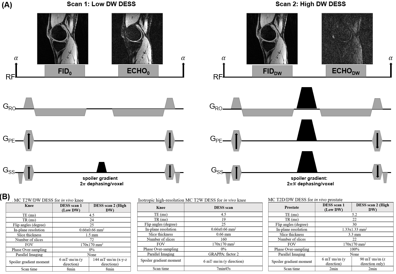
Fig 1: (A) Sequence diagrams for the multi-contrast T2 and Diffusion-weighted Dual Echo Steady State (DESS) MRI acquisitions, including a 1st scan acquiring FID0 (T1/T2 contrast) and ECHO0 (primarily T2 contrast) images with a small spoiler gradient, and another 2nd scan using larger spoiler gradient moment to generate diffusion weighted FIDDW and ECHODW images. A total of 2~4 multi-contrast DESS MRI images can be used to estimate quantitative T2 maps or quantitative T2+diffusivity (D) maps. (B) Imaging parameters for the in vivo knee and prostate MC DESS protocols.
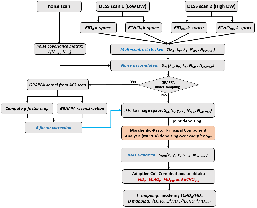
Fig 2: Reconstruction pipeline for SNR-enhanced Multi-contrast DESS MRI Using Random Matrix Theory-based (RMT) denoising. The FID and ECHO k-space data from the low diffusion weighting (DW) DESS and high DW-DESS scans were stacked into a multi-coil multi-contrast matrix, the noise covariance matrix estimated from a noise scan was used to de-correlate the signal to ensure each coil channel has same noise variance. In the presence of parallel imaging under-sampling, the gfactor map was analytically computed to renormalize the spatial noise statistics post GRAPPA reconstruction.
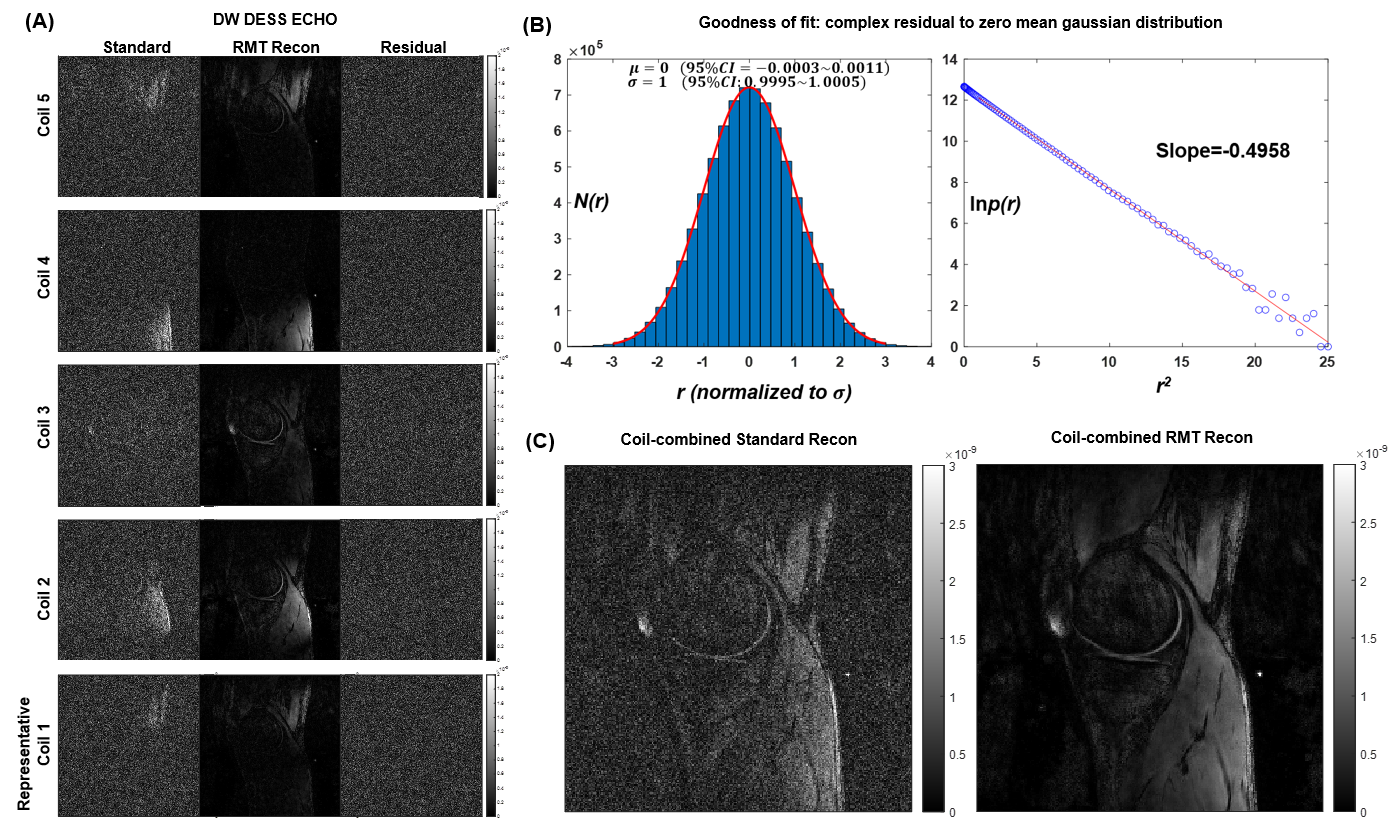
Fig3: (A) Comparisons between standard and RMT denoising reconstructed per channel coil DW DESS ECHO images and corresponding residuals. (B) Goodness of fit for the complex residual (real/imaginary part pooled together) across multi-channel coils for a zero-mean gaussian distribution. (C) Comparison between the post coil combined standard versus RMT reconstructed DW DESS ECHO at 0.66x0.66x1.5=0.65mm3 voxel resolution of in vivo knee.
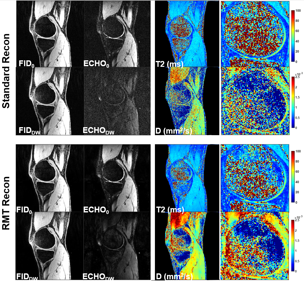
Fig4: Comparisons between standard and RMT denoising reconstructed DESS FID0, ECHO0 and diffusion weighted DESS FIDDW, ECHODW ; Quantitative T2 and diffusivity (D) maps derived from reconstructed DESS MRIs.
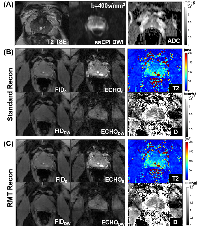
Fig 5: (A) Prostate anatomical reference with 2D TSE sequence, and standard spin-echo single-shot EPI DWI and corresponding ADC maps. (B-C) DESS FID0, ECHO0 and diffusion weighted DESS FIDDW, ECHODW; Quantitative T2 and diffusivity (D) maps derived from DESS MRIs using standard and RMT reconstructions.
DOI: https://doi.org/10.58530/2023/5023