5019
Diffusion diffraction in disordered systems1Department of Clinical Medicine, Aarhus University, Aarhus, Denmark, 2Department of Physics and Astronomy, Aarhus University, Aarhus, Denmark, 3Radiology, New York University School of Medicine, New York, NY, United States
Synopsis
Keywords: Microstructure, Diffusion/other diffusion imaging techniques, diffraction, disordered systems, exchange
We identify a new regime for the diffusion signal in non-confining disordered systems with locally varying diffusivity. It occurs at long times and large diffusion wavevectors, reminiscent of diffusion diffraction in closed pores. Remarkably, while for free diffusion the signal is exponentially strongly dephased, scattering off heterogeneities results in a much weaker, power-law decrease of the signal, thereby offering an enhanced sensitivity to the medium’s structure. In particular, we show how correlation functions of the medium’s microstructure to arbitrary order can be measured. We compare our theoretical predictions for the second order correlation function to numerical simulations with good agreement.Introduction
How much can one learn about the structure of a complex medium by manipulating the gradients of Larmor frequency in an NMR experiment? This fundamental question is at the heart of microstructure mapping with diffusion MRI. As a well-known example, for diffusion in closed pores at long diffusion times, the diffusion signal is explicitly related to the pore shape function and exhibits characteristic peaks at specific $$$q$$$-values -- the phenomenon of diffusion diffraction1.Here we extend the notion of diffraction onto a non-confining geometry of arbitrarily varying diffusivity $$$D(\mathbf{r})$$$ by considering a previously unexplored "diffraction limit" of strong gradients, when a typical spin phase along its Brownian path is large. Remarkably, while for free diffusion the signal is exponentially strongly dephased, scattering off heterogeneities results in a much weaker, power-law decrease of the signal, thereby offering an enhanced sensitivity to the medium's structure.
Theory
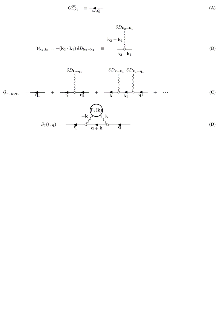
We consider arbitrary medium with the spatially varying diffusion coefficient $$$D(\mathbf{r})$$$ described by the diffusion equation $$\partial_t\psi_{t;\mathbf{r}}=\partial_\mathbf{r} \left( D(\mathbf{r}) \partial_\mathbf{r}\psi\right)\,,\quad D(\mathbf{r})=D+\delta D(\mathbf{r})\,.\qquad(1)$$Using the effective medium formalism2-4, we find the narrow-pulse signal to lowest order (2nd order) in the fluctuation $$$\delta D(\mathbf{r})/D$$$ (corresponding to the Feynman diagrams in Fig.1,
$$\delta S_2(t,\mathbf{q}) = \int_0^t \! \mathrm{d} t_2 \int_0^{t_2} \! \mathrm{d} t_1\, G^{(0)}_{t-t_2, \mathbf{q}} G^{(0)}_{t_1, \mathbf{q}} \int\! \frac{\mathrm{d}^d\mathbf{k}}{(2\pi)^d} \, \left[ \mathbf{q}(\mathbf{q}+\mathbf{k})\right]^2 \Gamma_2(\mathbf{k}) \, G^{(0)}_{t_2-t_1, \mathbf{q}+\mathbf{k}}\,.\qquad(2)$$
Here the power spectral density $$\label{Gamma-2}\Gamma_2(\mathbf{k}) = \frac1V\,\delta D(-\mathbf{k})\delta D(\mathbf{k})\qquad(3)$$$ is the Fourier transform of the correlation function $$$\Gamma_2(\mathbf{r}) = \left< \delta D(\mathbf{r}_0+\mathbf{r})\delta D(\mathbf{r}_0)\right>_{\mathbf{r}_0}$$$.
The cumulant expansion corresponds to both $$$\omega,q\to0$$$, with $$$q\to 0$$$ faster, formally defined as
$$\mbox{cumulant limit:} \quad \omega,q\to 0\,; \quad Dq^2/\omega \to 0 \,.$$
In this work, we are interested in the opposite, diffraction limit, where $$$q\to0$$$ slower than $$$\omega\to0$$$: $$\mbox{diffraction limit:}\quad \omega,q\to 0\,; \quad Dq^2/\omega \to \infty \,.$$ In this case, we are not operating with diffusion coefficient, kurtosis, etc, but we look at the whole signal, probing the system by large "momenta" (wavevectors) $$$\mathbf{q}$$$, whose associated length scale $$$1/q\ll L(t)=\sqrt{2Dt}$$$ is much smaller than the diffusion length $$$L(t)$$$.
In the diffraction limit, long $$$t$$$ effectively turns every free propagator inside momentum integrations, such as $$$G^{(0)}_{t_2-t_1, \mathbf{q}+\mathbf{k}}$$$ for the 2nd-order correction above, into a $$$\delta$$$-function of its momenta, i.e. $$$\sim \delta(\mathbf{q}+\mathbf{k})$$$. Assuming that $$$\Gamma_2(\mathbf{q})$$$ is slowly varying on the scale $$$1/L(t)$$$, we find our main result
$$S^{(1)}_2(t,\mathbf{q}) \simeq \frac{2\pi \Gamma_2(\mathbf{q})}{D^2q^2\, (4\pi Dt)^{d/2+1} }\,.\qquad(4)$$ Note that the correction (4) decreases merely as a power law in $$$t$$$ and $$$q$$$, in contrast to the usual exponential decay of the signal. Hence, remarkably, the nominally small correction to the free propagator becomes dominant at sufficiently large $$$Dq^2t\gg1$$$. This allows us to probe correlation functions such as $$$\Gamma_2(\mathbf{q})$$$ of the medium at finite $$$\mathbf{q}$$$.
Further analysis shows that higher order multiple diffusion encoding (MDE) sequences successively probe higher order correlation functions $$$\Gamma_n(\mathbf{q})$$$.
Monte Carlo simulations
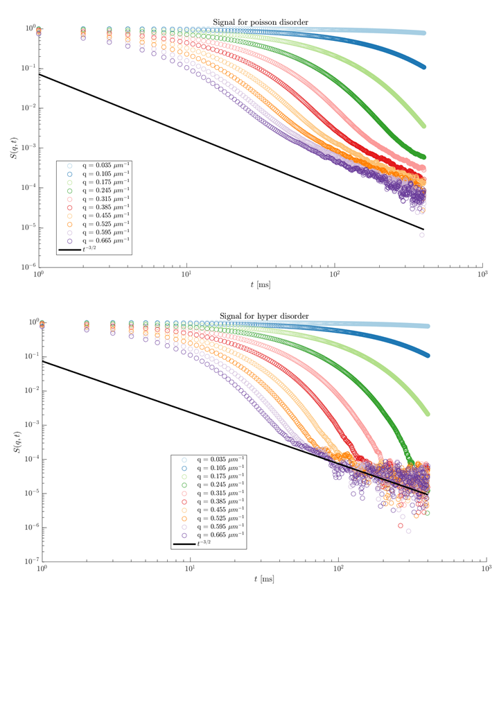
MC simulations were carried out in one dimension with permeable membranes placed according to 2 universality classes, short range (Poissonian) and hyperuniform3, with mean barrier spacing $$$\bar{a}=2\mu$$$m over a $$$4\cdot10^4\mu$$$m sample. The diffusion coefficient was $$$D_0=1\mu$$$m2ms, time step $$$1\mu$$$s, permeability of the membranes was $$$\kappa=0.5\mu$$$m/ms leading to mean residence and diffusion times between two barriers of $$$\tau_R=\bar{a}/2\kappa=2ms$$$ and $$$\tau_D=\bar{a}^2/D_0=4$$$, respectively. Total simulated time was $$$T=100\tau_D=400ms$$$ using $10^9$ particles. All simulations were run in C++ CUDA on a NVIDIA Tesla V100 GPU.Results
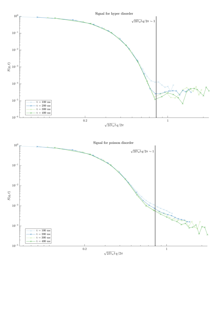
Figure 2 verifies that the exponential signal decay indeed changes to a much slower, power-law behavior as a function of diffusion time, Eq.(4), for sufficiently large $$$t$$$.In Figure 3 we plot the dependence on $$$q$$$ which also conforms to theoretical predictions $$$\sim \Gamma_2(\mathbf{q})/ q^2$$$ of Eq.(4) for the larger values of $$$\sqrt{2D_\infty t}\,q/2\pi\sim\sqrt{b}$$$.
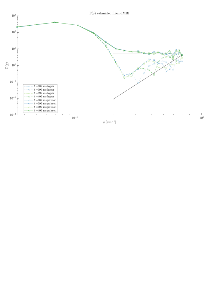
Finally, Figure 4 directly illustrates how Eq.(4) can be used to estimate $$$\Gamma_2(\mathbf{q})$$$ for a medium from the diffusion signal. Of course, such estimation works only for $$$q\gg 1/L(t)$$$.
Discussion and Conclusions
While we are used to, roughly, exponential decay of dMRI signal at strong diffusion weightings, it is not always so, and each example of specific, non-exponential functional form deserves special attention. Power-law dependencies always happen dueto specific reasons: diffusion-diffraction in a confined pore1, $$$1/b$$$ scaling due to randomly oriented zero-radius fibers5,6, Debye-Porod diffraction on a single barrier7,8, presence of the voids in a porous medium9, and the localization regime10.Here we uncover the diffraction-like phenomenon in a medium with smoothly varying local diffusivity $$$D(\mathbf{r})$$$. While perhaps less intuitive than coming from sharp boundaries, this phenomenon is due to scattering off a Fourier component $$$\delta D(\mathbf{q})$$$ that matches the $$$\mathbf{q}$$$-vector. In a disordered system, the spectrum of restrictions is continuous, and for any $$$\mathbf{q}$$$ there will exist the nonzero component $$$\delta D(\mathbf{q})$$$.
Recent developments of system with high-performance gradients are particularly exciting for exploring such physically compelling phenomena with the goal of deconvolving tissue micro geometry from the dMRI signal.
Acknowledgements
SJ acknowledges financial support from Aarhus University Research Foundation (AUFF), the Lundbeck Foundation (R291- 2017-4375), Augustinus Fonden (18-1456) and Kornings Fond. SJ is also supported by the Danish National Research Foundation (CFIN), and the Danish Ministry of Science, Innovation, and Education (MINDLab).
DN was supported by NIH under NINDS award R01 NS088040 and NIBIB awards R01 EB027075, and by the Center for Advanced Imaging Innovation and Research (CAI2R, https://www. cai2r.net), a NIBIB Biomedical Technology Resource Center (NIH P41- EB017183).
References
1 Paul T. Callaghan, A. Coy, D. MacGowan, K. J. Packer, and F. O. Zelaya, “Diffraction-like effects in NMR diffusion studies of fluids in porous solids,” Nature 351, 467–469 (1991).
2 Dmitry S. Novikov and Valerij G Kiselev, “Effective medium theory of a diffusion-weighted signal,” NMR Biomed 23, 682–697 (2010).
3 Dmitry S Novikov, Jens H Jensen, Joseph A Helpern, and Els Fieremans, “Revealing mesoscopic structural universality with diffusion.” Proceedings of the National Academy of Sciences of the United States of America 111, 5088–93 (2014).
4 S.N. Jespersen, E. Fieremans, and D. S. Novikov, “Effective medium theory of multiple diffusion encoding,” Proc. Int. Soc. Magn. Reson. Med. 27, 1009 (2019).
5 Emilie T McKinnon, Jens H Jensen, G Russell Glenn, and Joseph A Helpern, “Dependence on b-value of the direction-averaged diffusion-weighted imaging signal in brain,” Magnetic Resonance Imaging 36, 121–127 (2016).
6 Jelle Veraart, Els Fieremans, and Dmitry S Novikov, “On the scaling behavior of water diffusion in human brain white matter,” NeuroImage185, 379–387 (2019).
7 Pabitra N Sen, Martin D Hu ̈rlimann, and Thomas M De Swiet, “Debye-Porod law of diffraction for diffusion in porous media,” (1995).
8 Astrid F Frøhlich, Leif Ostergaard, and Valerij G Kiselev, “Effect of impermeable boundaries on diffusion-attenuated MR signal,” J MagnReson 179, 223–233 (2006).
9 Partha P. Mitra, Pabitra N. Sen, Lawrence M. Schwartz, and Pierre Le Doussal, “Diffusion propagator as a probe of the structure of porousmedia,” Physical Review Letters 68, 3555–3558 (1992).
10 S. D. Stoller, W. Happer, and F. J. Dyson, “Transverse spin relaxation in inhomogeneous magnetic fields,” Physical Review A 44, 7459–7477 (1991).
Figures