4980
Spherical harmonic shim analysis of B0 distributions in cardiac imaging planes from 921 subjects
Yun Shang1, Sebastian Theilenberg1, Boyu Peng2, Michael Hock3, Laura M. Schreiber3,4, Sachin Jambawalikar1,2, and Christoph Juchem1,2
1Department of Biomedical Engineering, Columbia University in the City of New York, New York, NY, United States, 2Department of Radiology, Columbia University in the City of New York, New York, NY, United States, 3Chair of Molecular and Cellular Imaging, Comprehensive Heart Failure Center (CHFC), Würzburg, Germany, 4Section of Medical Physics, Department of Radiology, Mainz University Hospital, Mainz, Germany
1Department of Biomedical Engineering, Columbia University in the City of New York, New York, NY, United States, 2Department of Radiology, Columbia University in the City of New York, New York, NY, United States, 3Chair of Molecular and Cellular Imaging, Comprehensive Heart Failure Center (CHFC), Würzburg, Germany, 4Section of Medical Physics, Department of Radiology, Mainz University Hospital, Mainz, Germany
Synopsis
Keywords: Heart, Shims
Cardiac functional scans adopting bSSFP sequences suffer from dark band artifacts due to B0 inhomogeneity. The best remedy to mitigate this issue is through cardiac B0 shimming. A limited understanding of the B0 conditions in clinical diagnostic orientations impedes the development of an optimal B0 shim strategy in the human heart. Here we perform a theoretical analysis of spherical harmonic B0 shim at 3 T using a static global approach and slice-specific dynamic shim updating in the short-axis views of human hearts from 921 subjects as a starting point for the development of optimized cardiac B0 shim strategies.Introduction
Cardiac functional scans adopting bSSFP sequences at 3 T suffer from dark band artifacts due to B0 inhomogeneity inside the human heart1, 2 (Figure 1). The most effective remedy to mitigate this issue is to homogenize the B0 distribution in the heart via B0 shimming3, typically achieved in MR scanners via spherical harmonic (SH) shim coils up to second- or third-order4. Mattar et al.5 theoretically accessed the potential of global SH shimming in the whole heart and dynamic shimming within local axial slices upon five in vivo B0 maps. Recent work at 7 T from Hock et al.6 compared experimental shim results of these types in the same orientation. Although both studies suggest better results for dynamic shimming, as expected, the associated shim performance on cardiac imaging planes, e.g., short-axis views, remains unknown. Furthermore, in vivo acquired B0 maps in these and other studies2, 4 are constrained by limited sample size, spatial resolution, and the number of slices due to short acquisition time under breath-hold conditions. The consequential limited knowledge of B0 conditions in cardiac imaging planes impedes the development of an optimal shim strategy. We recently presented a customized method for computing high-resolution cardiac B0 maps based on routine CT images7. Here we propose an oblique slicing method to obtain B0 distributions of short-axis views based on simulated B0 maps from a large and diverse population of human subjects. We analyze the B0 conditions in the heart with SH-based B0 shimming in clinically relevant slice orientations to set the stage for developing optimal B0 shim strategies tailored to cardiac MRI.Methods
Cardiac B0 maps with an isotropic spatial resolution of 1.5 mm were simulated for 921 subjects based on their CT images from examinations through a customized method7. To obtain cardiac imaging planes, we first co-registered a whole heart atlas model8 to the target CT images via an affine registration9, 10 followed by a diffeomorphic registration11, 12. The atlas labels were warped by corresponding transformations to obtain the segmentations of left ventricle and whole heart volume. The long axis of the left ventricle was determined by the eigenvector of its volume with the largest eigenvalue. The corresponding orthogonal planes are short-axis views, and their spatial coordinates were used to derive oblique magnitude images and B0 maps via 3D interpolation. The automated oblique slicing procedure (Figure 2) was implemented by an unsupervised pipeline written in Matlab (MathWorks, Natick, MA, USA).Zero through 5th-order spherical harmonic shim analyses were performed across the whole heart (global) and across sets of local oblique slices with a slab thickness of 10.5 mm, similar to the slice spacing in cardiac cine imaging (fully dynamic). The rapid updating of coil currents with dynamic shimming is known cause eddy currents and artifacts13. Clinical MRI scanners are typically capable of compensating eddy currents for their linear X, Y, and Z gradients. Considering the readily accessible means of dynamic shimming in experimental MRI systems, we investigated the specific hybrid case of static higher-order (i.e., ≥ 2nd) shimming combined with dynamic linear shimming (partially dynamic). All B0 field simulations and analyses were performed in B0DETOX software14, 15.
The overall and local B0 inhomogeneities in the heart were presented with standard deviation ($$$\sigma\left(B_0\right)$$$) and 99th percentile ($$$P_{99}\left(\vert B_0\vert\right)$$$). These inhomogeneities were calculated and analyzed statistically by the mean and standard deviation across subjects after global, fully dynamic, and partially dynamic shimming, respectively. Their shim performances, i.e., the average improvement of field homogeneity within whole heart referenced to the 1st-order global shim, were also calculated. To determine how different shims reduce dark band artifacts with bSSFP, we calculated the relative signal drops after each shim, i.e., the number of voxels in the heart in the frequency range causing significant signal drops (adopted from Weiben et al.1) divided by those after 1st-order global shim, and presented their averages across subjects.
Results
The B0 distributions in the heart become more homogeneous with higher-order SH shims, particularly reducing localized spots of B0-field inhomogeneities in the myocardium (Figure 3, arrows). As expected, dynamic shimming generally outperforms global shimming with lower average B0 inhomogeneity inside the heart and variations across subjects (Figure 4). A 2nd-order fully dynamic shim performs similarly to a 4th-order global shim. The calculated B0 fields show consistent location and shape of dark band artifacts with in vivo cardiac cine images, and such artifacts persist after 2nd-/3rd-order global shim in an exemplary subject (Figure 5A). 2nd- and 3nd-order global shim can reduce signal drops by 56% and 82% after 1st-order global shim, while fully dynamic can achieve 92% and 97%, respectively (Figure 5B).Discussion
We have performed a spherical harmonic shim analysis for B0 distributions in the short-axis views of the human heart from 921 subjects. Global shim at 2nd/3rd order, which is typical for clinical MRI systems, cannot fully mitigate dark band artifacts for all subjects. Fully dynamic shimming shows better shim performance than other types, suggesting its potential to further reduce such artifacts. The large sample of B0 distributions in short-axis views will be used for developing an optimal shim method based on the common SH shim system and the state-of-the-art multi-coil shim technology16.Acknowledgements
This work was supported by grant R01 EB030560 from the National Institutes of Health and a Research Initiatives in Science & Engineering (RISE) award from Columbia University.References
- Wieben O, Francois C, Reeder SB. Cardiac MRI of ischemic heart disease at 3 T: potential and challenges. Eur J Radiol. 2008;65(1):15-28.
- Schär M, Kozerke S, Fischer SE, Boesiger P. Cardiac SSFP imaging at 3 Tesla. Magn Reson Med. 2004;51(4):799-806.
- Juchem C, de Graaf RA. B0 magnetic field homogeneity and shimming for in vivo magnetic resonance spectroscopy. Anal Biochem. 2017;529:17-29.
- Kubach MR, Bornstedt A, Hombach V, Merkle N, Schär M, Spiess J, Nienhaus GU, Rasche V. Cardiac phase-specific shimming (CPSS) for SSFP MR cine imaging at 3 T. Phys Med Biol. 2009;54(20):N467.
- Mattar W, Juchem C, Terekhov M, Schreiber L. Multi-coil B0 shimming of the human heart: A theoretical assessment. 2016:1151.
- Hock M, Terekhov M, Stefanescu MR, Lohr D, Herz S, Reiter T, Ankenbrand M, Kosmala A, Gassenmaier T, Juchem C, Schreiber LM. B0 shimming of the human heart at 7T. Magn Reson Med. 2021;85(1):182-196.
- Shang Y, Theilenberg S, Terekhov M, Mattar W, Peng B, Jambawalikar SR, Schreiber LM, Juchem C. High resolution simulation of B0 field conditions in the human heart from segmented computed tomography Images. NMR Biomed. 2022:e4739.
- Zhuang X, Bai W, Song J, Zhan S, Qian X, Shi W, Lian Y, Rueckert D. Multiatlas whole heart segmentation of CT data using conditional entropy for atlas ranking and selection. Med Phys. 2015;42(7):3822-3833.
- Mattes D, Haynor DR, Vesselle H, Lewellyn TK, Eubank W. Nonrigid multimodality image registration. International Society for Optics and Photonics; 2001:1609-1620.
- Rahunathan S, Stredney D, Schmalbrock P, Clymer BD. Image registration using rigid registration and maximization of mutual information. 13th Annu Med Meets Virtual Reality Conf. 2005.
- Thirion J-P. Image matching as a diffusion process: an analogy with Maxwell's demons. Med Image Anal. 1998;2(3):243-260.
- Vercauteren T, Pennec X, Perchant A, Ayache N. Diffeomorphic demons: Efficient non-parametric image registration. Neuroimage. 2009;45(1):S61-S72.
- Juchem C, Nixon TW, Diduch P, Rothman DL, Starewicz P, de Graaf RA. Dynamic shimming of the human brain at 7 T. Concepts in Magnetic Resonance Part B: Magnetic Resonance Engineering. 2010;37(3):116-128.
- Juchem C. B0DETOX - B0 Detoxification Software for Magnetic Field Shimming. Columbia TechVenture (CTV), License CU17326. 2017;innovation.columbia.edu/technologies/cu17326_b0detox
- Juchem C, Herman P, Sanganahalli BG, Brown PB, Mclntyre S, Nixon TW, Green D, Hyder F, de Graff RA. DYNAmic Multi‐coIl TEchnique (DYNAMITE) shimming of the rat brain at 11.7 T. NMR Biomed. 2014;27(8):897-906.
- Juchem C, Nixon TW, McIntyre S, Boer VO, Rothman DL, de Graaf RA. Dynamic multi-coil shimming of the human brain at 7 T. J Magn Reson. 2011;212(2):280-288.
Figures
Fig. 1. Susceptibility-induced artifacts in the myocardium
caused by B0 variations in the heart. In vivo cardiac cine images were acquired using a bSSFP sequence
with a TR of 3.56 ms after a linear shim provided by the scanner at 3 T. Dark
band artifacts (white arrow) are presented in the inferolateral wall of the
myocardium in the systolic (left) and diastolic (right) cardiac phases. These
artifacts were induced by off-resonance frequencies in the heart.
Fig. 2. Automated procedure for oblique slicing. A whole
heart atlas intensity model was co-registered to target CT image using an
affine registration for matching overall shape followed by a diffeomorphic
registration for matching local structures. The left ventricle and the heart
volume were segmented by warped atlas labels. The long axis of the left
ventricle was then determined by the eigenvector of its volume with the largest
eigenvalue. The corresponding orthogonal planes and their spatial coordinates were
used to obtain short-axis (SA) image geometries via 3D interpolation.
Fig. 3. Spherical harmonic
(SH) shim analyses from zero to 5th-order were performed for B0 maps
of all subjects using shim types of global, partially dynamic, and fully
dynamic, respectively. Here present the results of an exemplary subject with
three short-axis slices, i.e. apex, middle, mitral slices, and a 3D heart volume with a right angle cut in left ventricle (magenta lines). Higher SH orders lead to more homogeneous B0 fields. Fully dynamic shim shows better shim performance with particularly reducing localized spots of B0-field inhomogeneities in the myocardium (arrows).
Fig. 4. Comparison of global (top row) and dynamic shimming (bottom row) as residual B0 inhomogeneity in the heart
for all 921 subjects (Mean +/- Standard deviation (SD)). Shim analyses of three
shim types were performed from zero to 5th SH order, individually. In general, fully
dynamic shim performs better than partially dynamic or global shim in terms
of lower average standard deviation and 99th percentile for each shim order, as
well as a higher average performance percentage. The performance of the 2nd-order
fully dynamic shim is comparable to the results of the 4th-order global shim.
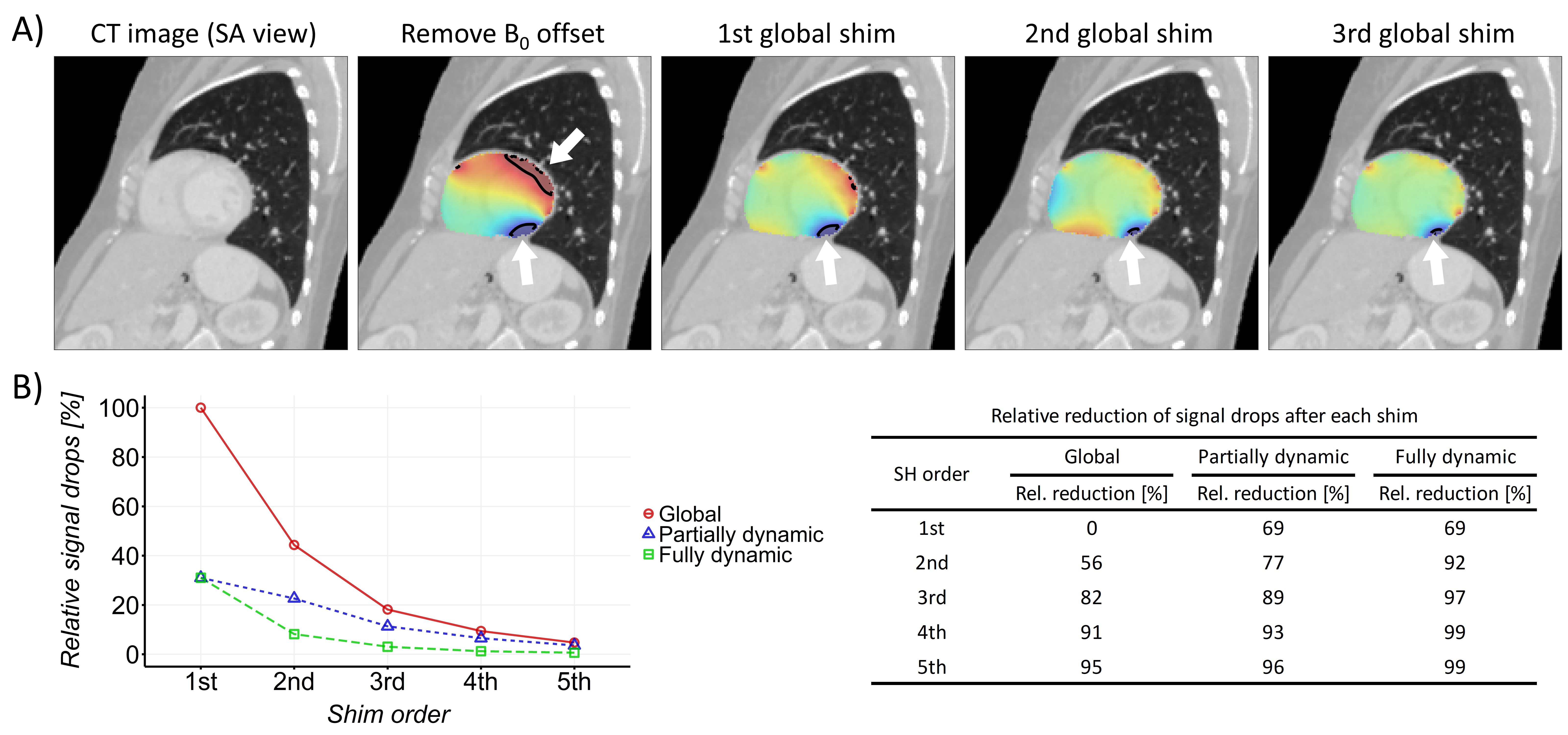
Fig. 5. A) Theoretical
prediction of dark band artifacts at 3 T from simulation. The marked black
contour lines have offsets of ±140 Hz (white arrows) consistent with
the locations of dark band artifacts with bSSFP (TR = 3.56 ms) in Fig. 1. B) The
impact of different shims on reducing signal drops. The number of voxels causing
significant signal drops after each shim was divided by those after 1st-order
global shim and averaged for all subjects. 2nd-order partially dynamic shim
reduces signal drops similarly to 3rd-order global shim, while fully dynamic
shim has an overall greater effect.
DOI: https://doi.org/10.58530/2023/4980