4978
Detection and tracking enhancement using 4-channels local standalone resonators for catheterized interventional MRI at 3T1Department of Biomedical Engineering, University at Buffalo, Buffalo, NY, United States
Synopsis
Keywords: Vessel Wall, Interventional Devices, Blood vessels
Catheter-based RF coils are preferred over conventional external coils due to the weak detection signal from intermediate field region. One of the major issues using catheter RF coils is the design payload (feeding network, preamplifier, balun) associated with the limited space of the catheter. The technique also lacks efficient way to track the catheter during the procedure. To overcome these challenges, we propose 4-channel standalone and wireless resonators to amplify the local B1 field while a body coil is used as the transceiver. The receive B1 fields with and without the local resonators are obtained for field enhancement analysis.
Introduction
Catheter-based RF coils1 has proved to be an alternative interventional MRI method over conventional external detecting RF coils. Superior detection capability is obtained since catheter-based RF coils can be placed very close to the target. While this technique is invasive, size constraint, patient safety and comfort have raised some concerns. Single passive local resonator or wireless detector resonator, without design payload2,3 can be used instead of the wired catheter-based coils to amplify the received signal. The SNR of the design using such method can be further enhanced using two stacked coils4 with decoupling network. However, the two stacked resonators subject to motion during intervention will be very sensitive to the MR signal with varying polarization. In order to minimize the polarization mismatch loss and further increase the SNR, we proposed an array of 4 decoupled elements that comprised of two rectangular conventional loop and two 8-shaped RF coils used as the passive local resonators. Such simple design doesn’t require any decoupling network and is less sensitive to the received signal with varying polarization thus increased the homogeneity and field strength in the targeted area.Methods
The array of 4-channels local resonators, designed to fit in specific vascular tubes is shown in Fig. 1. The proposed design is inserted at the center of a small size low pass birdcage coil designed for 3T. The shielded birdcage coil is composed of 8 legs with 10 cm length. The diameter of the end-ring is set to 10 cm. The lumped capacitors inserted within the leg are adjusted (CL = 13 pF) to tune the design at f0 = 127 MHz, the Larmor frequency of the proton 1H at 3T. The blood vessel is modeled as a cylindrical phantom with 2 cm outer diameter and 1.6 cm inner diameter. The local resonators RF coils are made of very thin copper tape of 0.2 mm width and the two 8-shaped coils are designed with a jumper at the junction to avoid any intersection between them. The local coils are standalone LC resonator operating at 3 T with no connection to the MRI system. The orientation of the 4-channels local resonators is very critical for field enhancement (assuming the static B0 in Z direction). Both the physical dimension and the electric parameters of the proposed model are illustrated in Fig.1. During the transmission phase, passive detuning network (not shown in the Fig.1) are needed so that the local resonators do not interfere with the transmission signal from the body coil. For verification purpose we integrated ports and matching capacitors (Cm) to the local RF coils to simulate their inter-coupling parameters. The potentiality of the received field enhancement within the blood vessel is investigated by driving only the volume coil system with and without the local resonators.Results
Numerical results are performed using High-Frequency Structure Simulator (HFSS) from the ANSYS Corporation. The simulated scattering parameters of the 4-channels local resonators in Fig. 2, shows excellent matching (50 ohms) at the input of each coil and strong decoupling among all of elements which is benefit for constructive interference. The birdcage coil designed to operate at 127 GHz, can be linearly driving using only a port (Port #1) or circularly driving using both ports with 90° phase difference as shown in Fig. 3. The standalone 4-channels local resonator (without ports and matching capacitors) are inserted within the birdcage and loaded with the blood vessel. Four different scenarios are assessed while computing the received B1 field ( ). The transversal received B1 field of the phantom obtained from exciting the birdcage without any passive local resonator show a very weak signal. By integrating the conventional coils, the received signal from the circular polarized birdcage coil is more enhanced compared to the one obtained from the linearly polarized birdcage. Moreover, by adding the 8-shaped local resonators to the system, the received B1 field is further amplified, and more signal coverage is obtained. The 4-channel local resonator, with no connection to the MRI system demonstrated local received amplification, thus SNR enhancement for enabling high resolution vessel wall imaging.Conclusion
An array of 4-channel local resonators with intrinsic decoupling for interventional procedures in the MR environment was designed and examined for 3T. Due to its versatile implementation, such design can fit within a limited space, and is adaptive to varying polarization for target detection and tracking. The proposed design proved to be a suitable approach to efficient catheter tracking and to enhancing the detection capability of target located deep inside the body to obtain high resolution imaging of the blood flow and/or vessel wall.Acknowledgements
This work is supported in part by the NIH under a BRP grant U01 EB023829 and by the State University of New York (SUNY) under SUNY Empire Innovation Professorship Award.
References
1. X. Zhang et al., "Design of catheter radio frequency coils using coaxial transmission line resonators for interventional neurovascular MR imaging," Quant Imaging Med Surg, vol. 7, no. 2, pp. 187-194, Apr 2017, doi: 10.21037/qims.2016.12.05.
2. X. Zhang, "Sensitivity enhancement of traveling wave MRI using free local resonators: an experimental demonstration," Quant Imaging Med Surg, vol. 7, no. 2, pp. 170-176, Apr 2017, doi: 10.21037/qims.2017.02.10.
3. X. Zeng, L. Chen, C. Wang, J. Wang, and C. Qian, "Wireless MRI Colonoscopy for Sensitive Imaging of Vascular Walls," Sci Rep, vol. 7, no. 1, p. 4228, Jun 26 2017, doi: 10.1038/s41598-017-03902-7.
4. M. Lu, S. Chai, H. Zhu, and X. Yan, "Low-cost inductively coupled stacked wireless RF coil for MRI at 3 T," NMR Biomed, p. e4818, Aug 22 2022, doi: 10.1002/nbm.4818
Figures
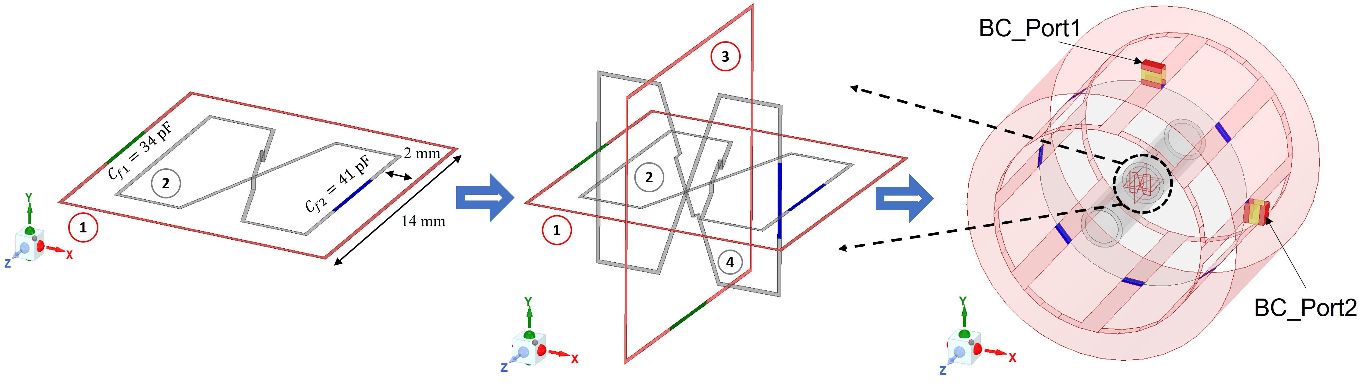
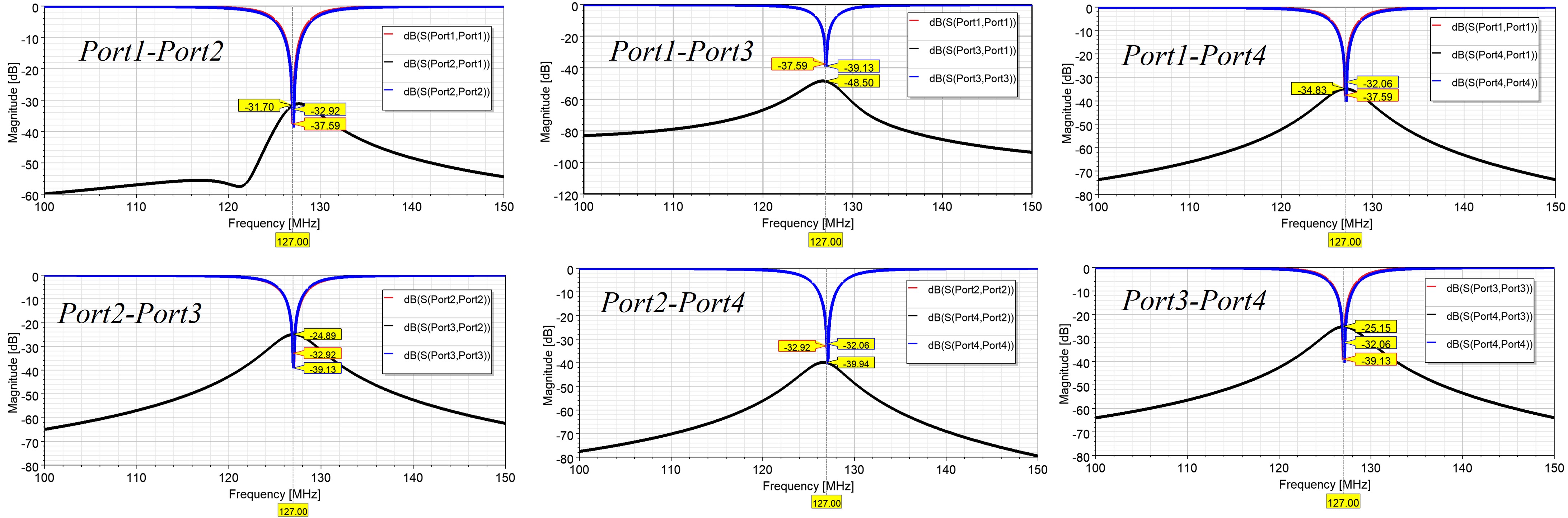
Fig. 2. Simulated scattering parameters of the 4-channels resonator (with ports for verification purpose) showing excellent input impedance matching and good isolation among all 4 elements.
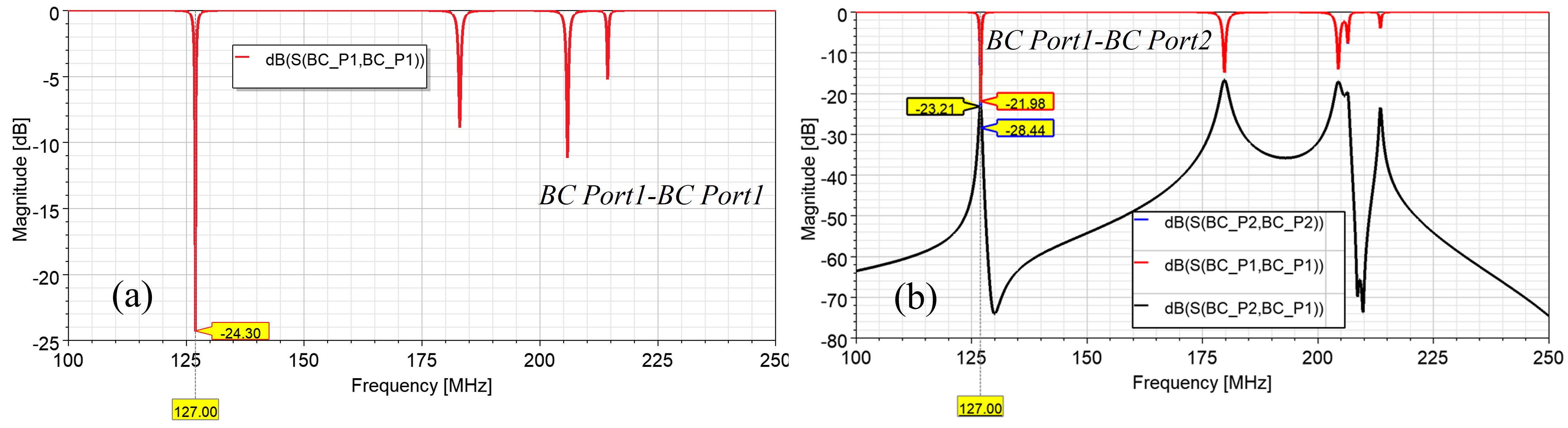
Fig. 3. Simulated scattering parameter of the birdcage coil operating at 127 MHz. (a) Linearly polarized driving using port 1; (b) Circularly polarized driving using both ports with 90° phase shift.
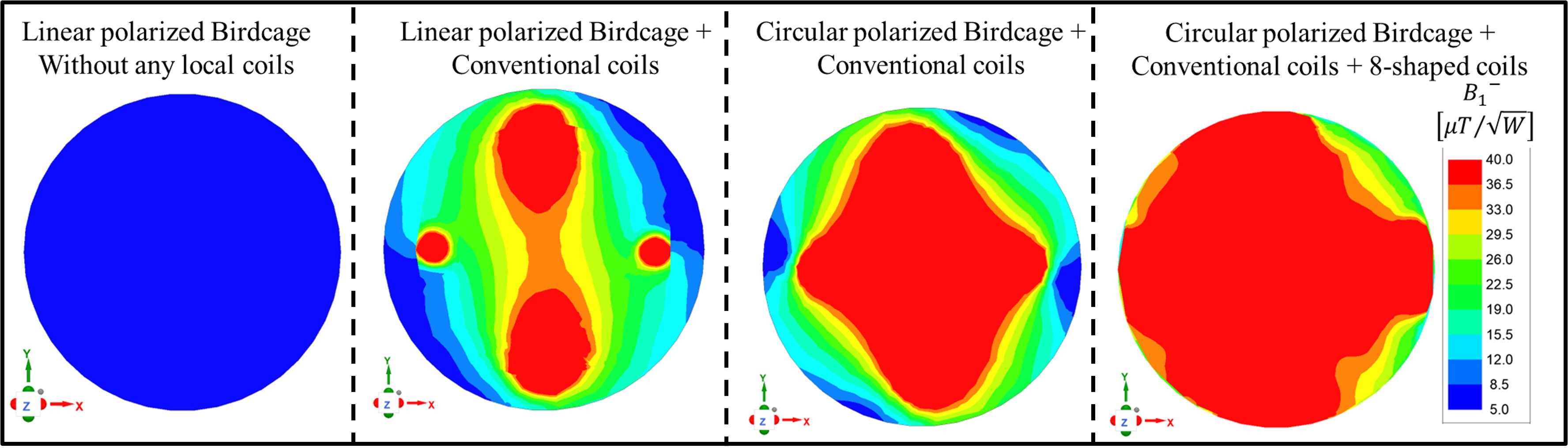
Fig. 4. Simulated receive B1 field distribution normalized to the accepted input power in the XY (transversal) plane through the center of the cylindrical phantom for 4 different scenarios.