4967
Mapping cardiac |B1+| field with B1+ prepared imaging1TU Delft, Delft, Netherlands, 2Department of Health, Medicine and Caring Sciences, Linköping University, Linköping, Sweden, 3Holland PTC, Delft, Netherlands
Synopsis
Keywords: Heart, Artifacts, cardiac B1+ mapping, transmit field imaging
Cardiac MRI at 3T offers gains in SNR, but is strongly affected by field inhomogeneities, including the transmit field B1+. Mapping |B1+| field around the heart is particularly challenging due to the presence of motion, and few techniques exist for robust imaging. Recently, Bloch-Siegert shift mapping was proposed for motion robust cardiac |B1+| mapping, however, it requires long echo times making it more susceptible to artifacts. Here, we introduce a new preparation module for efficient |B1+| mapping. The method shows 0.06% / 0.35% reduced noise variability in phantom and 0.61±0.35% / 0.89±0.63% in-vivo imaging, and visually improved map quality.Introduction
Cardiac MRI at high fields (≥3T) is increasingly gaining interest, due to the promise of increased SNR1. However, as the wavelength of the transmit field B1+ approaches body dimensions, standing waves form, resulting in strong B1+ inhomogeneities2. Accurate measurement of B1+ around the heart, as required in quantitative MR or for shimming, is hampered by respiratory and cardiac motion. Only few techniques have been proposed for robust B1+ mapping in the heart. Bloch-Siegert (BS) shift based transmit field mapping was recently introduced3 with promising results for motion resilient cardiac |B1+| mapping4. However, the technique requires long echo times, making it susceptible to B0 inhomogeneities or implant-induced artifacts, and frequent repetition of the Bloch-Siegert pulses yield a high SAR burden. In this work, we explore a new transmit field mapping technique, using a Bloch-Siegert based |B1+| preparation module for B1+ mapping in the heart with increased robustness and efficiency.Methods
The proposed technique is based on a |B1+| sensitive preparation module, which uses off-resonance pulses to sensitize the longitudinal magnetization to the |B1+|-dependent BS phase shift φBS. During the preparation, the magnetization is first flipped to the transverse plane with a 90° pulse. Next, off-resonant Fermi-pulses are used to induce the phase shift. Four such pulses, with different off-resonance polarity, are interleaved with refocusing pulses, creating 4φBS phase shift (see Fig. 1A). Finally, the magnetization is flipped back with a 90° pulse. To enable unbiased |B1+| map reconstruction, three kinds of images are acquired:1) One with 4φBS shift (Fig. 1 i4φ);
2) Identical pulse scheme image leading to a net 0 phase shift, to compensate for the magnetization transfer effects (Fig. 1 i0φ);
3) A saturation image without the |B1+| preparation module, included to record the effect of the acquisition readout (Fig. 1 isat).
In order to eliminate the effects from previous readouts, i4φ and i0φ are preceded by a saturation pulse played after detection of the R-wave. The duration of the Fermi pulse was set to 4 ms, to optimize the image contrast while avoiding negative cosine values (see Fig. 2).
The |B1+| maps were acquired in a homogenous phantom, as well as three healthy subjects (2 male, 1 female, age 28±4) using a 3T system (Philips Ingenia). Images were acquired using cardiac triggering and breath-holds. Resolution was 4.5x4.5x10 mm in phantom (FOV 450x450x10 mm) and 3x3x10 mm in-vivo (FOV 300x300x10 mm), with 9 heartbeat duration. |B1+|-prepared maps were acquired using a single-shot bSSFP readout, TR/TE = 3.2/1.5 ms in phantom and 2.5-3.1/1.2-1.6 ms in-vivo and N=3 averaging; Fermi pulse off-resonance fRF = 7 kHz and duration tBS = 8 ms. Reference Bloch-Siegert map used a spGRE acquisition, TR/TE = 17.6/8.9 ms in phantom and 17.6/9.4 in-vivo; Fermi pulse parameters fRF = 7 kHz and tBS = 8 ms. Multiple repetitions were acquired for each scan (10 times in phantom, 3 times in vivo) to derive standard deviation (SD) maps. Mean SD values were estimated using manually drawn ROI’s over the entire heart.
Results
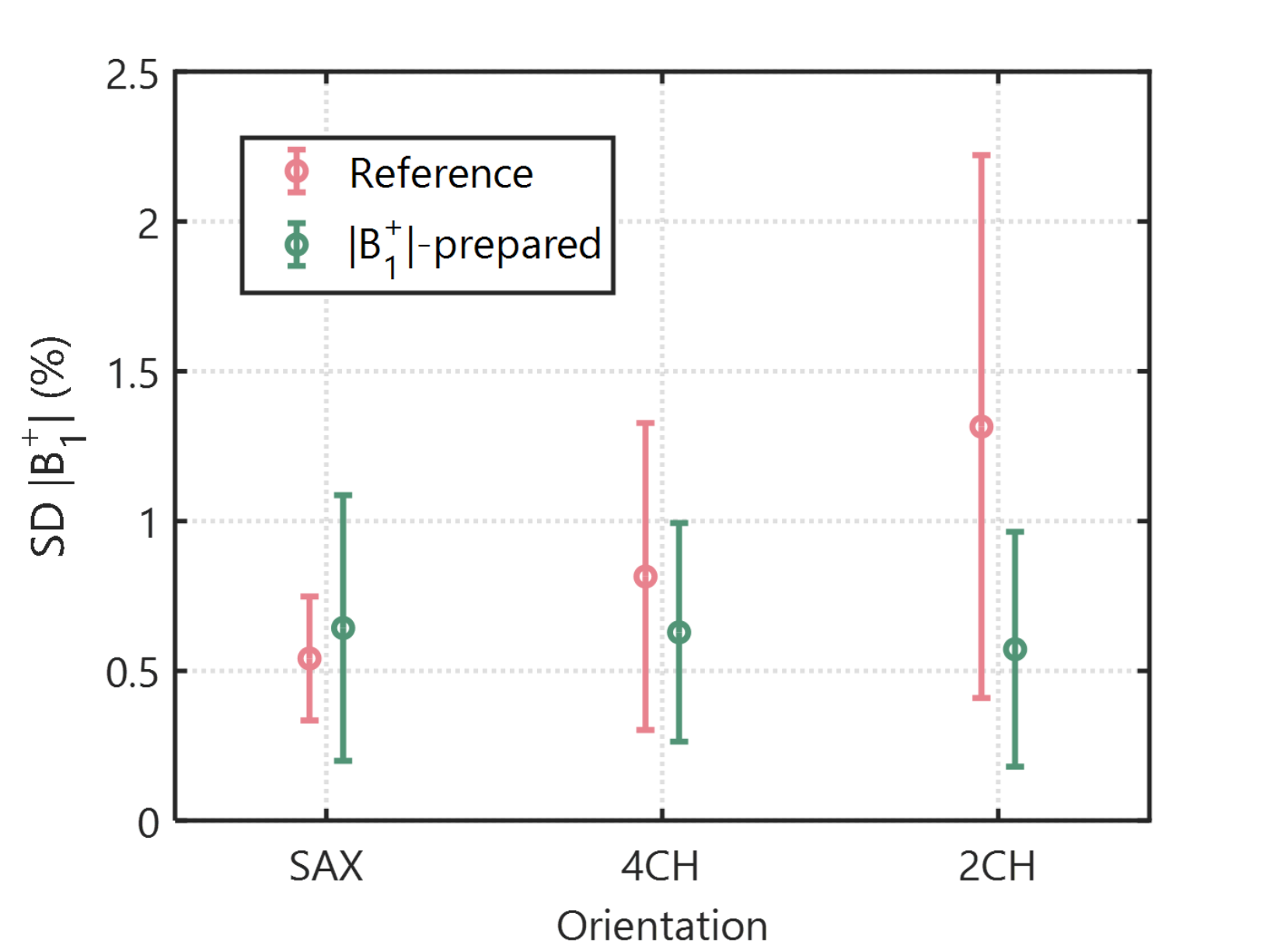
|B1+| maps show the expected pattern of central brightening in the phantom for both methods (Fig. 3). |B1+| prepared mapping achieved comparable map quality with fewer artifacts, when compared with the reference. Mean standard deviation in normalized |B1+| was reduced from 0.35% to 0.06%.In-vivo, both techniques show a typical inhomogeneity pattern, with a B1+ drop towards the right ventricle (Fig. 4). In visual assessment, the proposed |B1+| technique achieves better map quality in terms of noise performance compared with the reference technique. Average SD values in the heart across all 3 subjects were 0.54±0.21%, 0.82±0.51% and 1.31±0.91% for the |B1+| prepared technique in SAX/4CH/2CH orientations, respectively. The Bloch-Siegert shift technique yielded 0.64±0.44%, 0.63±0.36% and 0.57±0.39%, observing a reduction in the latter two orientations (Fig. 5).
Discussion
In this work, we propose a preparation-based cardiac |B1+| mapping technique, and demonstrate good map quality at 3T. Both phantom and in-vivo results show improved noise resilience, compared to previously proposed mapping.In the |B1+|-prepared mapping approach, the off-resonance pulses do not need to be played after the excitation, enabling highly reduced TE (from 9 to 1.5 ms). Thus, |B1+|-preparations can be used in combination with GRE imaging readouts with short TE, to achieve high off-resonance resilience, for example, when imaging in the presence of implants. Alternatively, bSSFP imaging readout, as used in this work, can be employed, resulting in improved SNR.
A low number of off-resonance pulses (four) is used in the proposed technique. This allows to tailor the strength of the pusles to achieve maximum sensitivity for an expected B1+ inhomogeneity range. Furthermore, the number of off-resonant pulses is independent of the imaging resolution or FOV. Thus, it can be straightforwardly extended to 3D imaging for efficient acquisition of whole-heart B1+ magnitude maps.
In the proposed preparation module, the increased duration of the off-resonance pulses leads to a longer preparation time and, thus, more T2 decay during the preparation. Simultaneously, increased pulse duration achieves lower SAR burden, allowing for a trade-off between mapping sensitivity and SAR.
Conclusion
|B1+| prepared mapping enables fast and efficient transmit field mapping around the heart with robust map quality at 3T.Acknowledgements
S.W. acknowledges funding from the 4TU Precision Medicine program, an NWO Start-up STU.019.024, and ZonMW OffRoad 04510011910073.References
1. Yun S, Park S, Park JG, Huh JD, Shin YG, Yun JH, Min JY, Ko SM, Song IY, Yi JG, Hwang HK. Comparison of the diagnostic accuracies of 1.5 T and 3T stress myocardial perfusion cardiovascular magnetic resonance for detecting significant coronary artery disease. Korean Journal of Radiology. 2018;19(6):1007-20.
2. Ferreira PF, Gatehouse PD, Mohiaddin RH, Firmin DN. Cardiovascular magnetic resonance artefacts. Journal of Cardiovascular Magnetic Resonance. 2013 Dec;15(1):1-39.
3. Sacolick LI, Wiesinger F, Hancu I, Vogel MW. B1 mapping by Bloch‐Siegert shift. Magnetic resonance in medicine. 2010 May;63(5):1315-22.
4. Weingärtner S, Zimmer F, Metzger GJ, Uğurbil K, Van de Moortele PF, Akçakaya M. Motion‐robust cardiac mapping at 3T using interleaved Bloch‐Siegert shifts. Magnetic resonance in medicine. 2017 Aug;78(2):670-7.
Figures