4952
Modified Homodyne reconstruction using a high-resolution phase in magnetic resonance images
Xinzeng Wang1, Daniel Litwiller2, Arnaud Guidon3, Tim Sprenger4, and Robert Marc Lebel5
1GE Healthcare, Houston, TX, United States, 2GE Healthcare, Denver, CO, United States, 3GE Healthcare, Boston, MA, United States, 4GE Healthcare, Stockholm, Sweden, 5GE Healthcare, Calgary, AB, Canada
1GE Healthcare, Houston, TX, United States, 2GE Healthcare, Denver, CO, United States, 3GE Healthcare, Boston, MA, United States, 4GE Healthcare, Stockholm, Sweden, 5GE Healthcare, Calgary, AB, Canada
Synopsis
Keywords: Image Reconstruction, Artifacts, Partial Fourier
A partial Fourier acquisition has been widely used for fast MR imaging. To reduce the truncation artifacts in partial Fourier image, Homodyne reconstruction is often used, and it exploits the conjugate symmetry in real-valued signal to recover the full k-space. However, the MR signal is complex-valued. Artifacts are commonly observed in Homodyne images in the regions of rapid phase change due to the interference of imaginary components of adjacent pixels. In this work, we proposed a modified Homodyne reconstruction to reduce the conventional Homodyne artifacts and truncation artifacts by using a high-resolution phase from a pre-trained deep-learning network.Introduction
A partial Fourier acquisition has been widely used for fast MR imaging. Many partial Fourier reconstruction methods have been proposed to minimize the truncation artifacts and image blurring due to the truncation in k-space, such as Conjugate Synthesis, Homodyne, POCS, etc (1-4). These reconstruction methods exploit the conjugate symmetry because real functions have conjugate symmetry in the frequency domain. However, inhomogeneous B0 field, motion, unwanted phase shift, etc. introduce phase into the MR images, violating the real-valued assumption. Therefore, partial k-space reconstruction always requires some type of phase estimation that is not affected by the aforementioned truncation artifacts. However, the conventional reconstruction methods use a low-resolution phase for phase correction, resulting in gross artifacts in regions of the image where the phase from the low-pass portion of kspace fails to adequately resolve rapid local variations.To overcome these challenges and mitigate these artifacts, this work describes a modified partial Fourier reconstruction method using a high-resolution phase generated by a pre-trained deep learning network. The proposed method was evaluated with out-of-phase image, functional MRI (fMRI) and diffusion weighted MRI (DWI) images.
Methods
The Conventional Homodyne reconstruction method uses a low-resolution phase for phase correction after weighting convolution. In regions with rapid phase variations, the phase correction could not completely suppress the interferences of the imaginary component in the nearby pixels (1), resulting in Homodyne artifacts.In contrast, the proposed modified Homodyne reconstruction uses a high-resolution phase map for phase correction before the weighting convolution. The high-resolution phase map was estimated using a deep-learning network, which was trained from a database of over 10,000 images. After a more accurate phase correction, the interference of the nearby pixels would be well suppressed, therefore reducing Homodyne artifacts. A digital phantom was used to demonstrate the feasibility and improvements of the modified Homodyne reconstruction method. Then, out-of-phase liver images, fMRI images and diffusion weighted images were acquired using half-NEX on a GE 3T MRI scanner (Discovery MR750, GE Healthcare, Waukesha, WI) with IRB approval and written informed consent to compare the conventional Homodyne reconstruction and the modified Homodyne reconstruction methods.
Results and Discussions
In the digital phantom, each component was assigned a different phase (Figure 1b) to simulate phase variations in real MR imaging. The k-space was truncated to simulate partial Fourier acquisition with a partial NEX of 0.625. The image reconstructed using zero-filling showed the ringing artifacts (Figure 1c), which are reduced by the Homodyne reconstruction (Figure 1d). The conventional Homodyne reconstruction generated artifacts (Homodyne artifacts) in the regions of rapid phase changes, as shown in Figure 1d. This is mainly caused by the interference of the imaginary component in the adjacent voxels. The modified Homodyne reconstruction corrects the phase with a high-resolution phase map generated from a pre-trained DL network, eliminating the interference of imaginary components in the adjacent voxels. Both the truncation artifacts and the traditional Homodyne artifacts were well minimized using the modified Homodyne reconstruction.To evaluate the Homodyne reconstruction methods in regions of rapid phase change, the out-of-phase images were acquired in a volunteer with a partial NEX of 0.65. Water and fat signals have ~180 degrees phase difference in out-of-phase images, generating the India ink artifact at the fat-water interface, as shown in Figure 2a. The India ink artifact could be used in diagnosis to confirm the fatty nature of some lesions. However, part of the dark boundary disappeared in the image that was reconstructed using conventional Homodyne due to the interference of the imaginary component in adjacent voxels, as shown in Figure 2b (red arrows). In contrast, modified homodyne reconstruction minimizes the interference with a robust high-resolution phase correction and improves the definition of water-fat boundaries (Figure 2c, green arrows).
Functional MRI (fMRI) images were often acquired with partial Fourier to accelerate the acquisition. However, bulk motion, pulsation motion and field inhomogeneities could introduce high-frequency phase in fMRI images. With the conventional homodyne artifacts, Homodyne artifacts with dark boundaries or pixels are commonly seen in fMRI images, as shown in Figure 3a (red arrows). These artifacts were well suppressed using the modified Homodyne reconstruction, as shown in Figure 3b.
Diffusion weighted imaging also uses partial Fourier to reduce the echo train length and distortion. However, the movement of the eyeball, CSF and blood during the diffusion preparation could introduce rapid phase change, resulting in a signal loss in these regions (red arrows, Figure 4a, 4c). However, this kind of artifact was minimized using the proposed modified Homodyne reconstruction.
Conclusion
A high-resolution phase generated from the pre-trained deep learning model enables robust phase correction for Homodyne reconstruction. With a robust and accurate phase correction, the proposed modified Homodyne swapped the order of phase correction and weighting convolution, reducing the truncation artifacts and the traditional Homodyne artifacts due to the interference of imaginary components in adjacent voxels.Acknowledgements
No acknowledgement found.References
[1] Partial k-space Reconstruction by John Pauly, https://users.fmrib.ox.ac.uk/~karla/reading_group/lecture_notes/Recon_Pauly_read.pdf
[2] Noll DC, Nishimura GD, Macoviski A. Homodyne detection in magnetic resonance imaging. IEEE Trans Med Imaging 1991; 10:154–163.
[3] Haacke EM, Lindskog ED, Lin W. A fast, iterative, partial-Fourier technique capable of local phase recovery. J Magn Reson 1991; 92:126–145.
[4] Cuppen J, van Est A. Reducing MR imaging time by one-sided reconstruction. Magn Reson Imaging 1987; 5:516–527
Figures
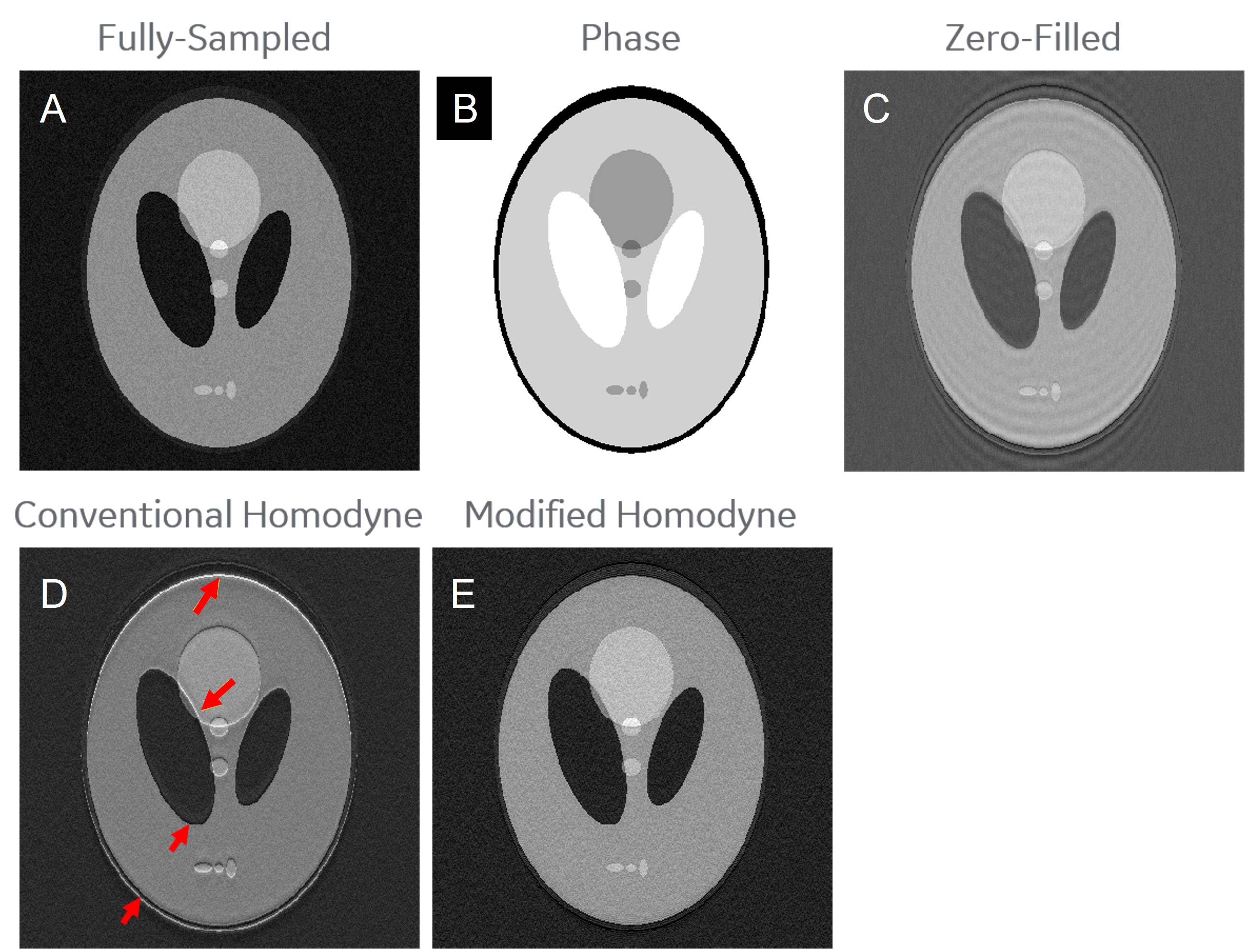
Figure 1. Comparison between the conventional homodyne
reconstruction and the proposed modified Homodyne reconstruction. The artifacts
(d, red arrows) in the conventional Homodyne image are corresponding to the
regions with rapid phase change, as shown in the phase map (b). These artifacts
were minimized using the proposed modified Homodyne reconstruction (e).
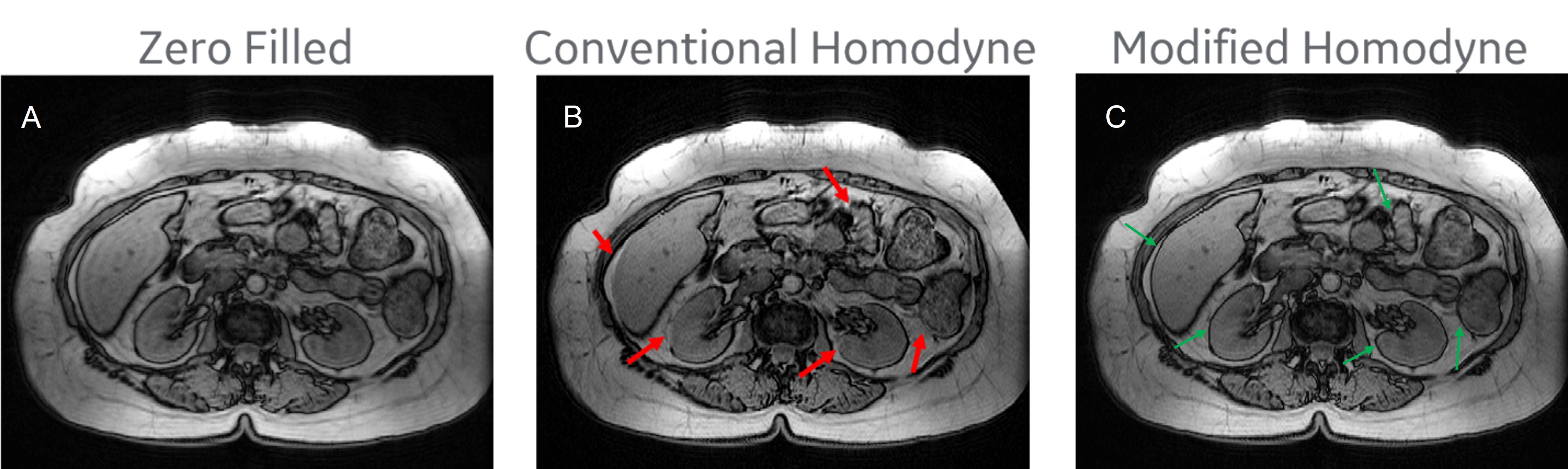
Figure
2. In-vivo out-of-phase images reconstructed using the conventional homodyne
reconstruction and the proposed modified homodyne reconstruction. Modified
homodyne reconstruction minimizes the artifacts in conventional homodyne images
(b, red arrows) and improve the definition of water-fat boundaries (c, green arrows).
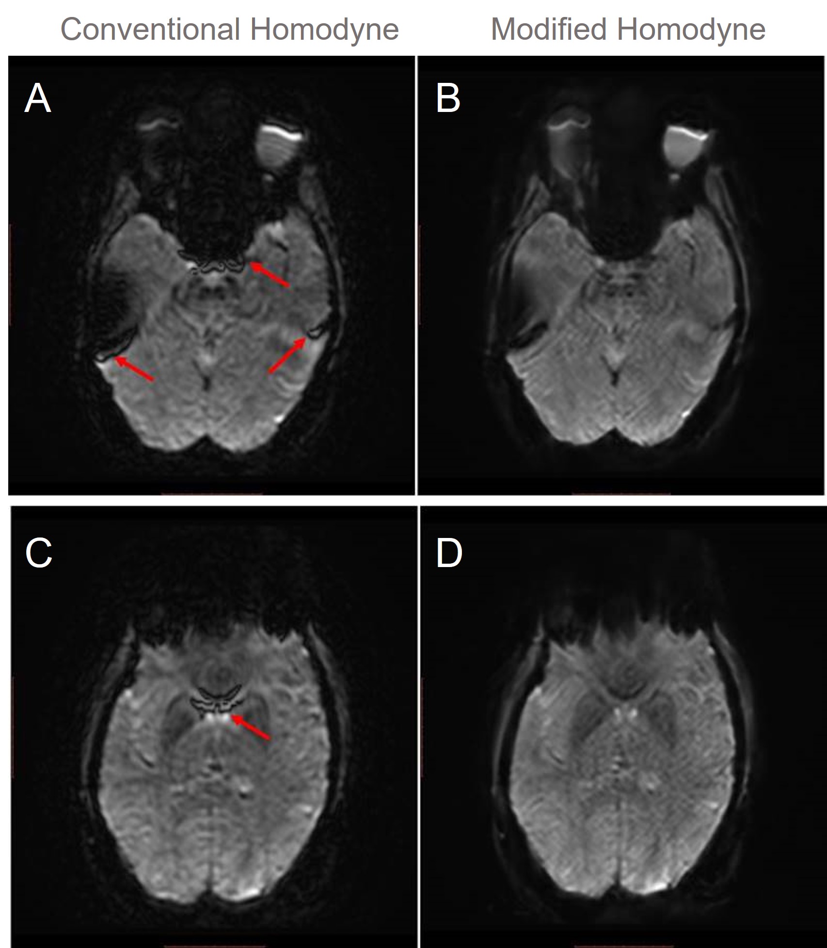
Figure 3. fMRI images reconstructed using the
conventional homodyne reconstruction and the proposed modified homodyne
reconstruction. Modified homodyne reconstruction minimizes the artifacts in
conventional homodyne images (a, c, red arrows).
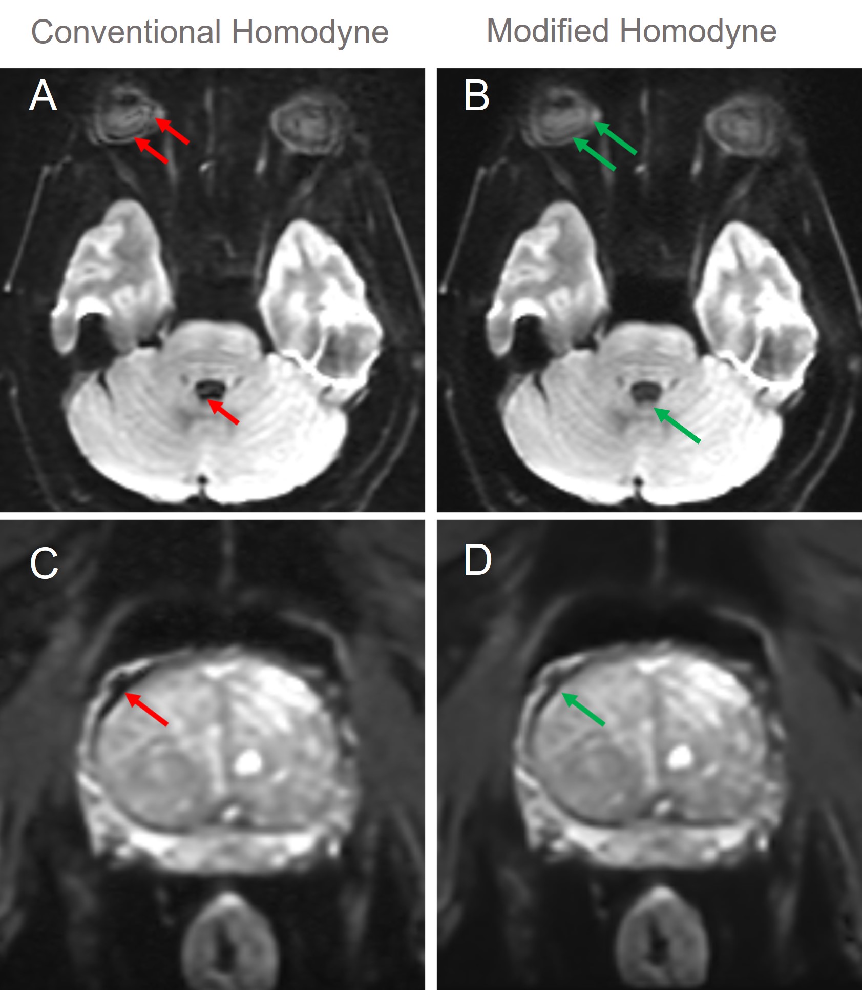
Figure 4. In-vivo DW images reconstructed using the
conventional homodyne reconstruction and the proposed modified homodyne
reconstruction. The movement of eyeball, CSF and blood could introduce rapid
phase change, resulting in signal loss in regions close to the ventricles of
the brain, eyeball (red arrows, a) and blood vessels (red arrow, c). This is one of the causes
of “wormhole” artifacts in DW images. However, this kind of artifact
was minimized using the proposed modified Homodyne reconstruction.
DOI: https://doi.org/10.58530/2023/4952