4940
Free water DTI for evaluating gray matter microstructure in major depressive disorder: A preliminary study1Medical Imaging Department, Nanfang Hospital, Guangzhou, China, 2MR Research, GE Healthcare, Beijing, China, Beijing, China, 3Department of Radiology, Zengcheng Branch of Nanfang Hospital, Guangzhou, China, 4PET/CT Center, Department of Nuclear Medicine, First Affiliated Hospital of Shenzhen University, Shenzhen Second People’s Hospital, Shenzhen, China
Synopsis
Keywords: Psychiatric Disorders, Brain, Major depressive disorder
Major depressive disorder (MDD) is a common mental disease with unclear pathophysiology. Herein, we explored the differences in the whole-brain gray matter free-water axial diffusivity (AD) and mean diffusivity (MD) values between MDD patients and healthy controls using free-water DTI. The results showed altered free-water AD and MD values in specific regions of the gray matter in MDD patients, which may be associated with alterations in the brain gray matter microstructure, such as microstructure damage or neurodegeneration. Thus, these quantitative variables could be used as valuable biomarkers that could further our understanding of the pathophysiological mechanisms of depression.Introduction
Major depressive disorder (MDD) is a clinical depression that affects about 280 million people worldwide. Still, its physiological and pathological mechanisms are not fully understood1. Diffusion tensor imaging is a noninvasive magnetic resonance imaging technique used to evaluate the integrity and fiber orientation of white matter, which have been widely used in the study of psychiatric diseases. However, due to the impact of cerebrospinal fluid (CSF) and partial volume effect on diffusion mapping2,3, the application of DTI in the analysis of gray matter (GM) microstructure alterations caused by MDD has been limited. Previous study has proposed a novel diffusion technique, named free-water diffusion tensor imaging (FW-DTI), to reduce the influence of partial volume factors, such as CSF, on diffusion signals and increase the interpretability of diffusion signals4. Therefore, this study aimed to explore the differences between the free-water mean diffusivity (MD) and axial diffusivity (AD) values in GM microstructure between MDD and healthy controls.Methods
The present study included 16 patients with MDD (2 males and 14 females, aged 18-34 years) and 24 healthy controls (9 males and 15 females, aged 21-29 years). Hamilton Anxiety Rating Scale (HAMA), 24-item Hamilton Depression Rating Scale (HAMD), and Beck Scale for Suicide Ideation (BSS) were used to evaluate all subjects. The protocol of this study was approved by the local ethics committee.All participants signed an informed consent form prior to participation in this study. All patients underwent the same brain MRI protocol on a 3.0T MRI scanner (Signa Architect 3.0T, GE, USA) with the 48-channel head coil. The T1-weight (T1w) images were acquired using sagittal 3D T1-weighted MP-RAGE sequence with an isotropic resolution of 1.00 mm. Then, DTI images were scanned. The main parameters of DTI were as follows: in-plane voxel size = 2 mm × 2 mm; slice thickness/gap = 2/0 mm; acquisition layers = 62; TR/TE = 7000/84 ms, diffusion sensitivity factor (b-value) = 1000 s/mm2.
The DTI images were first preprocessed using EDDY and TOPUP in FSL toolboxes (www.fmrib.ox.ac.uk/fsl) to correct the susceptibility-induced geometric distortions, eddy current distortions, and inter-volume subject motion. Preprocessed DTI data was fit to a regularized bi-component model5 using DIPY toolbox (https://dipy.org) and free-water AD and MD mapping images were calculated. Then, the linear transformation matrix between images with zero diffusion gradient (b0) images and T1w images and the non-linear warped images between T1w images and T1w template images in MNI space were obtained using the Advanced Normalization Tools (ANTs). Next, free-water AD and MD images were registered to MNI space by applying the linear transformation matrix and non-linear warped images. Finally, free-water AD and MD values in each cortical region were extracted using the Automated Anatomical Labeling atlas.
Group comparisons of the free-water AD and MD values were performed using an independent sample t-test and the multiple-comparison errors were controlled by the false discovery rate (FDR). Correlation analysis was performed using Spearman’s correlation coefficient. A two-tailed p-value < 0.05 indicated statistical significance.
Results
There was no significant difference in age and sex between groups (p > 0.05, Table 1). However, free-water AD and MD values of several brain regions were altered in patients with MDD compared to healthy controls (all p < 0.05, Table 2 and Figure 1). Among these regions, the free-water MD value of the left posterior cingulate gyrus was positively correlated with HAMD in patients with MDD (r = 0.542, p = 0.0321) (Figure 2). In addition, there was no significant correlation between no AD and MD values with HAMA or BSS (all p > 0.05).Discussion
In this study, decreases of free-water AD and MD caused by MDD were found in the widespread cortical regions, which may be associated with alterations in the brain gray matter microstructure, such as microstructure damage or neurodegeneration. Moreover, the posterior cingulate gyrus (PCC) is an important part of the limbic lobe, which is mainly responsible for emotion, action-outcome learning, memory, and other functions. Previous studies have shown functional and structural abnormalities in PCC in patients with depression6,7. In our study, abnormalities in the left PCC were found in MDD patients but not in healthy controls. Therefore, the decreasing free-water MD value in PCC in patients with MDD probably means that the gray matter microstructure of PCC has been damaged. Meanwhile, the MD value was correlated with the degree of clinical symptoms, which also confirmed the symptoms of MDD depression and memory decline combined with the main function of PCC.Conclusion
These preliminary data found that altered FW-DTI parameters in specific regions of the gray matter in first-visit MDD patients, which may be associated with alterations in the brain gray matter microstructure, such as reduced free-water MD value. These values could be potentially used as a biomarker to further understand the pathophysiological mechanisms of depression.Acknowledgements
This study was supported by the National Natural Science Foundation of China grant 82172012.References
1. Cuijpers P, Stringaris A, Wolpert M. Treatment outcomes for depression: challenges and opportunities. Lancet Psychiatry. 2020;7(11):925–927.
2. Koo B-B, Hua N, Choi C-H, Ronen I, Lee J-M, Kim D-S. A framework to analyze partial volume effect on gray matter mean diffusivity measurements. Neuroimage. 2009;44(1):136–144.
3. Ma X, Kadah YM, LaConte SM, Hu X. Enhancing measured diffusion anisotropy in gray matter by eliminating CSF contamination with FLAIR. Magn Reson Med. 2004;51(2):423–427.
4. Pasternak O, Sochen N, Gur Y, Intrator N, Assaf Y. Free water elimination and mapping from diffusion MRI. Magn Reson Med. 2009;62(3):717–730.
5. Hoy AR, Koay CG, Kecskemeti SR, Alexander AL. Optimization of a free water elimination two-compartment model for diffusion tensor imaging. Neuroimage. 2014;103323–333.
6. Rolls ET. The cingulate cortex and limbic systems for emotion, action, and memory. Brain Struct Funct. 2019;224(9):3001–3018.
7. Leech R, Sharp DJ. The role of the posterior cingulate cortex in cognition and disease. Brain. 2014;137(1):12–32.
Figures


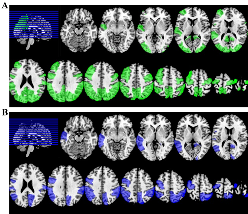
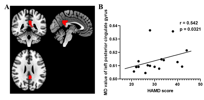
Fig.2. Relationship between clinical scales and free-water DTI parameters with significant group differences. The free-water MD values in the left posterior cingulate gyrus (A) show a significantly positive correlation (R = 0.542; P = 0.0321) with the HAMD scores (B).