4939
Altered Structural Connectivity Of Thalamocortical Connections in Major Depressive Disorder: A 7T Study
Ameen S. Al Qadi1, Gaurav S. Verma1, James S. Murrough2, and Priti S. Balchandani 1
1Biomedical Engineering And Imaging Institute, Icahn School Of Medicine At Mount Sinai, New York, NY, United States, 2Department Of Psychiatry, Icahn School Of Medicine At Mount Sinai, New York, NY, United States
1Biomedical Engineering And Imaging Institute, Icahn School Of Medicine At Mount Sinai, New York, NY, United States, 2Department Of Psychiatry, Icahn School Of Medicine At Mount Sinai, New York, NY, United States
Synopsis
Keywords: Psychiatric Disorders, Brain Connectivity, Tractography, 7T, Major Depressive Disorder
This study investigated the structural integrity of thalamic nuclei and their white matter connections in the brains of patients with Major Depressive Disorder (MDD) to help elucidate MDD etiology using diffusion tractography and structural MRI at 7T. Group differences were found with decreased FA and increased MD, RD, and AD in the tracts of multiple nuclei in the MDD cohort compared to controls. There were no significant differences in the average volumes of the nuclei between groups. These results suggest aberrant axonal integrity in multiple pathways responsible for visual & auditory processing, motor function, and emotional response networks.Introduction
Major Depressive Disorder (MDD) is a multi-factorial psychiatric disorder that is difficult to diagnose due to subjective criteria and variable presentation1. Several nuclei of the thalamus have been implicated in pathways affected by MDD, including the Mediodorsal (MDM and MDI) nuclei which are visual-processing nuclei2,3, and the Pulvinar nuclei4 which play a crucial role in emotional awareness and attention4. Understanding the potential changes in these nuclei and their thalamocortical circuits can aid in identifying structural biomarkers and potential etiological mechanisms of MDD. By taking advantage of the increased SNR and exquisitie resolution of structural and diffusion MRI at 7T, a structural and DTI study of thalamic nuclei and their white matter connections was investigated using diffusion tractography.Methods
T1-weighted MP2RAGE images were acquired for 41 MDD patients and 44 healthy controls were scanned on a Siemens Magnetom 7T scanner (Siemens Healthineers, Erlangen, Germany) using a SC72CD gradient coil with a 1TX/32RX head (Nova Medical, Wilmington, MA, USA). FreeSurfer was used for cortical reconstruction and thalamic nuclei segmentation. Thalamic nuclei volumes were normalized to the subject’s total brain volume (TBV) to account for inter-subject variability.Diffusion-weighted images were acquired for 33 MDD patients and 34 controls with the following parameters: b=1500s/mm2, TR=7200ms, TE=67.6ms, FOV=210x210mm, resolution=1.05mm isotropic, 64 diffusion directions and 5 b0 directions in forward and reverse phase encoding directions. Images were denoised and corrected for eddy-current distortion, subject motion, and B1 inhomogeneity using MRtrix5. Tractography was performed using the thalamic nuclei as seeding regions with MRtrix’s tckgen6, and spurious tracts were checked with SIFT27. Mean fractional anisotropy (FA), mean diffusivity (MD), radial diffusivity (RD), and axial diffusivity (AD) were sampled from the resultant tracts. All data was tested for normality using the Shapiro-Wilks test. An unpaired two-tailed t-test was used for normal distributions, and the Wilcoxon rank-sum test for non-normal distributions. The significance threshold for all tests was ⍺ = 0.05, and statistical analyses was performed in R8. These data were not corrected for age and gender.
Results
Diffusion Microstructure:Overall, 11 nuclei had tracts with significant differences in average diffusion microstructure between MDD patients and healthy controls. These nuclei were the left and right Medial Geniculate (MGN), left Centromedian (CM), left Parafascicular (Pf), left Ventral lateral anterior (VLa), left Pulvinar anterior (PuA), left Ventromedial (VM), left Lateral posterior (LP), left Lateral geniculate (LGN), right Ventral lateral posterior (VLp), right Ventral anterior magnocellular (VAmc) nuclei. Mean FA was decreased in the MDD cohort compared to controls for the left & right MGN, left CM and Pf, as shown in Figure 1, whereas mean MD was increased in the MDD cohort compared to controls for left & right MGN, left CM, VLa, PuA, VM, and LP as shown in Figure 2. Mean RD was increased in the MDD cohort for the left & right MGN, left LGN, CM, VLa, PuA, LP, and VLp, and mean AD was also increased in the MDD cohort for the left VAmc.
Volumetrics:
No significant differences found in volumes of thalamic nuclei between MDD patients and healthy controls. The mean right Inferior Pulvinar (PuI) and Medial Pulvinar (PuM) volumes trended near significance as shown in Table 1.
Discussion & Conclusion
This novel study provides an unprecedented view into the integrity of thalamocortical connectivity in the context of MDD. Decreased FA and increased mean, radial, and axial diffusivities suggest decreased axonal integrity in the tracts visualized in Figure 3. This is because reduced anisotropy in axonal fibers is associated with increased diffusivity given that water molecules can diffuse in more directions than just the direction of the axon. Functionally, these tracts participate in visual processing (PuA9, LGN9, LP10), auditory processing (MGN11), motor function (VLa12, VLp12), and mediate between the PFC and striatum (CM-Pf13, VM14), and substantia nigra (VAmc15). Aberrant axonal integrity in these white-matter pathways may be explained by increased stress-mediated inflammatory responses characteristic of cognitive distortions associated with the functions that these nuclei mediate. For example, connections between the amygdala and the Pulvinar nuclei have been strongly suggested to mediate emotional responses to visual stimuli as part of visual information pre-processing before being integrated into a person’s awareness16,17. A structural distortion of this pathway may explain overtly anxious behavior in for certain visual cues, which could in result increased anxiety.With regards to the thalamic nuclei volumes, this 7T study yielded no statistically significant differences in mean volume was found for the thalamic nuclei despite trends towards significance for the right PuI & PuM. Overall, the utilization of ROI-based tractography to study the structural connectivity of thalamic nuclei is feasible to identify DTI biomarkers in MDD at 7T.
Acknowledgements
This work was funded through NIH R01 MH109544.References
(1) Otte, C., Gold, S., Penninx, B. et al. Major depressive disorder. Nat Rev Dis Primers 2, 16065 (2016). https://doi.org/10.1038/nrdp.2016.65(2) Golden, Erin C., et al. "Mediodorsal nucleus and its multiple cognitive functions." Neurology 87.20 (2016): 2161-2168.
(3) Hamilton JP, Farmer M, Fogelman P, Gotlib IH. Depressive Rumination, the Default-Mode Network, and the Dark Matter of Clinical Neuroscience. Biol Psychiatry. 2015 Aug 15;78(4):224-30. doi: 10.1016/j.biopsych.2015.02.020. Epub 2015 Feb 24. PMID: 25861700; PMCID: PMC4524294.
(4) Hamilton, J. Paul, et al. "Functional neuroimaging of major depressive disorder: a meta-analysis and new integration of baseline activation and neural response data." American Journal of Psychiatry 169.7 (2012): 693-703.
(5) Iglesias, Juan Eugenio, et al. "A probabilistic atlas of the human thalamic nuclei combining ex vivo MRI and histology." Neuroimage 183 (2018): 314-326.
(6) Tournier, J‐Donald, Fernando Calamante, and Alan Connelly. "MRtrix: diffusion tractography in crossing fiber regions." International journal of imaging systems and technology 22.1 (2012): 53-66.
(7) Smith RE, Tournier JD, Calamante F, Connelly A. SIFT2: Enabling dense quantitative assessment of brain white matter connectivity using streamlines tractography. Neuroimage. 2015 Oct 1;119:338-51. doi: 10.1016/j.neuroimage.2015.06.092. Epub 2015 Jul 8. PMID: 26163802.
(8) R Core Team (2021). R: A language and environment for statistical computing. R Foundation for Statistical Computing, Vienna, Austria. https://www.R-project.org/.
(9) Cudeiro, Javier, and Adam M. Sillito. "Looking back: corticothalamic feedback and early visual processing." Trends in neurosciences 29.6 (2006): 298-306.
(10) Gale, Samuel D., and Gabe J. Murphy. "Distinct cell types in the superficial superior colliculus project to the dorsal lateral geniculate and lateral posterior thalamic nuclei." Journal of Neurophysiology 120.3 (2018): 1286-1292.
(11) Ferrara, Nicole C., et al. "Input from the medial geniculate nucleus modulates amygdala encoding of fear memory discrimination." Learning & Memory 24.9 (2017): 414-421.
(12) Bocchetta, Martina, et al. "Thalamic nuclei in frontotemporal dementia: Mediodorsal nucleus involvement is universal but pulvinar atrophy is unique to C9orf72." Human brain mapping 41.4 (2020): 1006-1016.
(13) Ilyas A, Pizarro D, Romeo AK, Riley KO, Pati S. The centromedian nucleus: Anatomy, physiology, and clinical implications. J Clin Neurosci. 2019 May;63:1-7. doi: 10.1016/j.jocn.2019.01.050. Epub 2019 Feb 28. PMID: 30827880.)
(14) Anastasiades PG, Collins DP, Carter AG. Mediodorsal and Ventromedial Thalamus Engage Distinct L1 Circuits in the Prefrontal Cortex. Neuron. 2021 Jan 20;109(2):314-330.e4. doi: 10.1016/j.neuron.2020.10.031. Epub 2020 Nov 13. PMID: 33188733; PMCID: PMC7855187.
(15) Ohye, Chihiro. "Thalamus and thalamic damage." (2002): 575-597.
(16) Brown, Stephanie SG, et al. "Ultra-high-resolution imaging of amygdala subnuclei structural connectivity in major depressive disorder." Biological Psychiatry: Cognitive Neuroscience and Neuroimaging 5.2 (2020): 184-193.
(17) Abivardi, Aslan, and Dominik R. Bach. "Deconstructing white matter connectivity of human amygdala nuclei with thalamus and cortex subdivisions in vivo." Human brain mapping 38.8 (2017): 3927-3940.
Figures
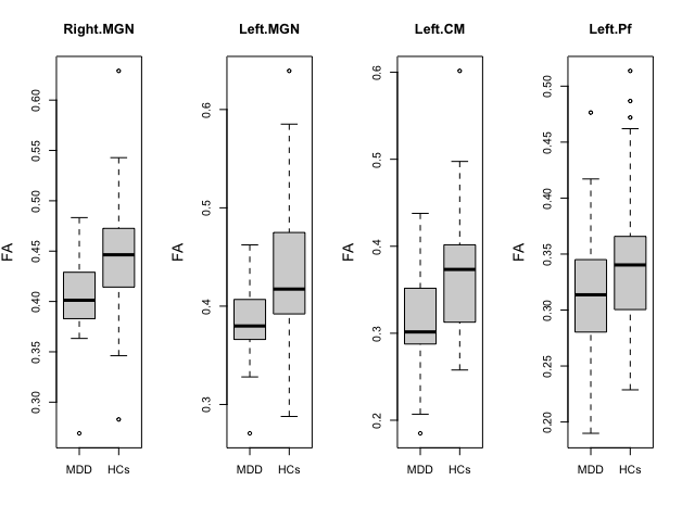
Figure 1. Box-plots of left & right MGN, left CM, and left Pf nuclei demonstrating decreased FA in MDD patients compared to healthy controls
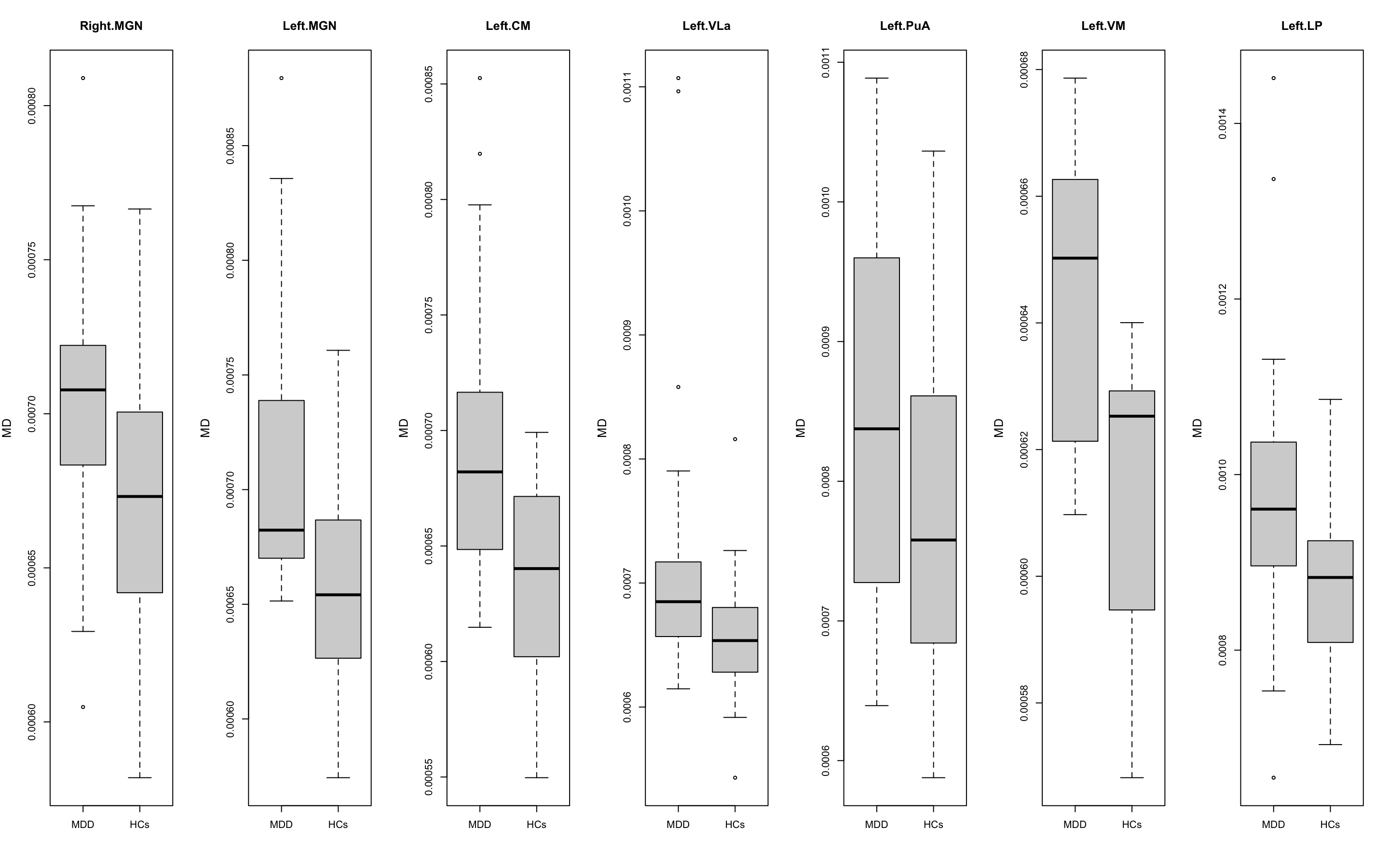
Figure 2. Box-plots of left & right MGN, left CM, VLa, PuA, VM, LP demonstrating increased MD in MDD patients compared to healthy controls
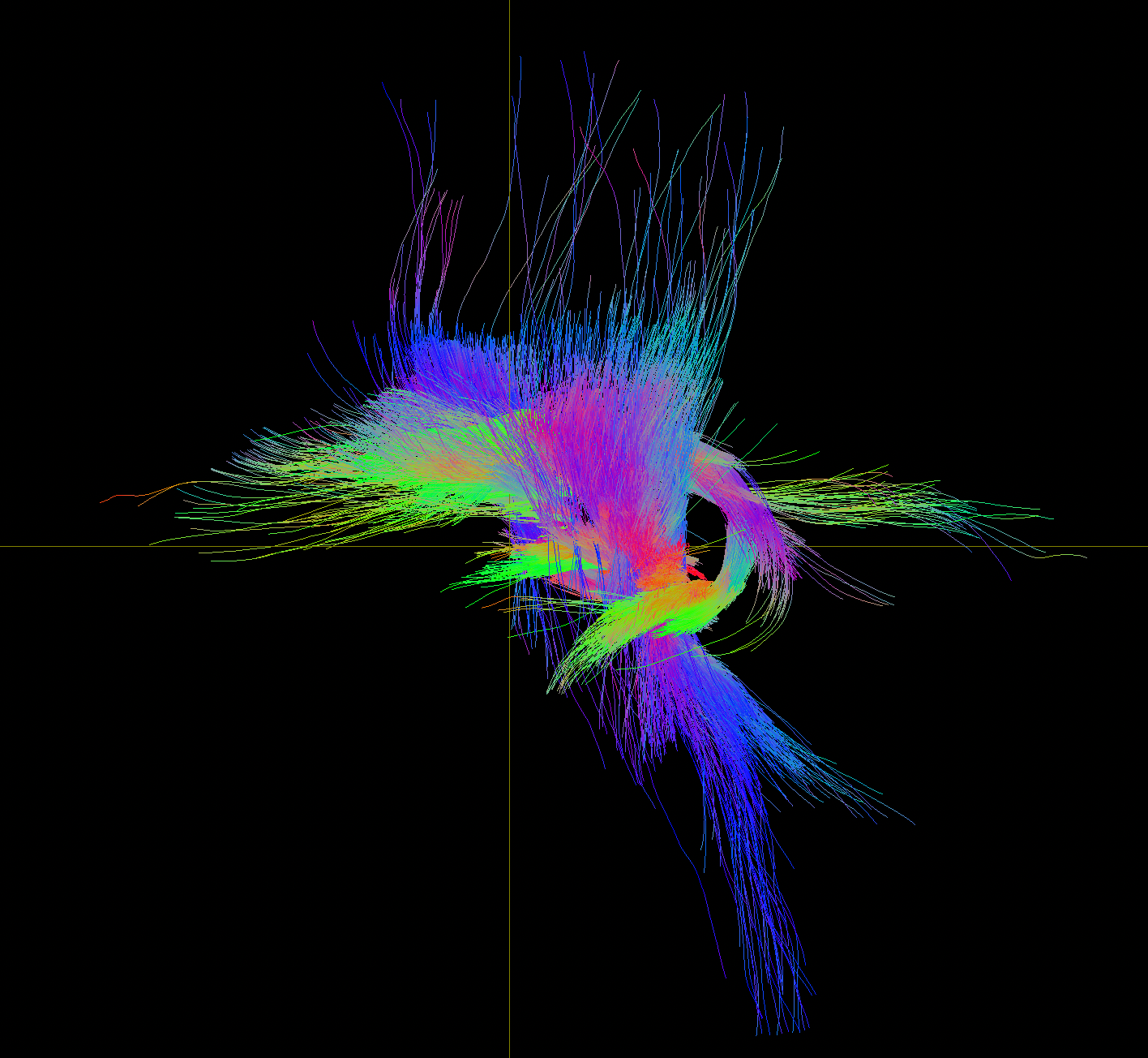
Figure 3. All statistically significant tracts isolated in a 3D render using MRView in the sagittal plane. The corticospinal tracts (long blue fibers) and thalamo-prefrontal tracts are clearly visible
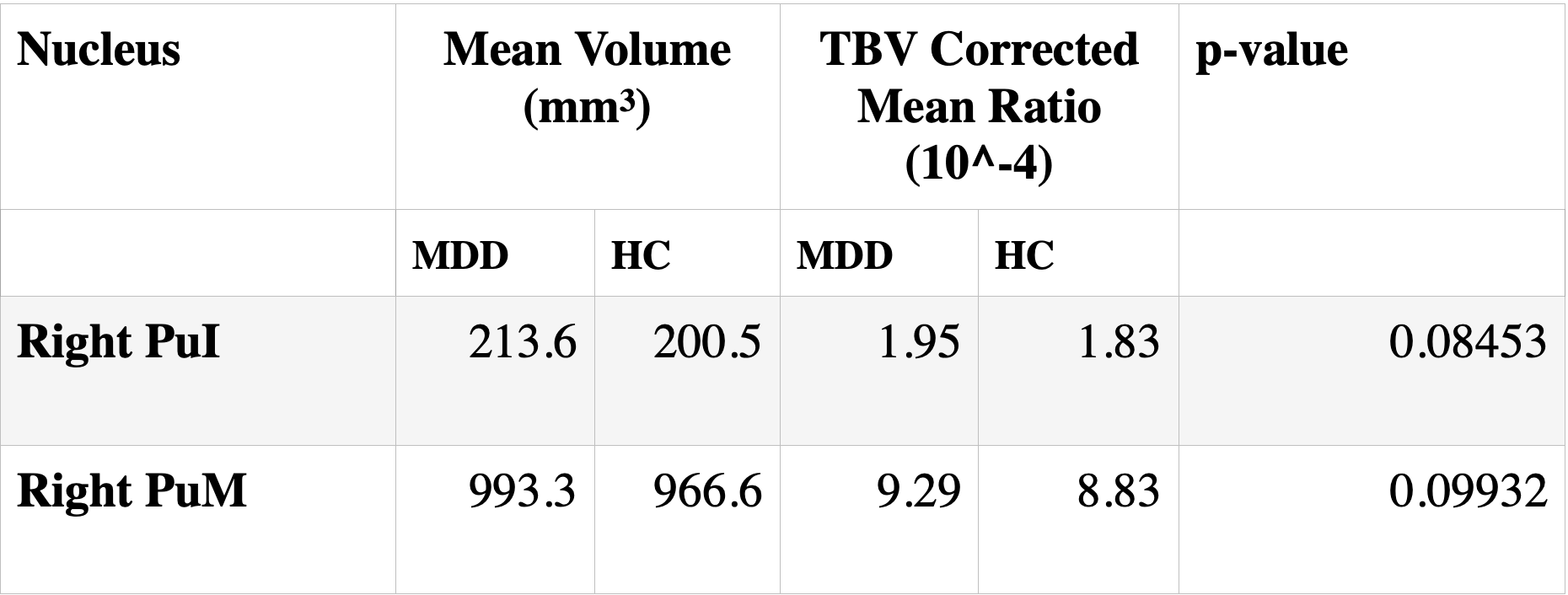
Table 1. Thalamic Nuclei that demonstrated group differences in mean volume between MDD patients and healthy controls close to the statistical significance threshold of ⍺ = 0.05. All other group comparisons yielded p-values > 0.1
DOI: https://doi.org/10.58530/2023/4939