4928
Sex Difference of Hippocampal Subfields in First-episode Drug-naive Patients with Major Depressive Disorder1Department of Radiology, Huaxi MR Research Center (HMRRC), Functional and Molecular Imaging Key Laboratory of Sichuan Province, Department of Radiology, West China Hospital of Sichuan University, Chengdu, China., Chengdu, China, 2Huaxi MR Research Center (HMRRC), Functional and Molecular Imaging Key Laboratory of Sichuan Province, Department of Radiology, West China Hospital of Sichuan University, Chengdu, China., Chengdu, China
Synopsis
Keywords: Psychiatric Disorders, Brain
We investigated hippocampal subfields volumes in a relatively large sample of first-episode drug-naive MDD and explored sex-related effects. We used Freesurfer software to segment the hippocampus with longitudinal axis and transverse axis automatically. We found there were different alterations of hippocampus along different axis. The sex-independent decreased volume of hippocampal and its subregions along the longitudinal, while the sex specific alteration only detected in the subfields along the transverse axis. These current findings may interpretate the hippocampus appears to have functional segregation along the different axis, which may contribute to developing new diagnostic and mechanism of sex difference for MDD.Introduction
The hippocampus, which can be segmented either into the head, body, and tail along with its longitudinal axis or into several subfields along the transverse axis, is important hubs in neurobiological model of major depressive disorder (MDD), and its subfields subserve different functions that may serve different roles in the neuropathophysiology of MDD[1]. Hippocampal volume reduction was reported to underlie depressive symptomatology, however, the evidence to date remains inconsistent[2-4]. Several MRI studies using automatic segmentation methods revealed hippocampal subfields volume reduction in MDD patients, but automatic segmentation was always used from one axis. Thus, it is crucial to assess the volumetric alteration in hippocampal subfields along with both longitudinal and transverse axes in individuals with MDD.Besides, sex differences in terms of incidence and symptoms were prominent in individuals with MDD[5], thus we aimed to explore whether there is sex difference in the hippocampal subfields and whether it is related to different segmentations axis in individuals with MDD.Methods
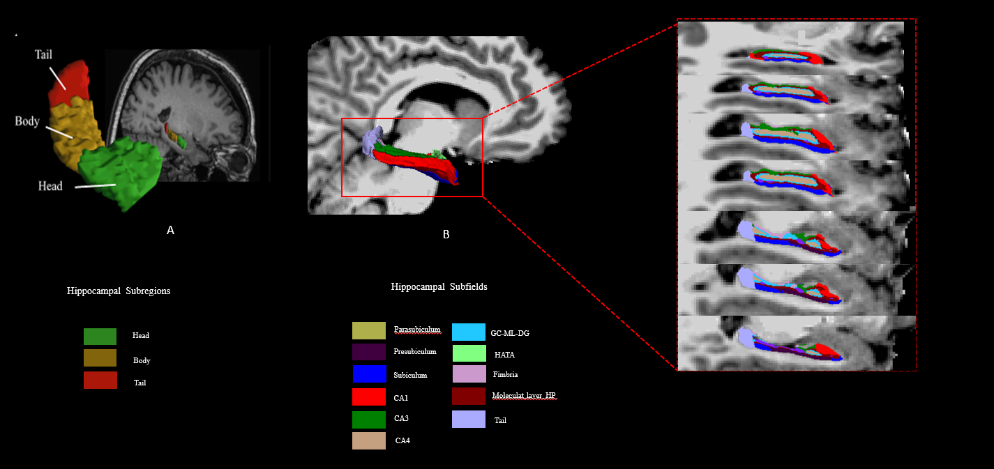
A total of 144 treatment-naive, non-comorbid individuals with MDD on early stage of illness progression and 135 age- and sex-matched HCs were recruited, and high-resolution T1-weighted MR images were obtained for all participants. The substructure segmentation was performed using FreeSurfer software automatically. The hippocampus were further divided into three subregions (head, body and tail) along its longitudinal axis(Fig.1A form Liu et al.,2021[6]) or eleven subfields (CA1,CA3,CA4, molecular layer, GC.ML.DG, subiculum, presubiculum, parasubiculum, fimbria, HATA, tail) according to its transverse axis(Fig.1B).We performed General linear model (GLM) to test the sex-by-diagnosis interactions and main effect of diagnosis. For these substructures with significant sex-by-diagnosis interaction, post-hoc analyses were conducted (female MDD vs. female HC, male MDD vs. male HC). Main effect of volumetric differences between MDD and HCs were also reported. Partial correlations were performed between HAMD/HAMA/memory related scores and altered hippocampal substructure volume with age and ICV as covariants in male/female individuals with MDD separately. The false discovery rate (FDR) method was applied to correct for multiple comparisons.
Results
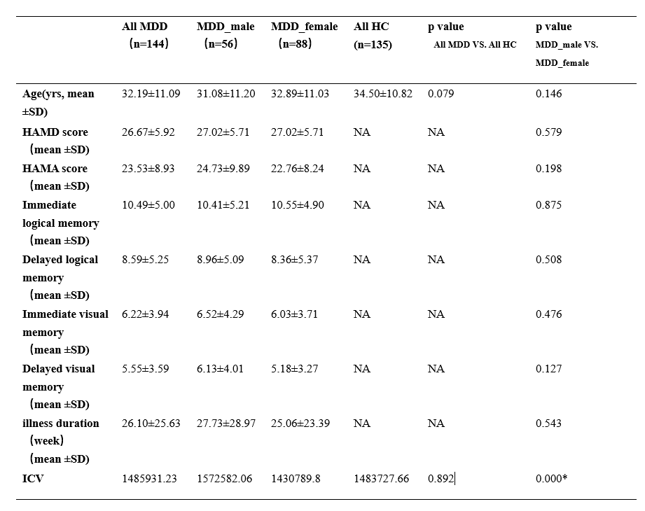
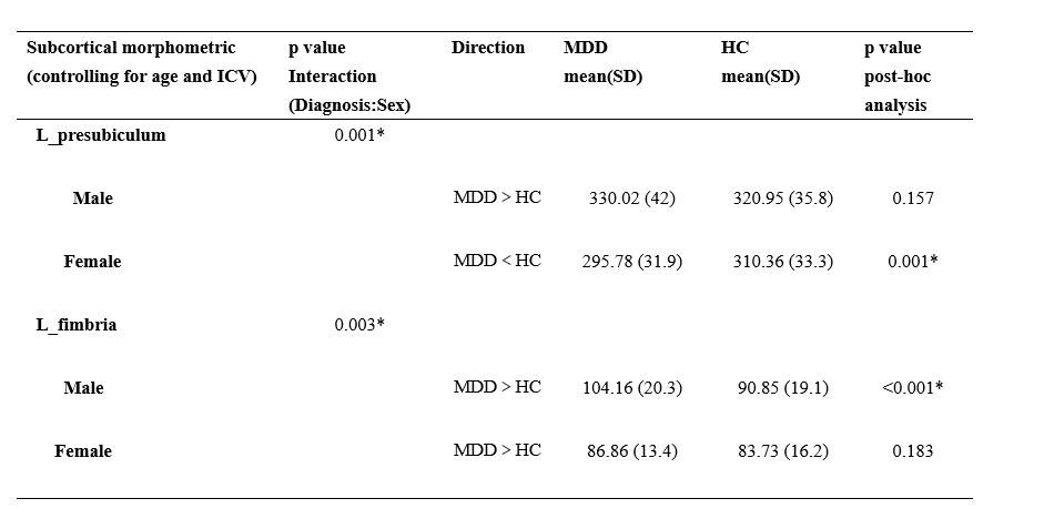
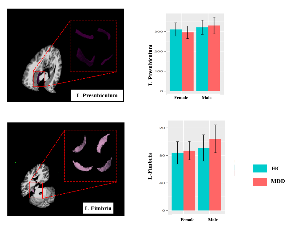
The demographic information and clinical characteristics of the participants are shown in Table1. There were no differences in clinical characteristics in the MDD group of both sexes. In the longitudinal segmentation, the overall hippocampal volume and hippocampal subregions didn’t show significant sex-by-diagnosis interaction but showed significant volume decrease of the bilateral overall hippocampus in individuals with MDD of both sexes, as well as bilateral hippocampal body and tail. In the transverse segmentation, the significant sex-by-diagnosis interactions were observed in the left presubiculum and left fimbria. Subsequent post-hoc analyses revealed that female individuals with MDD had smaller left presubiculum, while the male individuals with MDD had larger left fimbria when compared with their sex-matched counterparts(Table 2, Fig.2). The right fimbria didn’t show significant sex-by-diagnosis interaction but showed enlargered volume in individuals with MDD of both sexes when compared with HCs.In male individuals with MDD, the volumes of the bilateral hippocampal tail were positively correlated with HAMA scores (left: r=0.4, p=0.009; right: r=0.39, p =0.012). In female individuals with MDD, the volumes of the bilateral hippocampal tail were positively correlated with delayed logical memory scores (left: r=0.28, p =0.021; right: r=0.28, p =0.027) and the volumes of the left hippocampal body were positively correlated with delayed logical memory scores (r=0.26, p =0.021).Discussion and Conclusion
As far as we know, this is the first study to specifically investigate the volumetric alterations in hippocampus on subregions resolution and explore the sex difference along two axis. We found sex-independent decreased volume of the hippocampal and its subregions along the longitudinal axis, while the sex-specific alteration only detected in the subfields along the transverse axis. in the left presubiculum and left fimbria. Current findings may deepen our understanding of how hippocampus contribute to MDD psychopathology at subregional-level and suggesting the importance of exploring gender effects in imaging studies for MDD.Acknowledgements
This study is supported by grants from the 1.3.5 Project for Disciplines of Excellence, West China Hospital, Sichuan University (Grant No. ZYJC21041), the Clinical and Translational Research Fund of Chinese Academy of Medical Sciences (Grant No. 2021-I2M-C&T-B-097), and the Natural Science Foundation of Sichuan Province (Grant No. 2022NSFSC0052).
References
[1] Ho TC, Gutman B, Pozzi E,et al. Subcortical shape alterations in major depressive disorder: Findings from the ENIGMA major depressive disorder working group. Hum Brain Mapp. 2022 Jan;43(1):341-351.
[2]Han KM, Kim A, Kang W, et al Hippocampal subfield volumes in major depressive disorder and bipolar disorder. Eur Psychiatry. 2019 Apr;57:70-77.
[3] Cao B, Passos IC, Mwangi B,et al. Hippocampal subfield volumes in mood disorders. Mol Psychiatry. 2017 Sep;22(9):1352-1358.
[4] Nolan M, Roman E, Nasa A, et al. Hippocampal and Amygdalar Volume Changes in Major Depressive Disorder: A Targeted Review and Focus on Stress. Chronic Stress (Thousand Oaks). 2020 Sep 22;4:2470547020944553.
[5] Hu X, Zhang L, Liang K,et al. Sex-specific alterations of cortical morphometry in treatment-naïve patients with major depressive disorder. Neuropsychopharmacology. 2022 Oct;47(11):2002-2009.
[6] Liu MN, Pantouw JG, Yang KC,et al. Sub-regional hippocampal volumes in first-episode drug-naïve major depression disorder. Neurosci Lett. 2021 Oct 15:763:136178.
Figures