4926
Convolutional Neural Network based Stack-of-Star Imaging with Noise and Artifacts Removal
Xinzeng Wang1, Yedaun Lee2,3, Joonsung Lee4, Sagar Mandava5, Ty A. Cashen6, Xucheng Zhu7, and Arnaud Guidon8
1GE Healthcare, Houston, TX, United States, 2Department of Radiology, Haeundae Paik Hospital, Busan, Korea, Republic of, 3Inje University College of Medicine, Busan, Korea, Republic of, 4GE Healthcare, Seoul, Korea, Republic of, 5GE Healthcare, Atlanta, GA, United States, 6GE Healthcare, Madison, WI, United States, 7GE Healthcare, Menlo Park, CA, United States, 8GE Healthcare, Boston, MA, United States
1GE Healthcare, Houston, TX, United States, 2Department of Radiology, Haeundae Paik Hospital, Busan, Korea, Republic of, 3Inje University College of Medicine, Busan, Korea, Republic of, 4GE Healthcare, Seoul, Korea, Republic of, 5GE Healthcare, Atlanta, GA, United States, 6GE Healthcare, Madison, WI, United States, 7GE Healthcare, Menlo Park, CA, United States, 8GE Healthcare, Boston, MA, United States
Synopsis
Keywords: Cancer, Image Reconstruction, Liver, Pancreas
Stack of Star acquisition is one of the most frequently used non-Cartesian k-space sampling methods due to its fast speed and robustness to motion. To further reduce the scan time or increase the temporal resolution, Stack of Star is often down-sampled with fewer spokes and advanced sampling patterns, such as golden angle acquisition. However, this makes Stack of Star prone to noise and streak artifacts, limiting the in-plane resolution and degrading diagnostic quality. In this work, we evaluated a deep-learning based stack-of-star method for free-breathing abdominal imaging and it shows improved diagnostic quality.Introduction
Radial sampling is one of the most frequently used non-Cartesian k-space sampling methods due to its fast speed and robustness to motion. One of the 3D extensions of radial sampling is Stack of Star acquisition, which is often used in 3D breath-hold/free-breathing imaging and dynamic contrast enhanced imaging. (1-4)To further reduce the scan time or increase the temporal resolution, Stack of Star is often down-sampled with fewer spokes and advanced sampling patterns, such as golden angle acquisition. However, this makes Stack of Star prone to noise and streak artifacts. Many investigators have employed sophisticated reconstruction techniques to produce artifact-free images, such as using compressed sensing (CS). However, CS reconstruction is computationally intensive, and the optimization of reconstruction parameters is also challenging.
In addition to the under-sampled k-space, respiratory motions and other non-rigid motions in the abdomen also cause streak artifacts. These streak artifacts are more obvious in post-contrast Stack of Star images due to the high signal intensity in moving tissues, reducing the lesion conspicuity. Truncation artifacts are another common artifact in Stack of Star images due to the limited acquisition matrix size and scan times. Low-pass filters are also applied in the conventional reconstruction method to minimize the truncation artifacts, but at the cost of in-plane resolution. These artifacts limit the in-plane resolution and lesion conspicuity, resulting in degraded diagnostic quality.
In this work, we evaluated a deep-learning based reconstruction method (DL Star) to improve the stack-of-star image quality by removing streak artifacts, truncation artifacts and noise in MR images.
Methods
Pre- and post-contrast liver and pancreas images were acquired in 5 patients using free-breathing 3D LAVA Star on a GE 3T MRI scanner (SIGNA Architect, GE Healthcare, Waukesha, WI) with IRB approval and written informed consent. The images were acquired with the following parameters, Matrix Size = 320 x320, Slice Thickness = 3.4 mm, Flip Angle = 12o, Repetition Time/Echo Time = 4 ms/1.92 ms, Bandwidth = 325.5 Hz/px, and Number of Averages = 0.7. A deep-learning based network (DL Star) was trained from a database of over 10,000 images to reconstruct images with a high signal-to-noise ratio, high spatial resolution, and reduced streak artifacts. A tunable noise reduction factor was offered to accommodate user preference. The DL Star network was embedded into the conventional reconstruction pathway to generate two sets of image series (conventional reconstructed and DL reconstructed images) from a single set of MR raw data.Results and Discussions
Respiratory motion and other non-rigid motion in the abdomen often cause streak artifacts in the conventional stack-of-star images, especially in post-contrast images, as shown in Figure 1a. These streak artifacts reduce the conspicuity of lesions and anatomical details, degrading the diagnostic quality. To reduce these streak artifacts, DL Star reconstruction estimated and removed streak artifacts from the DL Star images, resulting in improved image quality and conspicuity of anatomical details, as shown in Figure 1b.Truncation artifacts are often visible in stack-of-star images (Figure 2a) due to limited matrix size and scan times. Truncation artifacts are often suppressed with low-pass filters, further reducing the in-plane resolution. In contrast, DL Star reconstruction could suppress truncation artifacts without using low-pass filters, improving the in-plane resolution, as shown in Figure 2b.
Besides removing streak and truncation artifacts, DL Star could also reduce noise and improve the SNR, as shown in Figure 3. At low-field MRI, pre-contrast Stack of Star image could suffer from low SNR, requiring the acquisition of more spokes and longer scan times. Since DL Star could reduce noise, it has the potential to be used to reduce the scan time of Stack of Star at low-field MRI.
With the removal of streaks, truncation artifacts and noise, DL Star improved in-plane resolution and the conspicuity of lesions and anatomical details compared to the conventional reconstruction method. As shown in Figure 4, DL Star improved the visualization of pancreas with higher in-plane resolution and fewer streak and truncation artifacts. As shown in Figure 5, lesions also showed better conspicuity in DL Star post-contrast liver images due to efficient streak artifact removal.
The improvement is not limited to LAVA Star images, DL Star could also be used in other Stack of Star applications, such as DISCO Star for dynamic contrast enhancement imaging. In the future, we will evaluate DL Star with more patients.
Conclusion
The in-plane resolution and lesion conspicuity of 3D Stack of Star images are well improved with DL Star due to the promising noise, truncation, and streak artifacts reduction. DL Star showed the potential to improve the diagnostic quality of Stack of Star applications.Acknowledgements
No acknowledgement found.References
[1] Hersh Chandarana, etc. Investigative Radiology 2011: 46(10):648-653
[2] Kai Tobias Block, etc. Journal of the Korean Society of Magnetic Resonance in Medicine 2014; 18(2): 87-106
[3] Shintaro Ichikawa, etc. Magnetic Resonance in Medical Sciences 2020; 19(2): 99–107
[4] Hersh Chandarana, etc. Investigative Radiology 2015: 50(11):749-756
Figures
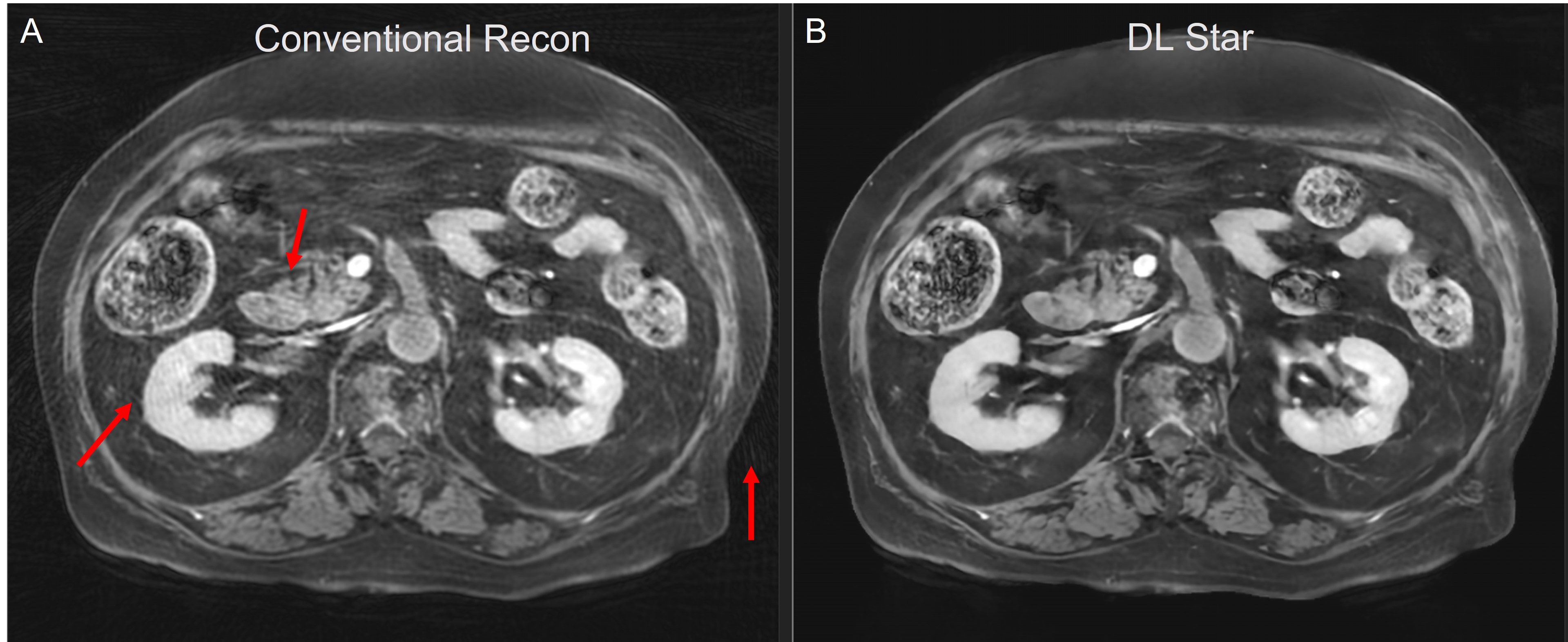
Figure 1. Post contrast
LAVA Star images. Due to motion, the original image (A) reconstructed using the
conventional reconstruction method has many streak artifacts, which impact the
conspicuity. DL Star reconstruction method suppressed the streak artifact in
the DL image (B), improving the conspicuity and in-plane resolution.
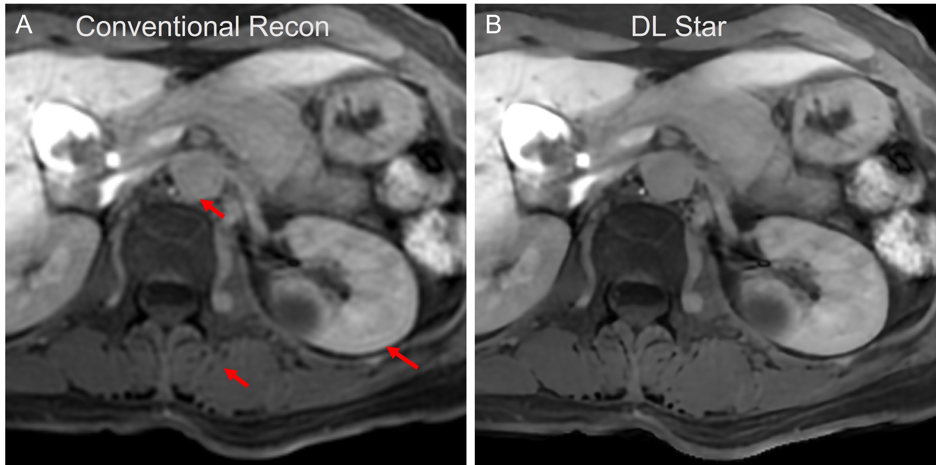
Figure 2. Post contrast
LAVA Star images. The original image (A) is blurring due to the presence of
truncation artifacts (red arrows). DL Star reconstruction method removed the truncation
artifact in the DL image (B) without using low-pass filters, improving the
in-plane resolution.
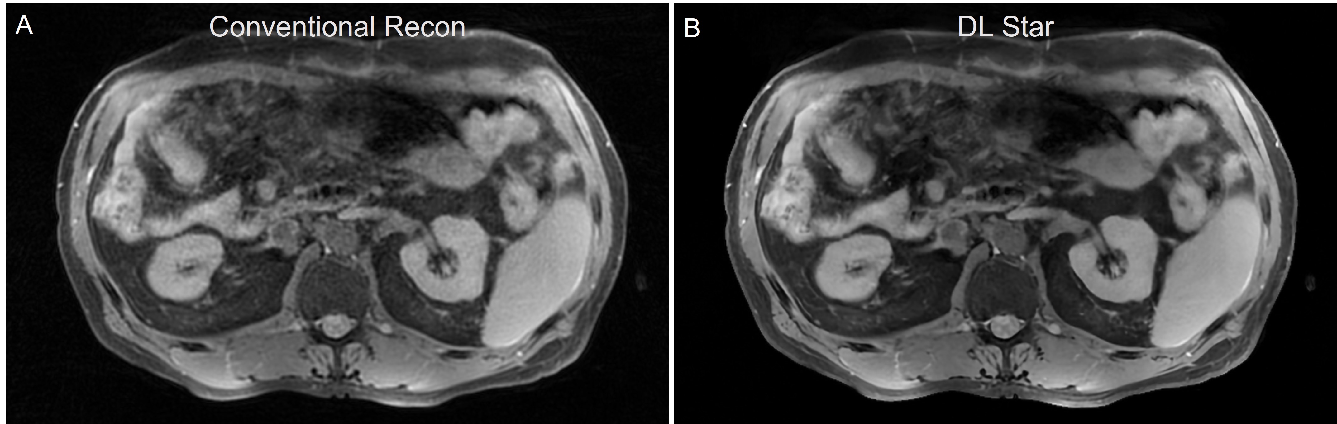
Figure 3. Pre contrast LAVA Star images. DL Star (B)
removed noise and improved the SNR compared to the conventional reconstruction
method.
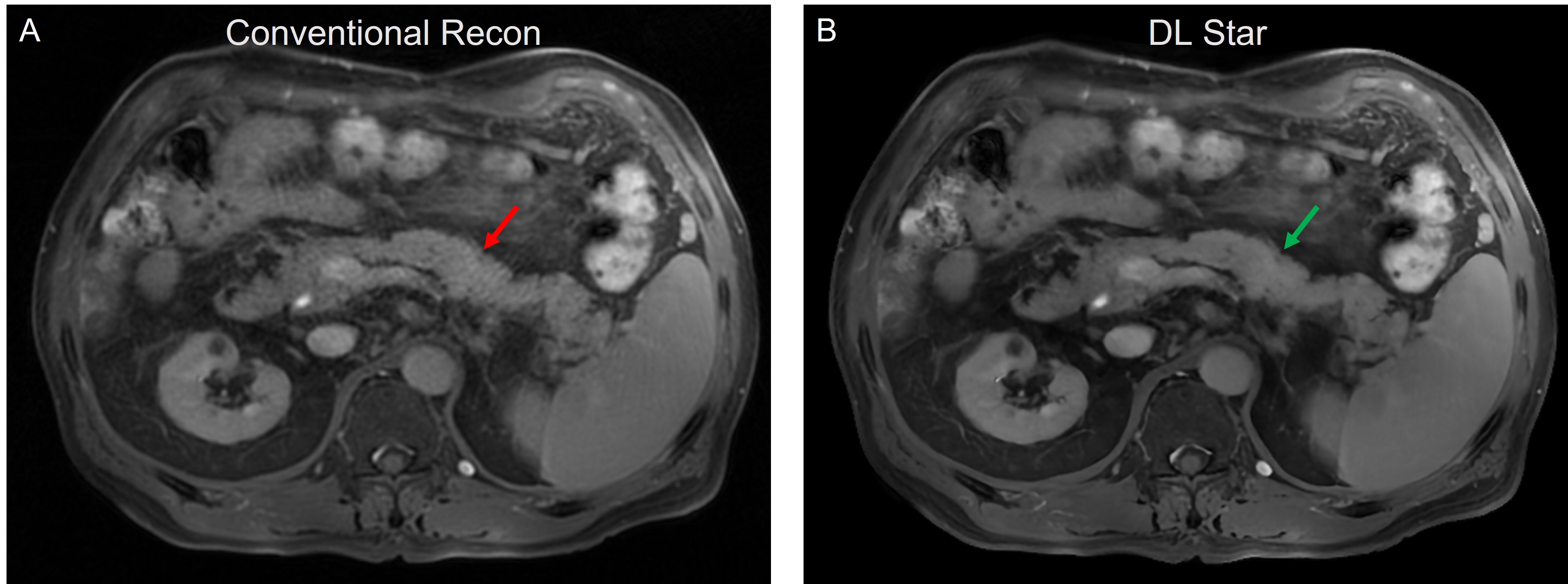
Figure 4. pancreas
imaging with LAVA Star. Steak artifacts caused by motion impact
the visualization of pancreas in
the original image (A). With DL Star, the streaks are well suppressed and the pancreas is will visualized, showing sharper edges and
high conspicuity of details.
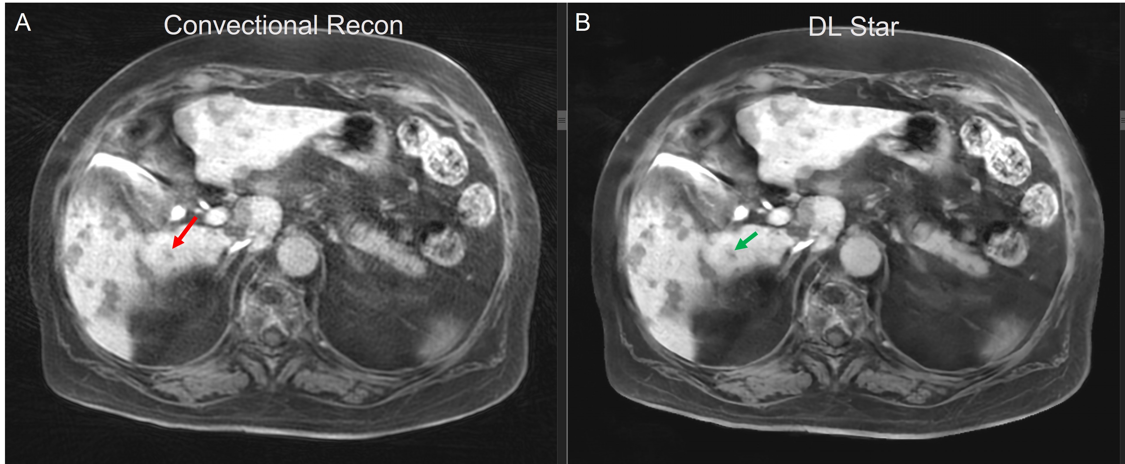
Figure 5. Post contrast liver images. In the original
image, streak artifacts reduced the conspicuity of lesions and in-plane
resolution. DL Star (B) showed promising removal of streak artifacts, improving
the diagnostic quality.
DOI: https://doi.org/10.58530/2023/4926