4901
FLAIR3Phase: a new synthetic MRI contrast for paramagnetic rim lesions detection in multiple sclerosis
Colin Vanden Bulcke1,2, Nicolas Delinte2, and Benoît Macq2
1Institute of Neurosciences, Université Catholique de Louvain, Brussels, Belgium, 2Institute for Information and Communication Technologies, Electronics and Applied Mathematics, Université Catholique de Louvain, Louvain-la-Neuve, Belgium
1Institute of Neurosciences, Université Catholique de Louvain, Brussels, Belgium, 2Institute for Information and Communication Technologies, Electronics and Applied Mathematics, Université Catholique de Louvain, Louvain-la-Neuve, Belgium
Synopsis
Keywords: Multi-Contrast, Multiple Sclerosis, Paramagnetic Rim Lesions
Chronic active multiple sclerosis (MS) lesions are visible on susceptibility-based MRI as paramagnetic rim lesions (PRL); these lesions are characterized by severe tissue damage and strongly correlate with disease severity. PRL assessment is usually performed manually on unwrapped-filtered phase images. However, PRL assessment is associated with high intra/inter-rater variability due to the poor visibility of PRL and to the lack of standardized imaging protocols/guidelines for their detection. Here, we propose a new synthetic contrast called FLAIR3Phase to augment PRL assessment workflow and to reduce rater variability.Introduction
Multiple Sclerosis (MS) causes focal inflammatory lesions in the brain white matter (WM), cortex and spinal cord,1 easily observable using a T2-weighted Fluid Attenuated Inversion Recovery (FLAIR) MRI.2 A subset of MS lesions features a persistent inflammatory edge of microglia/macrophages containing iron and can be detected on susceptibility-based MRI as paramagnetic rim lesions (PRL). PRL are characterized by severe tissue damage and are associated with an aggressive disease course, and are thus considered a novel and promising prognostic imaging biomarker in MS.3–5 PRL assessment requires an anatomical sequence for lesion visualization, usually a 3D T2-FLAIR, and a susceptibility-based MRI sequence to visualize the paramagnetic rim, such as unwrapped-filtered phase image. PRL detection is associated with high intra/inter-rater variability.6 The main reasons for this variability are the presence of frequent background phase artefacts, the lack of sufficient anatomical contrast on phase images and, more broadly, the lack of standardized MRI acquisition/post-processing guidelines for PRL detection. When analyzing the unwrapped-phase images for PRL detection, raters often adjust the phase contrast by hand, thus increasing the inter-rater variability. There is a need to ease and standardize the PRL detection workflow to reduce this variability. In this work, we propose a new synthetic contrast, called FLAIR3Phase, that combines 3T FLAIR and 3T susceptibility-based unwrapped-phase images to ease the PRL detection workflow.Methods
FLAIR is the standard contrast to investigate WM disease because of its high sensitivity to WM abnormalities and its suppression of cerebrospinal fluid, but it is not specific to lesion pathology.2 On the other hand, susceptibility-based MRI can detect tissue iron accumulation, in particular iron-laden phagocytes.7 FLAIR3Phase contrast non-linearly combines FLAIR and unwrapped-phase images to take advantage of the hyperintense parenchymal lesion contrast of the FLAIR and the hypointense plaque-edge paramagnetic contrast of the unwrapped-phase. Non-linear combination is used to achieve a higher contrast combination inside WM lesions, and a lower contrast combination outside the lesions to avoid deterioration of the combined image by the background artifacts of the unwrapped-phase. FLAIR3Phase computation method is described hereunder.The FLAIR image is registered to the susceptibility-based magnitude image, and the unwrapped-phase image is skull-stripped using the magnitude image. Then, both FLAIR and unwrapped-phase images are preprocessed independently with N4 bias correction (simpleitk python package) and normalization between 0 and 1 (FLAIRnorm). The contrast of the FLAIR is then enhanced using contrast-limited adaptive histogram equalization (FLAIRpre). The contrast of the unwrapped-phase is enhanced, first using a histogram equalization method, followed by a spatially adaptive non-local means denoising step (from ANTs),8 with a final contrast enhancement using a histogram stretching method (Phasepre). All contrast enhancements are performed using the exposure module of the scikit-image python package.9 Then the combination of the two images follows the scheme below:
- Non-linear activation of the FLAIR: $$\textit{FLAIR}_{activated}=1-\frac{1}{1+e^{(-20\cdot \textit{FLAIR}_{pre}-0.85)}}$$
- Multiplication of FLAIR and phase images: $$\textit{FLAIRPhase}=\textit{FLAIR}_{activated}\cdot \textit{FLAIR}_{pre}+(1-\textit{FLAIR}_{activated})\cdot FLAIR_{pre}\cdot Phase_{pre}$$
- Final combination: $$\textit{FLAIR}^3\textit{Phase}=\textit{FLAIR}_{norm}+\textit{FLAIR}_{norm}\cdot \textit{FLAIRPhase}$$
To test this new synthetic contrast, three independent raters were asked to compare FLAIR3Phase and unwrapped-phase (processed with an automatic contrast adjustment) images for the assessment of 88 PRLs. For each PRL, raters were asked whether the paramagnetic rim was more visible, less visible or about the same on the FLAIR3Phase image when compared to the unwrapped-phase image.
Results
The three raters agreed on 45 lesions (51%), of which one is shown in Figure 2. Of the 45 lesions, 16 (35%) PRLs were considered more visible on the FLAIR3Phase, while 12 (27%) PRLs have been considered less visible on the FLAIR3Phase. The rest (38%) were rated to be similarly visible on the two contrasts. Therefore, FLAIR3Phase has been considered better or the same in 73% of the lesions.Discussion
The proposed FLAIR3Phase contrast combines FLAIR and unwrapped-phase images to visualize at the same time both the hyperintense lesion parenchyma and the hypointense lesional paramagnetic rim of PRL. Since this FLAIR3Phase is automatically computed and does not require further manual contrast adjustment, it could help reduce the intra/inter-rater variability during PRL assessment. Although not all PRLs were more visible on FLAIR3Phase, our preliminary results suggest that the FLAIR3Phase synthetic contrast is useful to help PRL assessment as an additional contrast to the unwrapped-phase. Compared to a previously published work combining both 3T and 7T images to obtain a synthetic contrast for PRL detection,10 the FLAIR3Phase contrast is obtained using exclusively 3T MRI data and can be thus more easily integrated in a future clinical workflow. Moreover, although an MRI-histopathological validation of this novel MRI contrast is still needed, FLAIR3Phase seems to detect potential PRL that are nearly invisible on the phase-unwrapped images alone, thus potentially increasing our visualization of chronic active lesions with MRI (see Figure 3).Conclusion
In this work, we have implemented a new synthetic contrast, FLAIR3Phase, based on the non-linear combination of FLAIR and unwrapped-phase images. This novel contrast, when used in-addition to the original unwrapped-phase one, could allow a more systematic PRL assessment workflow.Acknowledgements
The authors thank the Radiology departments of the Cliniques universitaires Saint-Luc (UCLouvain, Brussels, Belgium) for helping with the MRI data acquisition. The authors also want to thank Daniel S. Reich, Martina Absinta and Alessandro Cagol for completing the survey about this new contrast and giving valuable feedback. Finally, we thank Pietro Maggi for his feedback on the writing of the abstract and Anna Stolting for coming up with the name "FLAIR3Phase" for this new synthetic contrast.References
1. Reich, D. S., Lucchinetti, C. F. & Calabresi, P. A. Multiple Sclerosis. N. Engl. J. Med. 378, 169–180 (2018).2. Barkhof, F. & Scheltens, P. Imaging of white matter lesions. Cerebrovasc. Dis. Basel Switz. 13 Suppl 2, 21–30 (2002).
3. Maggi, P. et al. Paramagnetic Rim Lesions are Specific to Multiple Sclerosis: An International Multicenter 3T MRI Study. Ann. Neurol. 88, 1034–1042 (2020).
4. Absinta, M. et al. Persistent 7-tesla phase rim predicts poor outcome in new multiple sclerosis patient lesions. J. Clin. Invest. 126, 2597–2609 (2016).
5. Absinta, M. et al. Association of Chronic Active Multiple Sclerosis Lesions With Disability In Vivo. JAMA Neurol. 76, 1474–1483 (2019).
6. Barquero, G. et al. RimNet: A deep 3D multimodal MRI architecture for paramagnetic rim lesion assessment in multiple sclerosis. NeuroImage Clin. 28, 102412 (2020).
7. Haacke, E. M. et al. Imaging iron stores in the brain using magnetic resonance imaging. Magn. Reson. Imaging 23, 1–25 (2005).
8. Avants, B., Tustison, N. & Song, G. Advanced normalization tools (ANTS). Insight J 1–35, (2008).
9. Walt, S. van der et al. scikit-image: image processing in Python. PeerJ 2, e453 (2014).
10. Grabner, G. et al. Analysis of multiple sclerosis lesions using a fusion of 3.0 T FLAIR and 7.0 T SWI phase: FLAIR SWI. J. Magn. Reson. Imaging JMRI 33, 543–549 (2011).
Figures
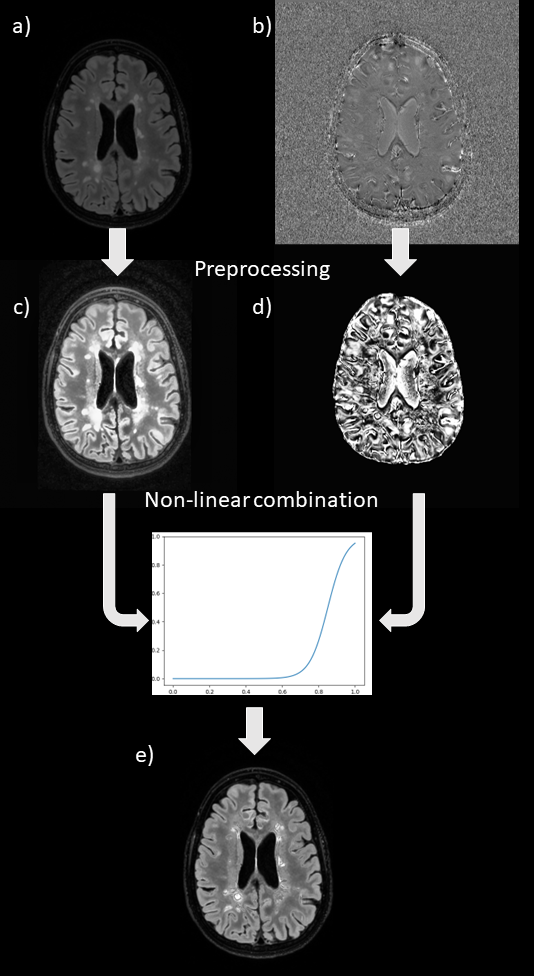
Figure 1: FLAIR3Phase computation pipeline.
First, FLAIR (a) and unwrapped-phase (b) images are preprocessed with N4 bias correction and normalization between 0 and 1 and different methods of contrast enhancement, giving enhanced images respectively in (c) and (d). Finally, these preprocessed images are non-linearly combined to create the FLAIR3Phase
synthetic contrast (e).
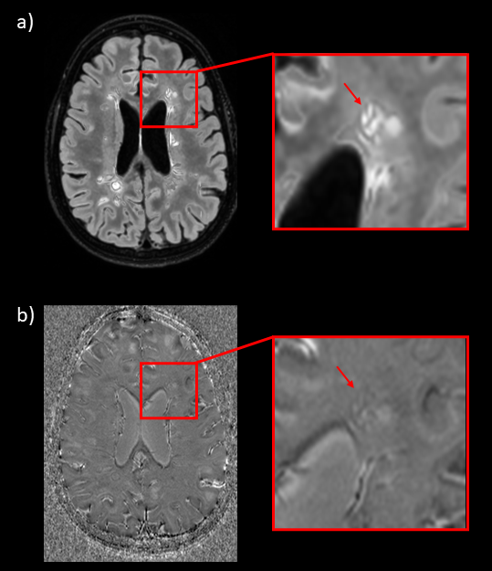
Figure
2: Comparison of the FLAIR3Phase contrast (a) and unwrapped-phase contrast (b)
for the detection of a paramagnetic rim lesion.
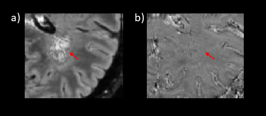
Figure 3:
Potential paramagnetic rim lesion, observable on the FLAIR3Phase contrast (a), while nearly invisible on the unwrapped-phase image (b).
DOI: https://doi.org/10.58530/2023/4901