4889
MULTIPLEX with water-fat imaging (WFI) capacity for body imaging1United Imaging, Houston, TX, United States
Synopsis
Keywords: Multi-Contrast, Body
A multi-parametric method with WFI imaging capacity, i.e. MULTIPLEX-WFI, was developed and demonstrated. Routine two-point Dixon WFI images are additionally achieved, and a fat-suppression mask is extracted for use on the MULTIPLEX images to improve the overall image quality for body imaging. The cost of adding the WFI capacity to the MULTIPLEX method was negligible. The MULTIPLEX-WFI method was tested on phantom and in vivo knee and pelvis scans, showing much improved imaging quality for body imaging.Introduction
Recently, a MULTI-parametric MR imaging with fLEXible design method, namely MULTIPLEX (1), was proposed for full brain 3D high resolution multi-parametric imaging. By incorporating the dual-FA (flip angle), dual-TR and multi-echo GRE features, a single scan of the MULTIPLEX offers numerous contrasts and mappings, covering T1, T2*, proton density (PD), aT1W and QSM, a name a few. The MULTIPLEX design also supports certain degree of flexibility for accommodating additional contrast mechanisms, as the method collects 2x2 (i.e. 2FA x 2TR) = 4 individual ‘sections’ of data, and in each section, different signal modulation may be flexibly added. For example, one can add flow-refocusing and flow-dephasing modules to different sections to additionally generate MR angiography contrast (1).Currently, the majority of reported MULTLIPLEX applications have been focused on the brain (2-4). In order to employ MULTIPLEX on other body parts such as the knee or the pelvis, challenges arising from the presence of fatty tissues must be addressed. Therefore, the purpose of this work is to introduce the capacity of water-fat imaging (WFI) to MULTIPLEX, i.e. MULTIPLEX-WFI, with negligible costs on scan time and imaging quality, for 3D high resolution, high-quality multi-parametric imaging on the body.
Methods
The two-point Dixon strategy was adopted for WFI. In the routine MULTIPLEX, the first echo times (TE1) in both TR1 and TR2 are the same. In MULTIPLEX-WFI, on the other hand, the echo time in TR1 is shifted by an amount of δ to being TE1+ δ, while the first echo in TR2 remains TE1, as shown in Figure 1. As the result, the first-echoes in TR1 and TR2 provide the two-point Dixon signals with a Dixon phase angle θ = γΔωδ, where γ is the gyromagnetic ratio and Δω is the water-fat resonance frequency shift.For demonstration, MULTIPLEX-WFI data were collected on a proton-density fat-fraction (PDFF) phantom (Calimetrix, Madison, WI) and on the knee and pelvis of a healthy volunteer (male, 38y/o), both on a 3T system (uMR790, UIH, Shanghai, China), using 24-channel head-neck coil, a 12-channel knee coil and the combination of spine/body coils, respectively. The common parameters for MULTIPLEX-WFI scans were:TE1=4.48ms , δ=-0.84ms,TR1/TR2=8.3/33.0ms, echo#=5, α1/α2=4°/16°. In all three scans, TE1 was set to the shortest in-phase echo time of 4.48ms, and the -0.84ms echo time shift corresponded to a Dixon phase angle θ of 135°. The WFI calculation was performed using the asymmetric two-point Dixon method as described previously (5), between the two first-echoes collected under α1 with PD weightings.
A fat-fraction (FF) map was estimated and used to extract a fat suppression (FS) mask for the MULTIPLEX images. The FS mask was first generated as 1-FF (i.e. water fraction), which can be directly applied to qualitative contrast images such as aT1W. For quantitative maps such as T1 or T2* maps, the mask was further processed with a threshold and set to 1 for voxels with 1-FF values >75%.
Results
Figure 2 shows some representative images of a MULTIPLEX-WFI scan on the PDFF phantom, demonstrating reliable water-fat separation.Figure 3 compares the measured and the manufactureir reference FF values, suggesting a linearly well-correlated FF from the two-point WFI calculation, albeit with a slight overall underestimation.
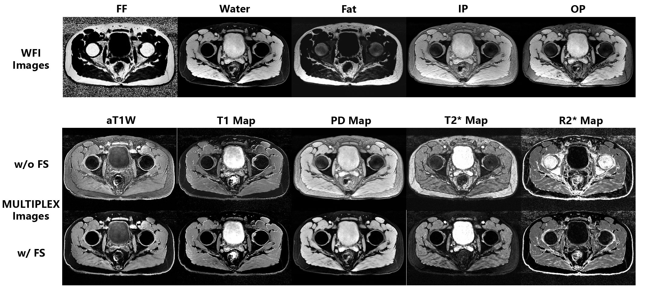
Figures 4 & 5 show the MULTIPLEX-WFI knee and pelvis images, respectively. The WFI images are all generally satisfying. And by employing the FS mask, the visual quality of the original MULTIPLEX images and maps are much improved over those without.
Discussion & conclusions
In this work, we have implemented the WFI capacity to the MULTIPLEX method, employing a two-point Dixon acquisition and reconstruction scheme.Compared to the original MULTIPLEX method (1), the only implementation difference in this work is the insertion of a small TE shift (i.e. 0.84ms) to the TR1 echo, leading to a negligible cost in total scan time (i.e. δ/(TR1+TR2)≈2%) and negligible effects on the MULTIPLEX image quality. And since the two first echoes in TR1 and TR2 are acquired in an interleaved manner, no additional restriction would be imposed to the imaging resolution and readout bandwidth.
The MULTIPLEX-WFI scan offers 5 additional WFI image sets, i.e. FF, water- & fat-images, as well as in-phase (IP) & out-of-phase images (OP). With the two-point acquisition, the estimated FF values are slightly underestimated, which is expected (6). However, the phantom results indicate that the measured FF values are still linearly well-correlated to the actual FF (Fig.3), such that higher/lower fat content always correspond to higher/lower FF values. This makes the FF map still reliable for generating a linear FS mask to suppress fatty tissues in other MULTIPLEX images (Figs. 4&5). Since all MULTIPLEX images are strictly spatially aligned, the FS mask is applied in a direct voxel-wise manner to generate visually much improved image quality for MULTIPLEX images.
In conclusion, we have implemented the MULTIPLEX-WFI method to address the fat-related challenges in body imaging, offering additional WFI images and excellent multi-parametric imaging quality with minimal costs.
Acknowledgements
No acknowledgement found.References
1.Ye Y, Lyu J, Hu Y, Zhang Z, Xu J, Zhang W. MULTI-parametric MR imaging with fLEXible design (MULTIPLEX). Magn Reson Med 2021.
2.Zhong J, Ye Y, Yang H, et al. Multi-parametric imaging on cerebral infarction using MULTIPLEX. Proceedings of the joint annual meeting ISMRM-ESMRMB 2022 & ISMRT annual meeting. London, UK; 2022. p. 4464.
3.Wang H, Ye Y, Liu C, et al. Highly Accelerated Multiple Parametric MR Imaging with Wave-CAIPI and MULTIPLEX. Proceedings of the joint annual meeting ISMRM-ESMRMB 2022 & ISMRT annual meeting. London, UK; 2022. p. 4979.
4.Ye Y, Liu X, Wu Y, Zhang Z, Xu J. An initial multi-parametric imaging experience of MULTIPLEX at 5T whole body system. Proceedings of the joint annual meeting ISMRM-ESMRMB 2022 & ISMRT annual meeting. London, UK; 2022. p. 2855.
5.Xiang QS. Two-point water-fat imaging with partially-opposed-phase (POP) acquisition: an asymmetric Dixon method. Magn Reson Med 2006;56(3):572-584.
6.Bernard CP, Liney GP, Manton DJ, Turnbull LW, Langton CM. Comparison of fat quantification methods: a phantom study at 3.0T. J Magn Reson Imaging 2008;27(1):192-197.
Figures