4882
Monitoring triple negative breast cancer therapy against lncRNA MANCR with MT218, a targeted MRMI contrast agent1Department of Biomedical Engineering, Case Western Reserve University, Cleveland, OH, United States, 2Case Comprehensive Cancer Center, Case Western Reserve University, Cleveland, OH, United States
Synopsis
Keywords: Molecular Imaging, Cancer, Breast
Developing new molecular targeted therapies for triple negative breast cancer remains an obstacle. MANCR is a long noncoding RNA overexpressed in TNBC that sustains aggressive breast cancer growth. Silencing its expression with siRNA nanoparticles suppresses tumor growth, and treatment efficacy can be assessed by MRMI with a targeted contrast agent, MT218. Reduction in contrast-to-noise ratio following treatment confirmed effectiveness of siRNA therapy and allowed regular monitoring of tumor inhibition. RNA therapy shows promise as a new approach for treating aggressive breast cancer, and MRMI with the targeted contrast agent is a non-invasive imaging tool for TNBC detection and therapeutic surveillance.Introduction
Targeting tumor-specific biomarkers with gadolinium-based contrast agents allows for more accurate cancer detection with significantly lower doses. MT218 is a targeted contrast agent specific to extradomain-B fibronectin (EDB-FN), an alternatively spliced isoform of fibronectin involved in cancer epithelial-to-mesenchymal transition and found to be upregulated in aggressive cancers with minimal expression in healthy tissue.1 It has been used for detection of multiple cancer types, including aggressive triple negative breast cancer (TNBC).2-4 Here, we demonstrate the efficacy of MT218 at monitoring siRNA therapy against mitotically associated long noncoding RNA (MANCR). MANCR is upregulated in TNBC relative to normal breast tissue and promotes increased cell proliferation and viability.5Methods
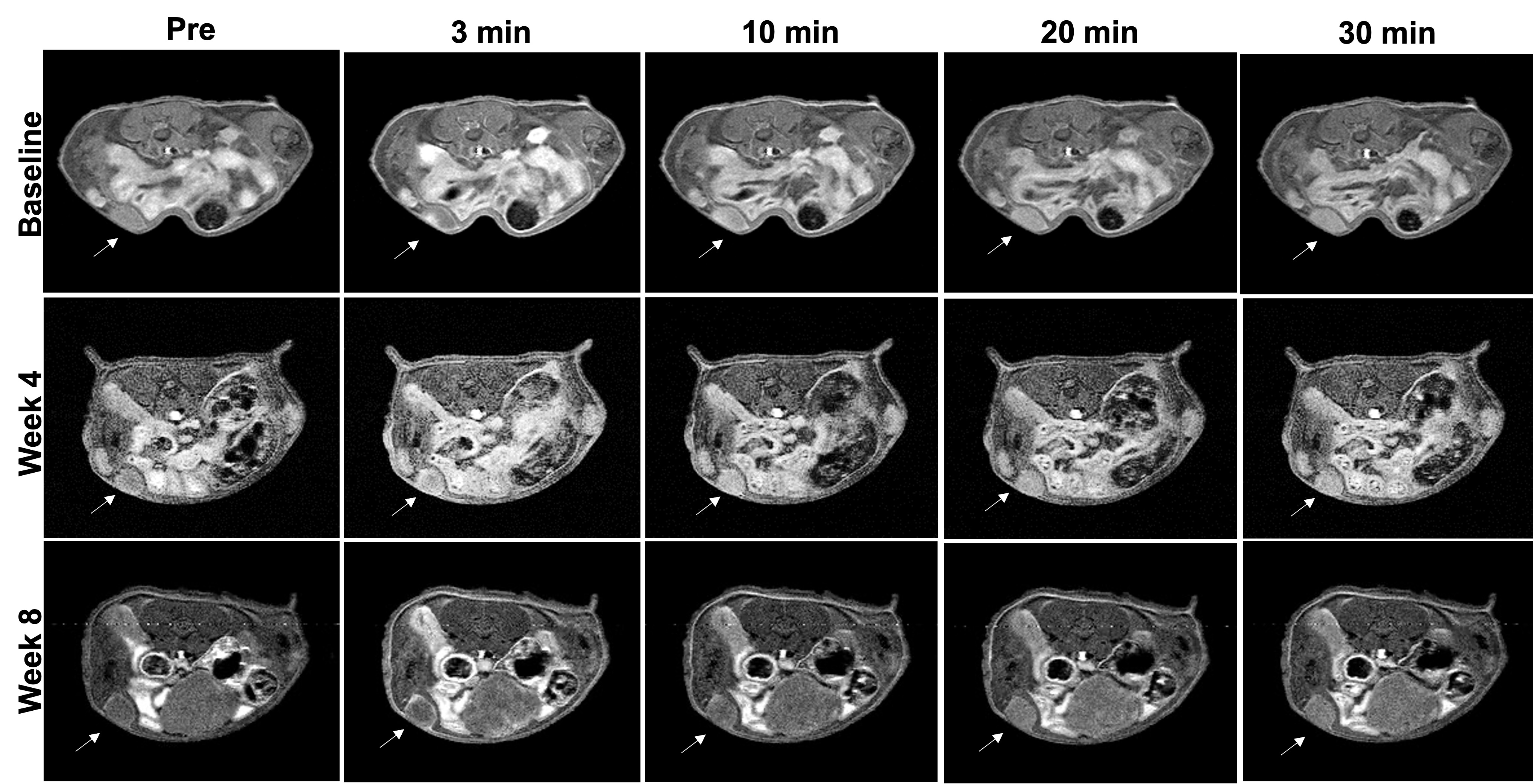
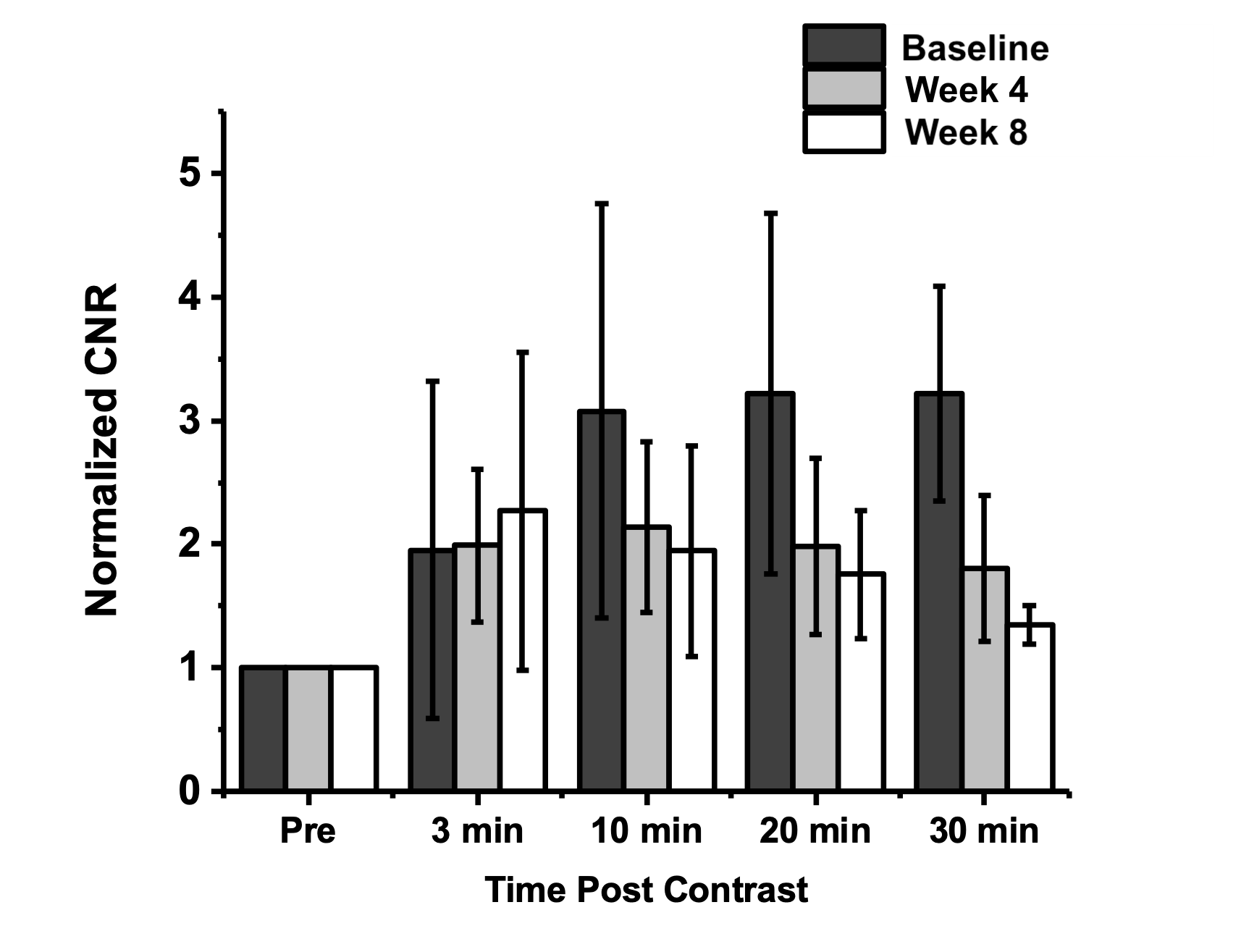
Human triple negative breast cancer cells (MDA-MB-231) were acquired from ATCC. Expression of EDB-FN and MANCR was detected on the mRNA level using qRT-PCR. 2x10⁶ cells suspended in Matrigel were injected into the mammary fat pad of athymic nude mice, and tumors were grown to a size of 100 mm3. MR images were obtained on a 3T MRS 3000 scanner with a mouse short quad coil. The targeted contrast agent ZD2-N3-Gd(HP-DO3A) (MT218) was intravenously administered at a dose of 0.04 mmol Gd/kg in saline. T1-weighted images were acquired using a fast spin echo sequence pre- and 20 minutes post-contrast injection. Image contrast-to-noise ratio (CNR) was analyzed by measuring the difference signal between tumor and muscle, divided by standard deviation of noise. Mice were treated weekly by i.v. injection with siRNA against MANCR in nanoparticles made with multifunctional pH-sensitive ionizable lipid, ECO.Results
Silencing MANCR in vitro with ECO nanoparticles lowers expression of EDB-FN by 40% relative to no treatment and (Figure 1). MR molecular imaging using MT218 revealed contrast uptake as soon as 3 minutes post-injection, with peak signal intensity reached by 20 minutes (Figure 2). Tumor sizes over the course of treatment remained stable. Image analysis showed a reduction in CNR from 3-fold enhancement at baseline that dropped to 2-fold and 1.6-fold enhancement following 4 and 8 weeks of siRNA therapy, respectively (Figure 3).Discussion
Reducing expression of the oncogenic lncRNA MANCR with siRNA nanoparticles prevents cancer cell growth and concurrently downregulates expression of EBD-FN, the target for MT218 contrast agent. The results suggest that siRNA therapy is effective as early as 4 weeks to suppress tumor growth, and that reduction in contrast enhancement indicates therapeutic efficacy. The reduction in EDB-FN that results from MANCR silencing and inhibited tumor growth is observed by a decrease in the uptake of MT218 in tumors measured by CNR. The drop in CNR enhancement confirms successful MANCR silencing, which otherwise cannot be assessed during the course of treatment.Conclusion
MRMI allows for noninvasive monitoring of breast cancer progression and response to therapy. This approach can be used along with neoadjuvant chemotherapy prior to surgical tumor resection, and allows clinicians to better identify aggressive tumors along with assessing which are responding to therapy. The targeted contrast agent can be used at a dose substantially lower than existing clinically approved GBCAs while providing superior contrast enhancement localized to the tumor and not in normal tissues.Acknowledgements
No acknowledgement found.References
[1] Patten, J., & Wang, K. Fibronectin in development and wound healing. Advanced Drug Delivery Reviews. 2021;170, 353-368.
[2] Han Z, Zhou Z, Shi X, et al. EDB fibronectin specific peptide for prostate cancer targeting. Bioconjug Chem. 2015;26(5):830-838.
[3] Han Z, Li Y, Roelle S, et al. Targeted contrast agent specific to an oncoprotein in the tumor microenvironment with the potential for detection and risk stratification of prostate cancer with MRI. Bioconjug Chem. 2017;28(4):1031-1040.
[4] Hall, R.C., Ayat, N.R., Qiao, P.L. et al. Preclinical Assessment of the Effectiveness of Magnetic Resonance Molecular Imaging of Extradomain-B Fibronectin for Detection and Characterization of Oral Cancer. Mol Imaging Biol. 2020;22, 1532–1542.
[5] Tracy, K. M., Tye, C. E., Ghule, P. N., Malaby, H., Stumpff, J., Stein, J. L., Stein, G. S., & Lian, J. B. (2018). Mitotically-Associated lncRNA (MANCR) Affects Genomic Stability and Cell Division in Aggressive Breast Cancer. Molecular cancer research : MCR, 16(4), 587–598.
Figures