4874
A Novel Iron-oxide-based T1 Contrast Agent outperforms Gd-DOTA for Magnetic Resonance Lymphography1Department of Convergence Medicine, Asan Medical Center, University of Ulsan College of Medicine, Seoul, Korea, Republic of, 2Department of Radiation Science & Technology, Jeonbuk National University, Jeonbuk, Korea, Republic of, 3Trial Informatics. Inc, Seoul, Korea, Republic of, 4Convergence Medicine Research Center, Asan Institute for Life Sciences, Asan Medical Center, Seoul, Korea, Republic of, 5Department of Radiology, Asan Medical Center, University of Ulsan College of Medicine, Seoul, Korea, Republic of, 6Medical Research Institute, Gangneung Asan Hospital, Gangneung-si, Gangwon-do, Korea, Republic of
Synopsis
Keywords: Contrast Agent, Contrast Agent, Magnetic resonance lymphography, Iron-oxide-based T1 contrast agent
The novel iron-oxide-based T1 contrast agent may be an optimal contrast agent for MR lymphangiography, because it enhances the lymphatics only without venous contamination for more than 1 hour. It can overcome the limitations of gadolinium-based contrast agents such as venous contamination and rapid washout.INTRODUCTION
Magnetic resonance lymphography (MRL) is a non-invasive method to analyze the lymphatic system and has been increasingly used nowadays in patients with lymphedema. Visualization of the lymphatic system is essential to identify the location of lymphatic obstruction and to determine treatment methods1. The use of gadolinium-based contrast agents for MRL is hampered because they enhance both lymphatics and veins (so called venous contamination) and the lymphatics enhancement does not last more than 30 minutes. The novel iron-oxide-based T1 contrast agent was developed for MRL with following two aims: (1) for visualization of lymphatics only without venous contamination and (2) for prolonged enhancement of lymphatics more than 1 hour.MATERIALS & METHODS
The novel T1 contrast agent is produced from iron oxide nanoparticles with a mean hydrodynamic diameter of approximately 4.0 nm. A gadolinium-based contrast agent (Gd-DOTA, Dotarem) was used as a control contrast agent. Seventy-five microliters of the contrast agent preparation were injected intradermally in the fourth phalanx of both hind limbs using a 31-gauge syringe in a nine-week-old male Sprague-Dawley (SD) rats. The Gd-DOTA and the novel iron-based T1 contrast agent by administering 30 mM of each. MRL was performed every 16 minutes on coronal 3D time-of-flight (TOF) sequence with saturation bands using a 9.4-T magnet. MRI parameters were as follows: TR/TE=10/2.54 ms, flip angle=40°, matrix size=256×256×192, and FOV=70×70×30 mm2. The signal-to-noise ratio (SNR) was calculated as SILN/Mean SDbackground, and the contrast-to-noise ratio (CNR) was calculated as SILN–SImuscle/Mean SDbackground2.RESULTS AND DISCUSSION
In MRL using the Gd-DOTA, the lymph nodes and lymphatic vessels reached a peak enhancement 16 minutes after injection from the injection site, and then rapidly washed out (SNR at 0 min: 4.86 ± 2.54, 16min: 21.16 ± 3.74, 32min: 15.31 ± 3.16, 48min: 11.67 ± 2.79, 64min: 9.25 ± 1.66, 80min: 8.62 ± 1.33, 96min: 7.65 ± 1.58, and CNR at 0 min: 1.49 ± 0.49, 16min: 16.32 ± 1.47, 32min: 15.04 ± 3.22, 48min: 13.13 ± 4.72, 64min: 11.68 ± 4.44, 80min: 8.03 ± 2.26, 96min: 7.26 ± 2.06). In addition, enhancement of normal veins was prominent, which hampered accurate evaluation of lymphatics. On the other hands, the MRI using the novel contrast agent, peak enhancement was reached 16 minutes after injection and lasted longer than at least 64 minutes (SNR at 0 min: 5.82 ± 1.46, 16min: 21.27 ± 2.28, 32min: 19.89 ± 4.29, 48min: 18.56 ± 6.48, 64min: 16.60 ± 5.77, 80min: 13.09 ± 3.61, 96min: 12.13 ± 3.48, and CNR at 0 min: 0.90 ± 0.50, 16min: 16.54 ± 2.78, 32min: 10.71 ± 2.21, 48min: 6.48 ± 1.95, 64min: 3.73 ± 0.89, 80min: 3.27 ± 0.74, 96min: 2.45 ± 0.66). Unlike MRL with Gd-DOTA, the MRL with novel agent did not show venous contamination (Fig. 1, 2). Two days after MRL, the location of the lymphatic structures was visually confirmed after the intradermal injection of methylene blue in the foot (Fig. 3).CONCLUSION
In MRL, the novel iron-based T1 contrast agent outperformed Gd-DOTA as for the prolonged enhancement of lymphatics without venous contamination.Acknowledgements
This study was supported by the National Research Foundation of Korea (NRF-2021M3A9G102660).References
1. Polomska, A. K, Proulx, S. T. Imaging technology of the lymphatic system. Adv Drug Deliv Rev. 2021;170:294-311.
2. Müller, A, Fries, P, Jelvani, B, et al. Magnetic resonance lymphography at 9.4 T using a gadolinium-based nanoparticle in rats: investigations in healthy animals and in a hindlimb lymphedema model. Invest Radiol. 2017;52(12):725-733.
Figures
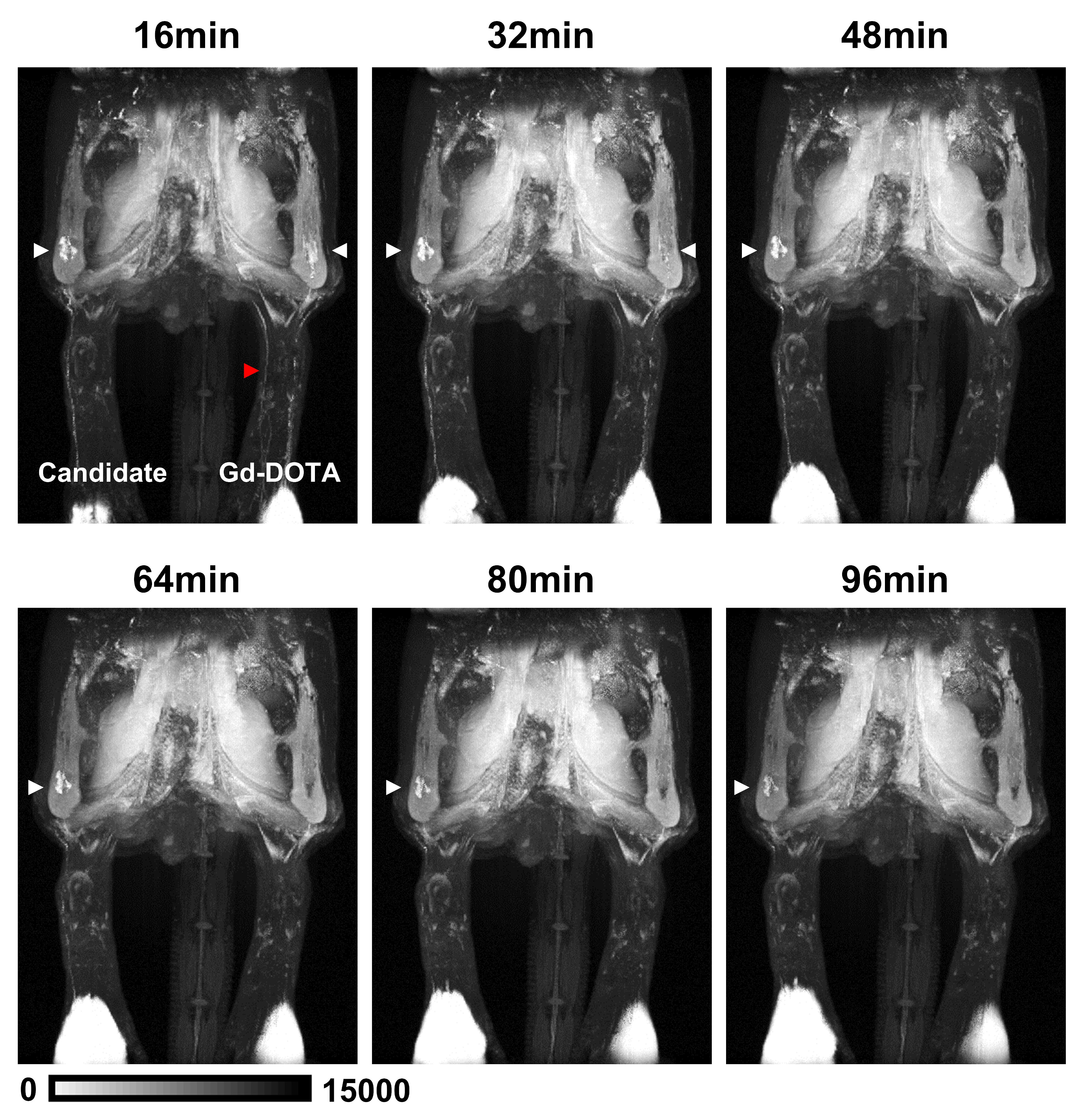
Fig. 1. Lymph nodes and lymphatic vessels detection on 3D time-of-flight (TOF) Magnetic Resonance Lymphography (MRL).
MRL was performed every 16 min after the injection of contrast agents (R; Gd-DOTA, L; a novel candidate). The lymph nodes are indicated by white arrows and the saphenous vein is indicated by a red arrow.
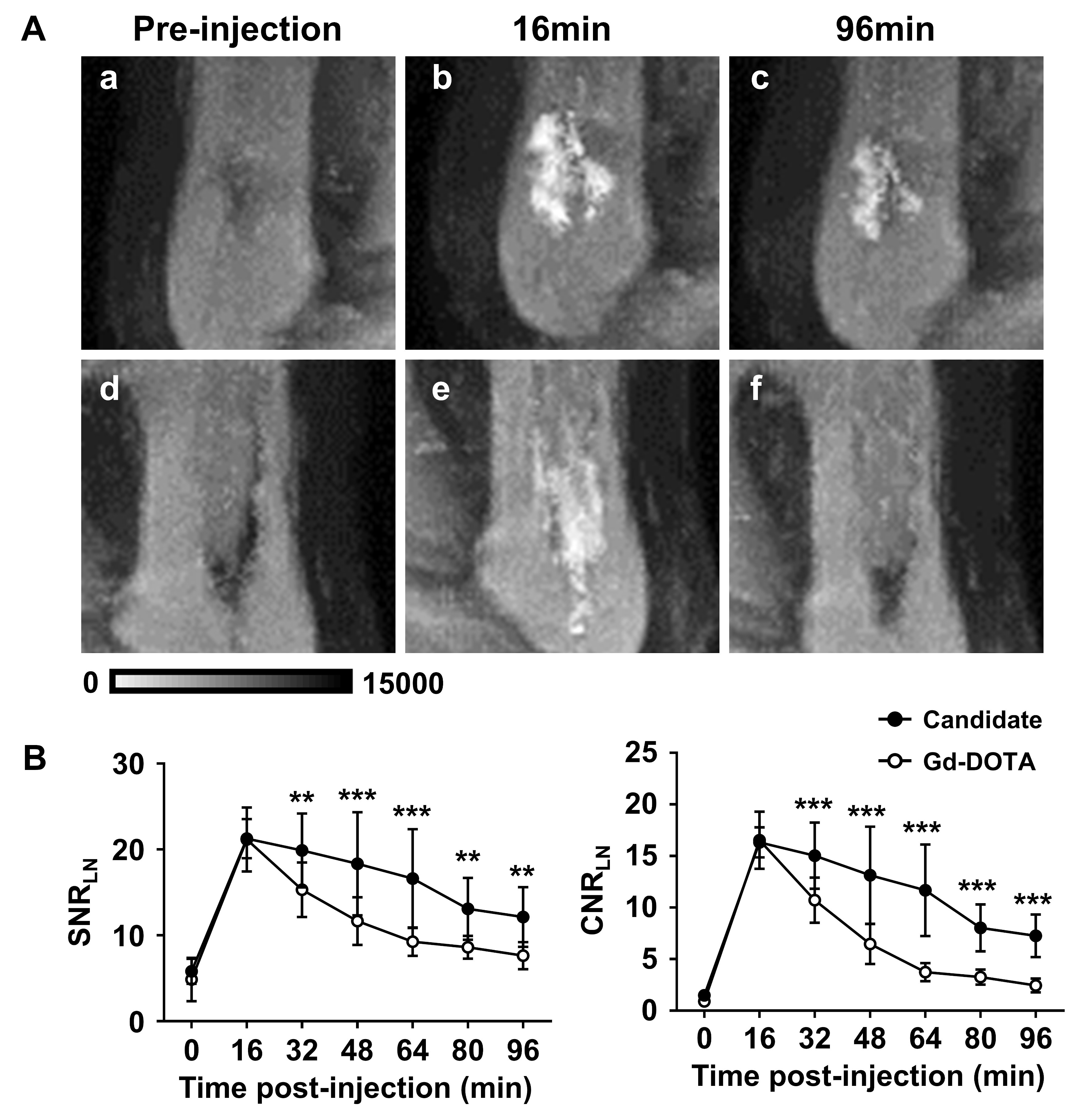
Fig. 2. Contrast effects of the novel contrast agent and gadolinium-based contrast agent on the lymph node.
(A) Coronal 3D TOF images pre-injection (a, d) and after injection at 16 min (b, e) and at 96 min (c, f). (B) Comparison of the signal-to-noise ratio (SNR) of lymph nodes and contrast-to-noise ratio (CNR) of lymph nodes (*p<0.05, **p<0.01, ***p<0.001 vs. Gd-DOTA).
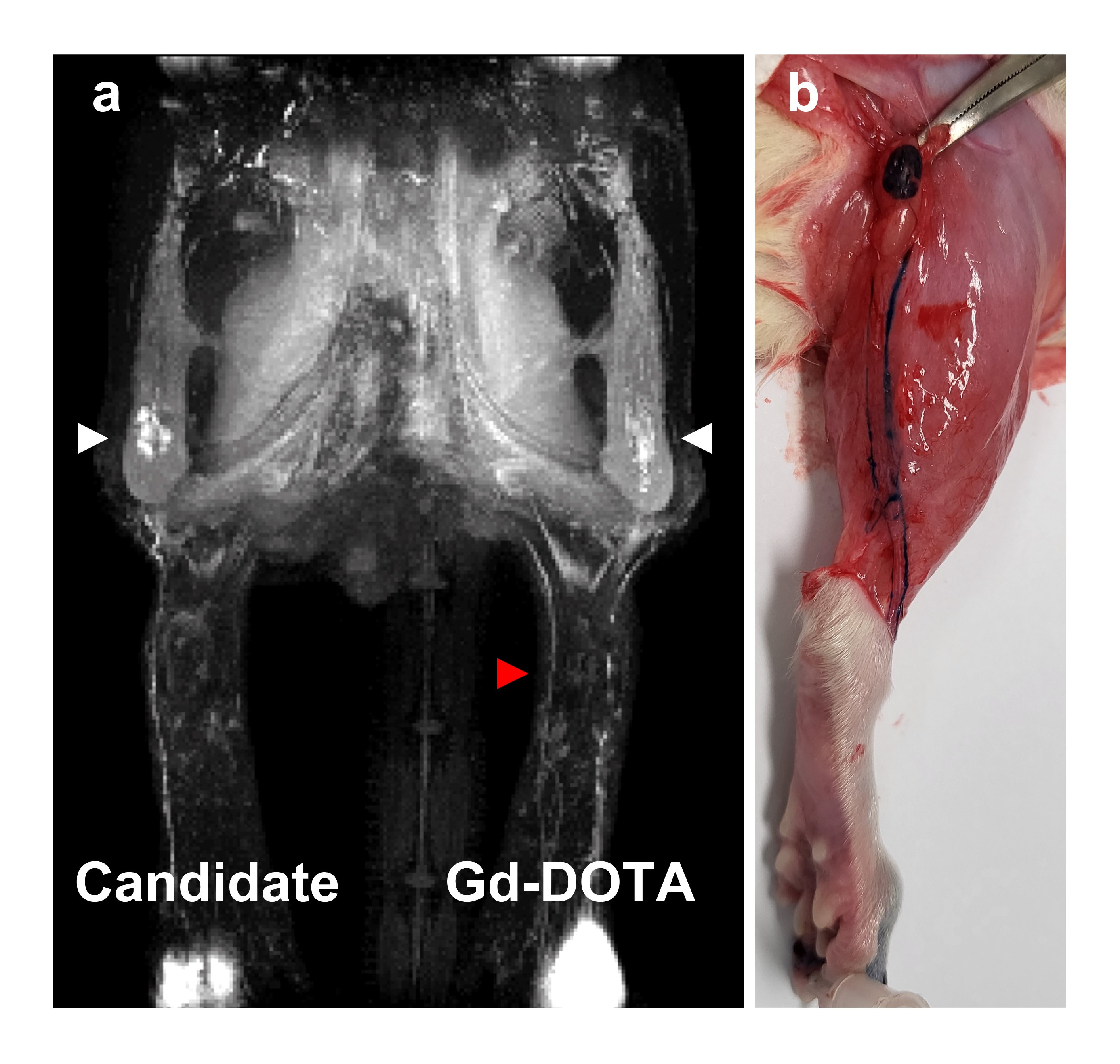
Fig. 3. Verification of MRL by methylene blue staining.
The coronal 3D TOF image 16 min after injection (a) and methylene blue staining 2 days after MRL (b).