4867
Cine MRI-derived radiomics features of the blood pool in pulmonary hypertension–heart failure with preserved ejection fraction (PH-HFpEF)1Northwestern University, Chicago, IL, United States
Synopsis
Keywords: Heart, Radiomics, blood pool
Radiomics features acquired from the blood pool in different chambers (LV/RV/LA/RA) at various periods within a cardiac cycle present diverse property in characterizing PH-HFpEF. Multiple blood pool radiomics features were significantly related to PCWP and mPAP (r: 0.4 – 0.643). Compared to the LV wall, the blood pools provide more efficient features (individual AUC: 0.7 – 0.821) to discriminate PH-HFpEF from controls (p < 0.05). Cine MRI-derived radiomics features of the blood pool can be used to characterize PH-HFpEF. Radiomics features of the blood pool have the potential to become novel quantitative imaging biomarkers for presenting cardiovascular pathology.Objective
To test the hypothesis that cine MRI-derived radiomics features in the blood pool can present hemodynamic characteristics of pulmonary hypertension-heart failure with preserved ejection fraction (PH-HFpEF).Materials and methods
Nineteen PH-HFpEF patients (9 male, 57.8 ± 14.7 years) and 20 healthy controls (13 male, 50.3 ± 13.6 years) were enrolled. All participants underwent a cardiac MRI scan. One hundred and seven radiomics features (7 classes) of the left and right ventricles/atrium (LV/RV/LA/RA) and LV wall were extracted from 4-chamber cine at the stages of systole, rapid filling, diastasis and atrial contraction within a cardiac cycle. For PH-HFpEF patients, features acquired from LV/LA were related to the pulmonary capillary wedge pressure (PCWP); features acquired from RV/RA were related to the mean pulmonary artery pressure (mPAP) using the Pearson correlation coefficient (r). The receiver operating characteristic (ROC) curve and the area under the curve (AUC) were used to test the capability of radiomics features in discriminating 2 subject groups.Results
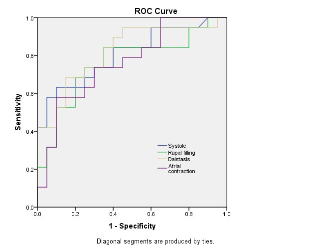
Cine MRI images from all participants were eligible for quantitative analysis. Radiomics features were successfully acquired at 4 time points in a cardiac cycle. Figure 1.Images of the blood pool acquired at systole, rapid filling, diastasis, and atrial contraction provided different numbers of radiomics features related to RHC results. The images of the blood pool of the LV acquired at 4 time points provided 3 (r: 0.468 – 0.485), 2 (r: 0.481 – 0.501), 1 (r: 0.456) and 0 features that were significantly related to PCWP. The LV blood pool provided 7 (AUC: 0.7 – 0.776), 2 (AUC: 0.712 – 0.745), 1 (AUC: 0.746), and 5 (AUC: 0.711 – 0.771) features at 4 time points that can predict the existence of PH-HFpEF. The images of the blood pool of the LA acquired at 4 time points provided 0, 1 (r: 0.573), 3 (r: 0.401- 605) and 2 (r: 0.495 – 0.506) features that are significantly related to PCWP, respectively. The LA blood pool provided 12 (AUC: 0.703 - 0.75), 1 (AUC: 0.703), 11 (AUC: 0.716 – 0.784), and 11 (AUC: 0.708 – 0.753) features at 4 time points that can predict the existence of PH-HFpEF. The images of the blood pool of the RV acquired at 4 time points provided 4 (r: 0.457 – 0.565), 3 (r: 0.468– 0.643), 2 (r: 0.457 – 0.527) and 7 (0.469 – 0.555) features that were significantly related to mPAP. The RV blood pool provided 6 (AUC: 0.713 – 0.792), 0, 1 (AUC: 0.718), and 3 (AUC: 0.704 – 0.762) features at 4 time points that can predict the existence of PH-HFpEF. The images of the blood pool of the RA acquired at 4 time points provided 2 (r: 0.475 – 0.53), 3 (r: 0.482 – 0.643), 0 and 1 (r: 0.59) features that were significantly related to mPAP. The RA blood pool provided 11 (AUC: 0.7 – 0.807), 29 (AUC: 0.703 – 0.761), 32 (AUC: 0.705 – 0.821), and 17 (AUC: 0.7 – 0.776) features at 4 time points that can predict the existence of PH-HFpEF. The images of the LV wall at 4 time points provided 0, 4 (r: 0.457 – 0.499), 0, and 3 (r: 0.465 – 0.533) features that were significantly related to PCWP. The LV myocardium provided 1 (AUC: 0.703), 0, 1 (AUC: 0.791), and 1 (0.778) feature at 4 time points that can predict the existence of PH-HFpEF. Tables 1 and 2. Figures 2 and 3.
Compared to the LV wall, the blood pool seemed to provide more radiomics features that can be used to discriminate PH-HFpEF patients from healthy volunteers (p < 0.05, Fisher’s tests).
There was good intraobserver and interobserver agreement on 88% and 85% radiomics features of the blood pool (defined as ICC > 0.75 or CoV < 20%).
Conclusion
Cine MRI-derived radiomics features of the blood pool can be used to characterize PH-HFpEF. Radiomics features of the blood pool have the potential to become novel quantitative imaging biomarkers for presenting cardiovascular pathology.Acknowledgements
N/AReferences
N/AFigures
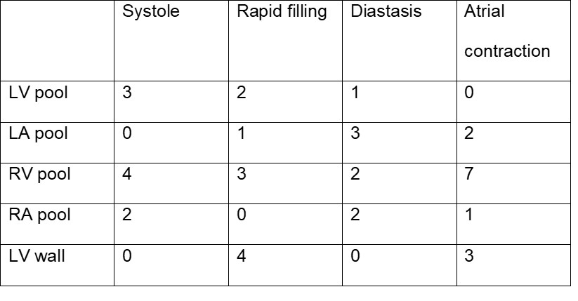
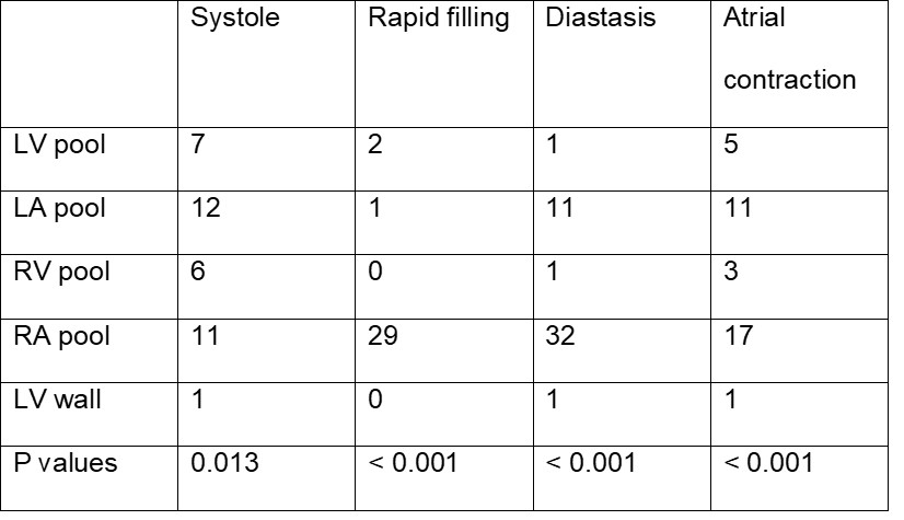
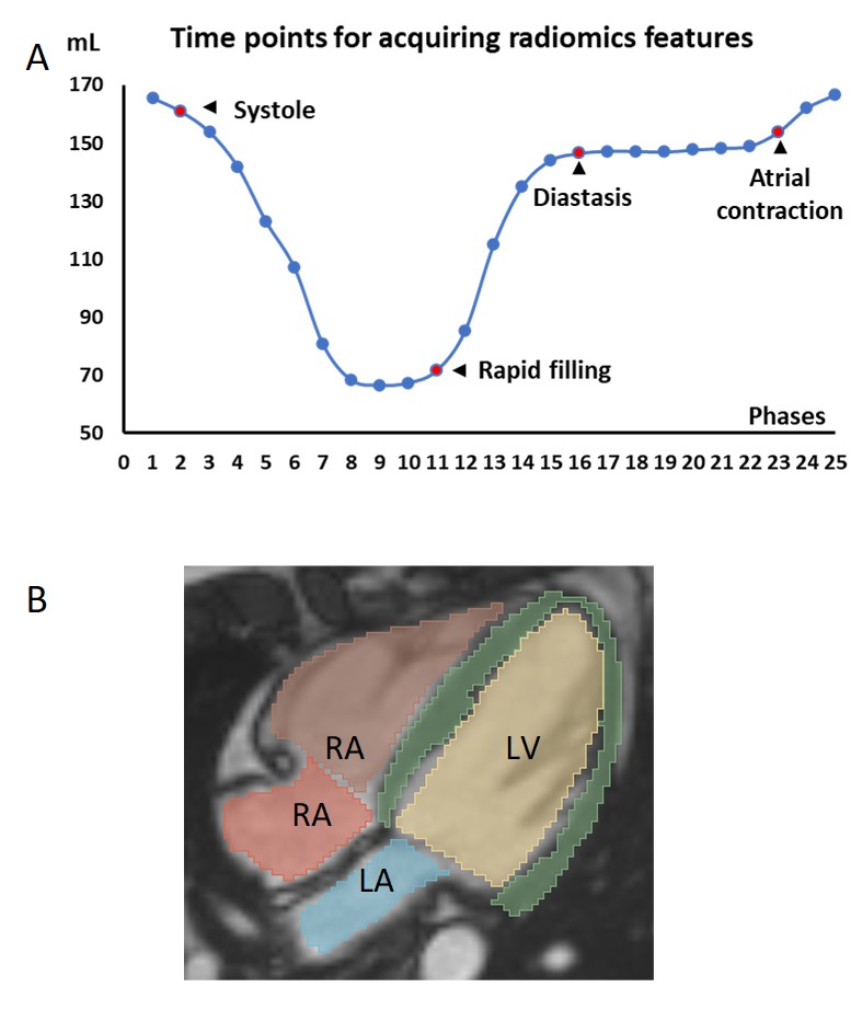
Figure 1 The workflow of extracting radiomics features of the blood pool at selected periods within a cardiac cycle.
A Times points of systole, rapid filling, diastasis, and atrial contraction were identified based on the volumes of the LV (measured on short-axis cine MRI).
B Regions of interest (ROIs) were manually drawn on corresponding cine images (4-chamber view) to extract radiomics features.
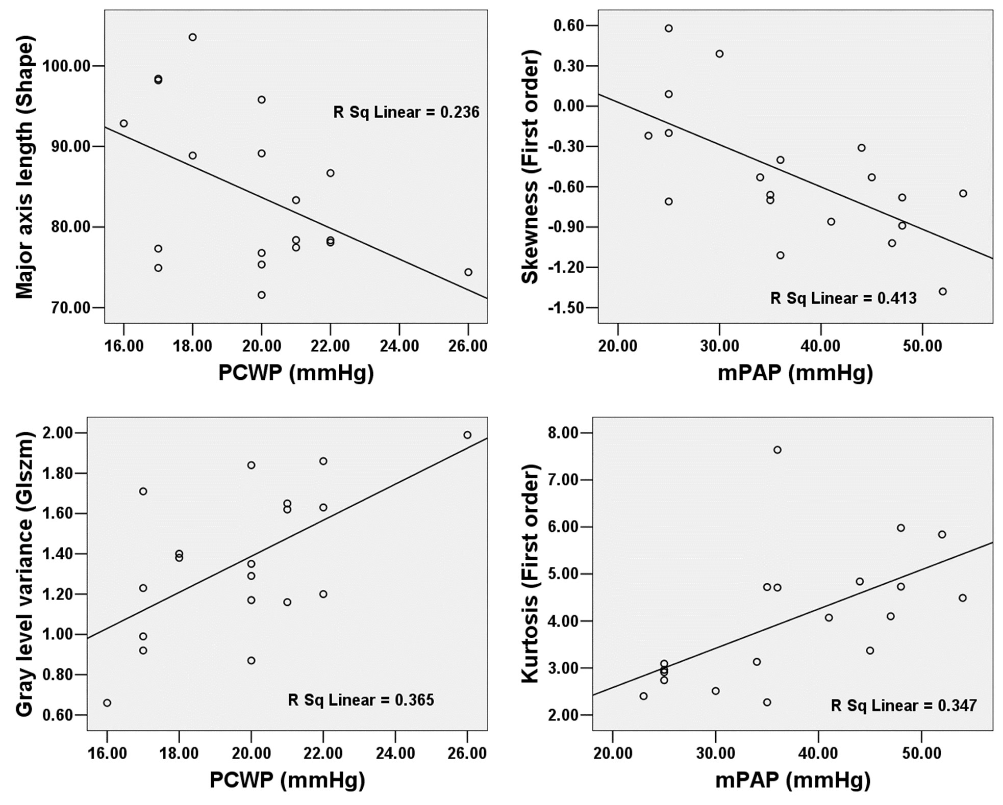
Figure 2 Multiple classes of radiomics features of the blood pool in the left and right heart acquired at different time points were related to PCWP and mPAP, respectively.
A At systole, the “major axis length” of the LV blood pool was related to PCWP (r = -0.485, p = 0.035).
B At rapid filling, the “skewness” of the RV blood pool was related to mPAP (r = -0.643, p = 0.003).
C At diastasis, the “gray level variance” of the LA blood pool was related to PCWP (r = -0.604, p = 0.006).
D At atrial contraction, the “Kurtosis” of the RA blood pool was related to mPAP (r = 0.589, p = 0.008).