4861
Cardiac MRI left ventricular and left atrial parameters predict myocardial late gadolinium enhancement in hypertrophic cardiomyopathy1Department of Radiology, The First Affiliated Hospital of Dalian Medical University, Dalian, China, Dalian, China
Synopsis
Keywords: Cardiomyopathy, Cardiomyopathy
Myocardial fibrosis causes functional impairment in patients with hypertrophic cardiomyopathy (HCM). The clinical gold standard for imaging myocardial fibrosis is cardiac magnetic resonance imaging (MRI) with late gadolinium enhancement (LGE), which is commonly used for the diagnosis and prognostic evaluation of patients with HCM. However, LGE imaging is an invasive test and some patients have adverse reactions to contrast agents. The aim of this study is to predict the presence of fibrosis in the left ventricular myocardium by cardiac magnetic resonance (CMR) non-invasive assessment of left atrial and left ventricular parameters in patients with HCM.Purpose
The aim of this study is to predict the presence of fibrosis in the left ventricular myocardium by cardiac magnetic resonance (CMR) non-invasive assessment of left atrial and left ventricular parameters in patients with HCM.Introduction
Myocardial fibrosis causes functional impairment in patients with hypertrophic cardiomyopathy (HCM). The clinical gold standard for imaging myocardial fibrosis is cardiac magnetic resonance imaging (MRI) with late gadolinium enhancement (LGE), which is commonly used for the diagnosis and prognostic evaluation of patients with HCM [1-2]. However, LGE imaging is an invasive test and some patients have adverse reactions to contrast agents. The aim of this study is to predict the presence of fibrosis in the left ventricular myocardium by cardiac magnetic resonance (CMR) non-invasive assessment of left atrial and left ventricular parameters in patients with HCM.Material and Methods
Seventy-two patients with HCM were retrospectively included, all of whom underwent 3.0T CMR and were stratified according to LGE. Cine images of the LV short axis were acquired for analysis of LV parameters. LA volume (LAV) is calculated using the two-plane area-length method[3]. LAV includes minimum LAV (LAVmin), maximum LAV (LAVmax), and pre-contraction LAV (LAVpre-ac). LA strain parameters were derived from CMR two- and four-chamber cine images by a semiautomatic method. LA strain parameters include global longitudinal strain (GLS) and global circumferential strain (GCS). The LA GLS includes reservoir strain (GLS reservoir), conduit strain (GLS conduit), and booster strain (GLS booster). Three LA GLS strain rate (SR) parameters were derived: SR reservoir, SR conduit, and SR booster. Left ventricular short-axis late gadolinium enhancement (LGE) images were obtained after 7-10 minutes of gadopentetate glucosamine injection.RESULTS
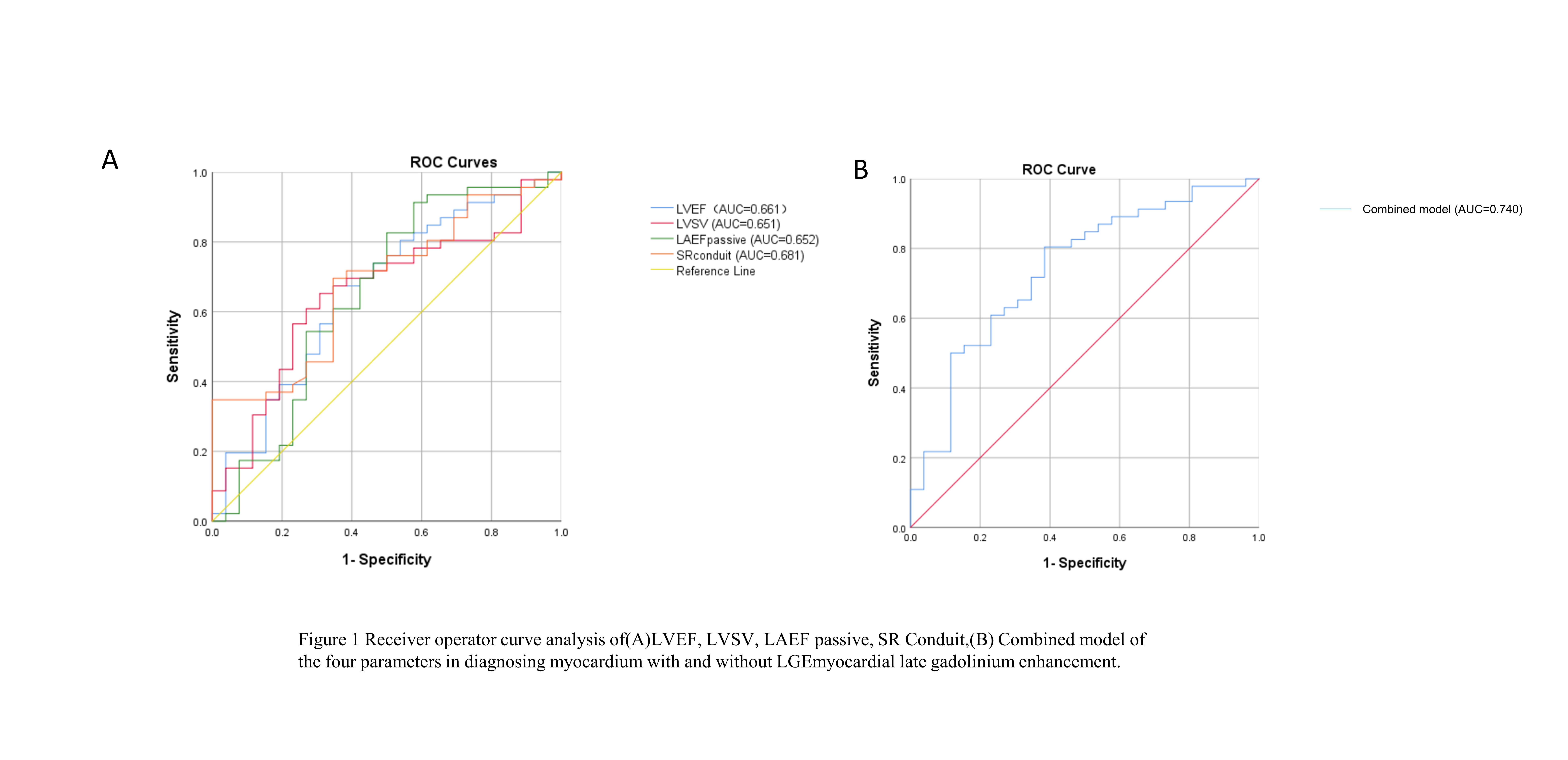
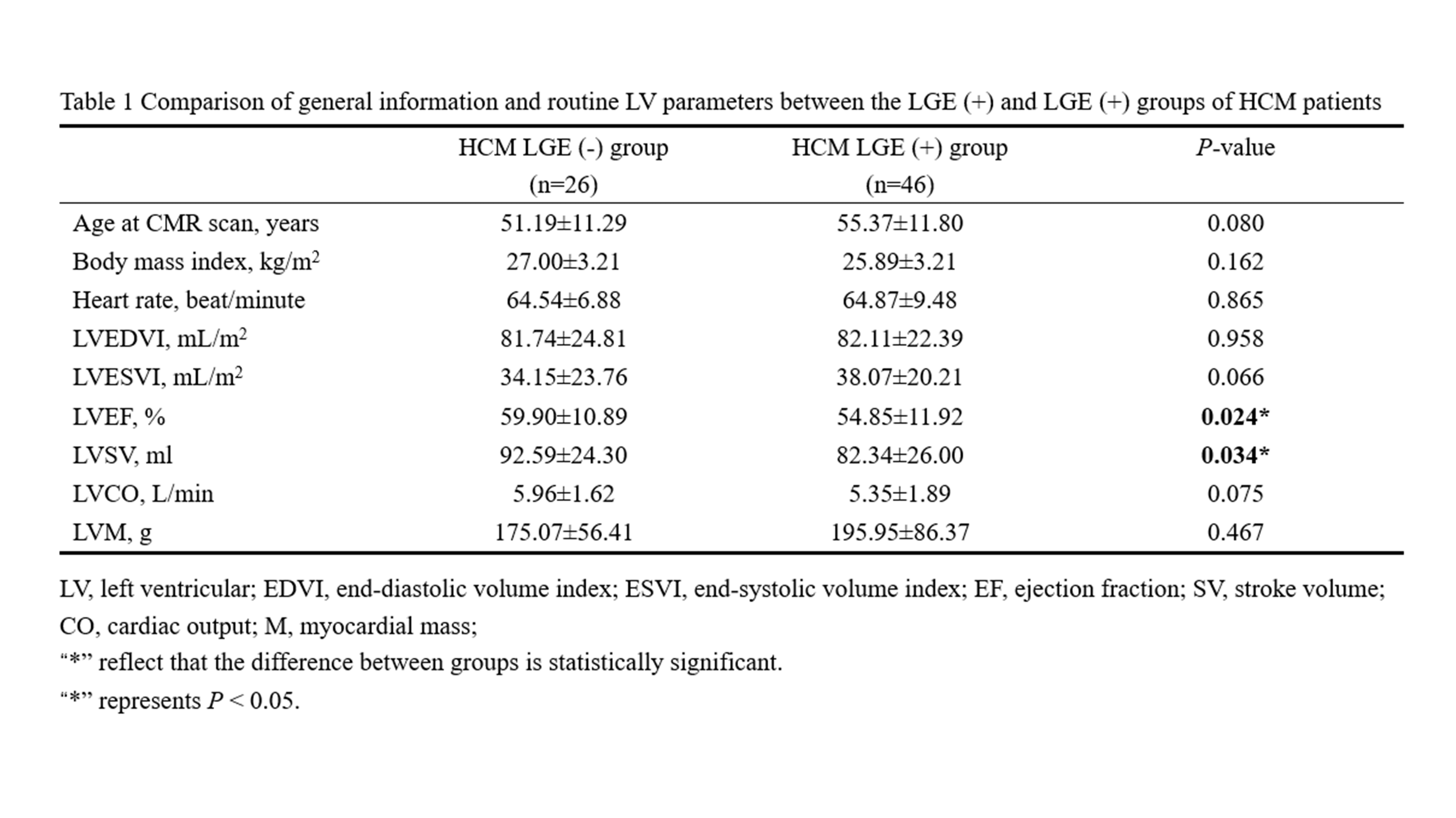
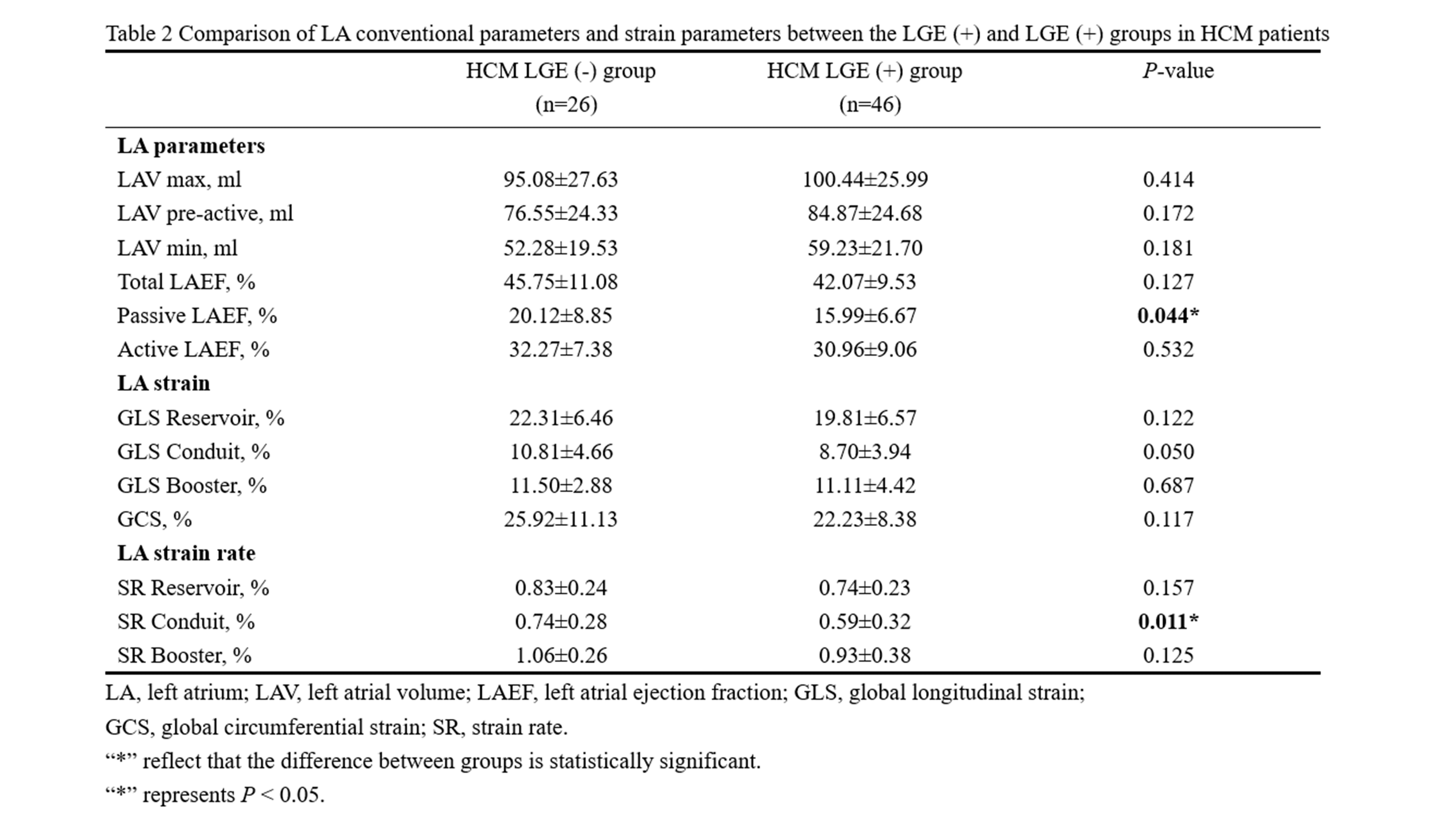
LVEF, LVSV, Passive LAEF, and SR Conduit were significantly lower in the LGE (+) group compared with the HCM LGE (-) group (P < 0.05) (Tables 1 and Table 2). The diagnostic performance of LA SR conduit to differentiate the presence or absence of fibrosis in HCM myocardium (AUC = 0.681) was superior to conventional LV and LA parameters (Figure 1A). The combined LVEF, LVSV, LAEF passive and LA SR conduit model had the best diagnostic performance with an AUC of 0.740 (Figure 1A and B).CONCLUSION
Noninvasive assessment of LV and LA parameters by CMR in patients with HCM may predict the presence or absence of fibrosis in the LV myocardium.Acknowledgements
No acknowledgementsReferences
[1]Moravsky G, Ofek E, Rakowski H, Butany J, Williams L, Ralph-Edwards A, Wintersperger BJ, Crean A. Myocardial fibrosis in hypertrophic cardiomyopathy: accurate reflection of histopathological findings by CMR. JACC Cardiovasc Imaging. 2013 May;6(5):587-96. doi: 10.1016/j.jcmg.2012.09.018.
[2]Song Y, Bi X, Chen L, Yang K, Chen X, Dong Z, Wang J, Kong X, Zhao K, Wang H, Duru F, Lu M, Ma L, Qiao S, Zhao S. Reduced myocardial septal function assessed by cardiac magnetic resonance feature tracking in patients with hypertrophic obstructive cardiomyopathy: associated with histological myocardial fibrosis and ventricular arrhythmias. Eur Heart J Cardiovasc Imaging. 2022 Jul 21;23(8):1006-1015. doi: 10.1093/ehjci/jeac032.
[3]Peters DC, Lamy J, Sinusas AJ, Baldassarre LA. Left Atrial Evaluation by Cardiovascular Magnetic Resonance: Sensitive and Unique Biomarkers. Eur Heart J Cardiovasc Imaging (2021) 23(1):14-30. Epub 2021/11/01. doi: 10.1093/ehjci/jeab221.
Figures