4828
Mitigation of water-fat swap caused by off-resonance effects in Dixon-based non-contrast-enhanced MR angiography1Diagnostic and Interventional Radiology, University Hospital RWTH Aachen, Aachen, Germany, 2Philips Japan, Tokyo, Japan, 3Philips GmbH Market DACH, Hamburg, Germany
Synopsis
Keywords: Vessels, Vessels, non-contrast-enhanced angiography, REACT
Recently introduced Dixon-based MR angiographic techniques have shown promise for non-contrast enhanced vascular imaging, though off-resonance effects induced by B0 inhomogeneities may result in water-fat separation errors (i.e., swaps) during Dixon water fat separation, typically seen in a large field of view at high field strength. In this work, we investigate possible swaps via numeric simulations and in vivo measurements with various shimming techniques as well as reconstruction options for B0 demodulation. Initial results may suggest practical methods to mitigate water-fat swaps and improve technical robustness even in challenging applications.Introduction
Flow-independent non-contrast-enhanced MR angiography (NCE-MRA) techniques are typically based on relaxation time differences of the tissues, for example, between blood and muscles. While balanced SSFP-based methods often suffer from banding artifacts at high field strengths or across large fields-of-view (FOV), a recently introduced non-balanced multi-echo Dixon method, termed REACT (Relaxation-Enhanced Angiography without Contrast and Triggering), has overcome these issues1,2 and yields good SNR and high blood-tissue contrast for assessment of vascular disorders2,3. However, off-resonance effects induced by local B0 inhomogeneity can result in water-fat separation errors, or swaps more specifically, which cause signal voids in the Dixon water images4,5. Hence, we aimed to mitigate such signal swaps during Dixon acquisition and reconstruction by including improved B0 shimming information.Methods
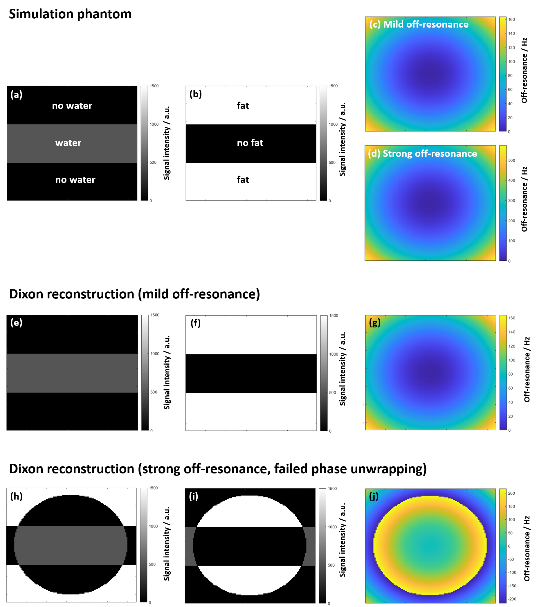
Both numerical simulations and in vivo experiments were conducted. Simulations were performed in MATLAB (R2021b, MathWorks, Natick, MA) following Ma6. Water and fat tissue distributions MW(x,y) and MF(x,y) as well as a mild and a strong parabolic off-resonance distribution Δf0(x,y) were defined as shown in Figure 1(a-d). Complex in-phase/out-of-phase/in-phase MR images of the simulation phantom were calculated for TE1/2/3=2.3/3.45/4.6 ms, using$$$ M_i(x,y,TE_i)=[M^W(x,y)+M^F(x,y)·e^{2iπ·Δf_{cs}·TE_i}]·e^{2iπ·Δf_0(x,y)·TE_i} $$$, (Eq.1)
with Δfcs=1/(2·ΔTE), ΔTE=1.15 ms and i={1,2,3}6. After calculating the Dixon-reconstructed off-resonance distribution as
$$$Δf_0^{rec}=\frac{φ^{rec}(x,y)}{2π·ΔTE}$$$, (Eq.2)
where the phase advance over ΔTE is
$$$φ^{rec}(x,y)=\frac{1}{2}·\arg\left(\frac{M_3(x,y)}{M_1(x,y)}\right)$$$. (Eq.3)
Dixon water and fat images were reconstructed as
$$$W^{rec}(x,y),F^{rec}(x,y)=\frac{1}{4}·\left(M_1(x,y)+M_3(x,y)·e^{-2iφ^{rec}(x,y)}\right)±\frac{1}{2}·M_2(x,y)·e^{-iφ^{rec}(x,y)}$$$. (Eq.4)
Importantly, Eq.3 defines the phase on an interval [0,2π). If the phase exceeds this interval, a phase wrap occurs, which has to be removed by a phase unwrapping algorithm. To simulate failed phase unwrapping, phase wraps in φrec were deliberately not removed before its insertion into Eq.2.
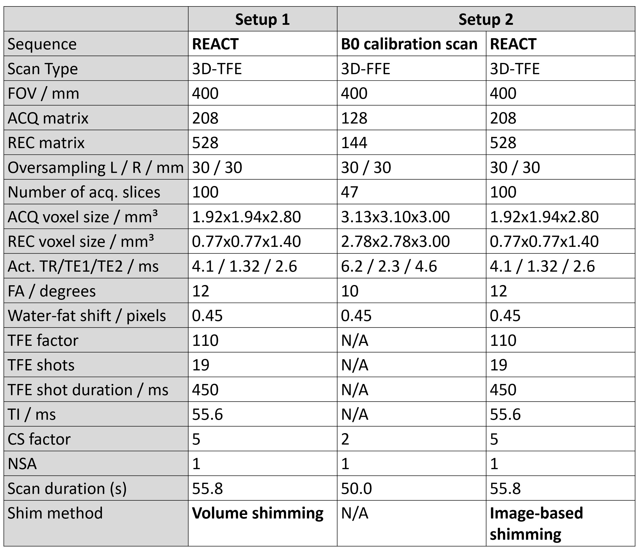
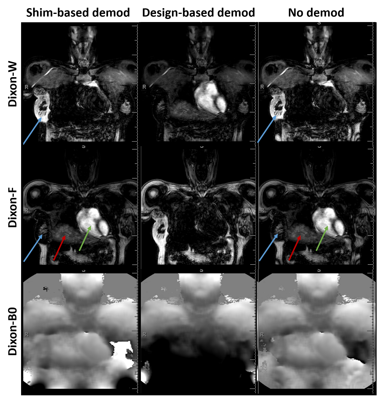
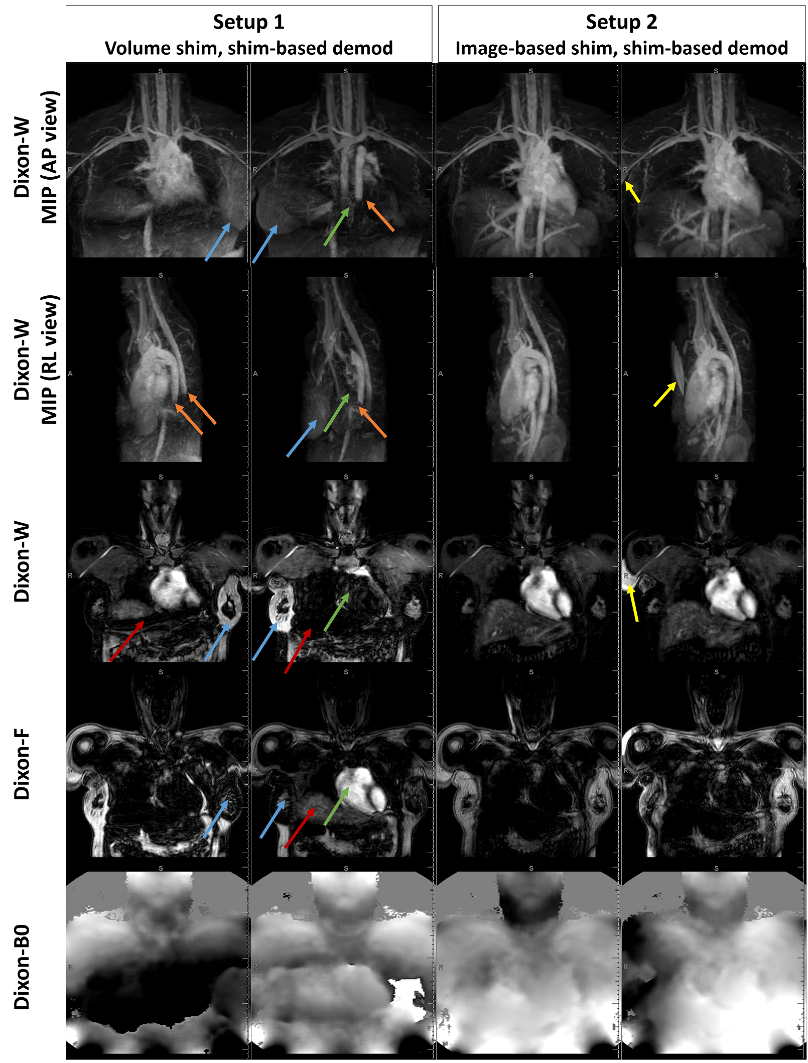
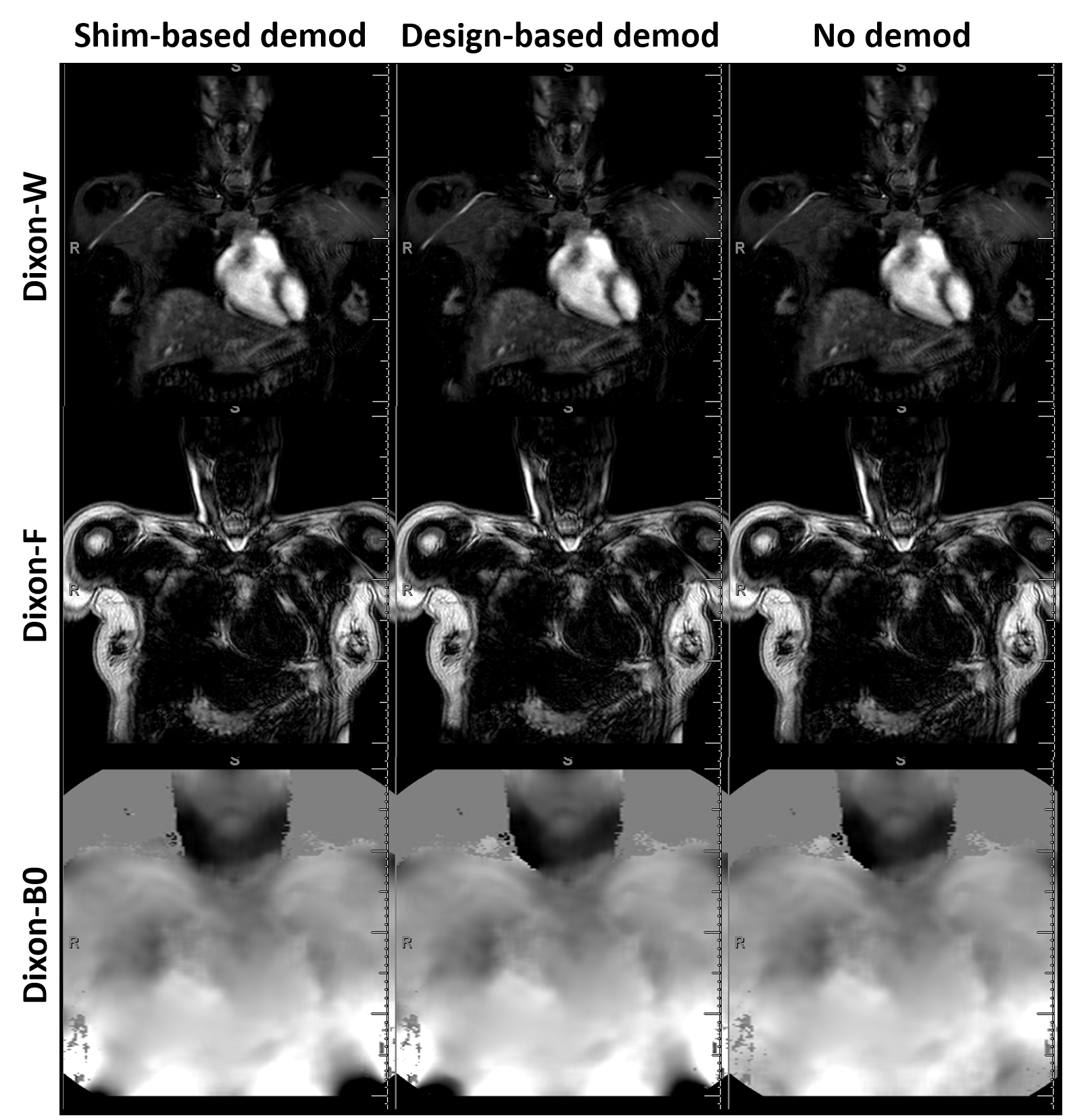
In vivo measurements were performed in a healthy female human subject on a whole-body clinical 3.0T MRI system (Elition X, Philips, Best, The Netherlands) using the torso and posterior coils. REACT was applied in two setups, based on a 2-echo Dixon gradient echo sequence preceded by T2-preparation and inversion-recovery magnetization preparation pulses without motion compensation1,2. Main scan parameters are summarized in Figure 2. Per setup, REACT was performed twice. In setup 1, REACT was carried out with volume-based B0 shimming based on a manually defined shim volume that represented a non-optimal shimming condition. In setup 2, REACT was performed with image-based shimming that considered a dedicated B0 calibration scan acquired prior to the REACT sequences. For both setups, different demodulation methods for B0 map estimation were performed during Dixon reconstruction, namely, conventional shim-based demodulation integrating B0 field information pre-determined at system installation, design-based demodulation incorporating a priori knowledge of the magnet design to improve B0 inhomogeneity correction7, and no demodulation. All images were reconstructed on the scanner.
Results
The simulation predicted the occurrence of swaps in the absence of phase unwrapping for the strong, but not for the mild off-resonance distribution, as shown in Figure 1(e-j). For in vivo scans for setup 1, design-based demodulation was able to retrospectively correct swaps in the heart, large thoracic vessels, breast and liver, that were present for shim-based demodulation. This was not the case for no demodulation, although both options resulted in local changes in the Dixon-reconstructed B0 map (Figure 3). Comparing setup 1 and 2, image-based shimming including the B0 calibration scan yielded images free of swaps and smoother B0-maps also for shim-based demodulation (Figure 4). For setup 2, all reconstruction options resulted in images free of swaps and visually identical B0 maps (Figure 5).Discussion
For large FOV scans including the head and neck area, strong off-resonance may be present towards the edges of the FOV, and manual positioning of a shim box can deteriorate field homogeneity. By comparing our findings in vivo, i.e., the reduced occurrence of swaps alongside with the smoother appearance of B0 maps under image-based shimming, to the simulations, it can be inferred that image-shimming based on a dedicated B0 calibration scan resulted in an overall milder off-resonance distribution. This, in turn, reduced the chance for residual phase wraps in the off-resonance maps and hence for swaps on the acquisition side. However, the calibration scan required additional scan time. Besides, swaps under volume shimming were reduced by the design-based demodulation reconstruction option, which takes large-FOV field inhomogeneity information into account. This indicates a strategy for improved phase unwrapping on the scanner without additional scan time. While this work assessed the influence of off-resonance on the occurrence of swaps, it did not touch upon the influence of noise or of gross motion as potential other causes for off-resonance errors.Conclusion
Water-fat swaps occurring in large-FOV NCE-MRA based on REACT can be avoided by a dedicated calibration scan for optimal B0 estimation and improved field demodulation during reconstruction.Acknowledgements
No acknowledgement found.References
1 Yoneyama M, et al. Free-breathing non-contrast-enhanced flow-independent MR angiography using magnetization-prepared 3D non-balanced dual-echo Dixon method: A feasibility study at 3 Tesla. Magn Reson Imaging 2019;63:137–146.
2 Tan EJ, et al. REACT – A novel flow-independent non-gated non-contrast MR angiography technique using magnetization-prepared 3D non-balanced dual-echo Dixon method: Preliminary clinical experience. Eur J Radiol Open 2020; 7:100238.
3 Dillman JR, et al. Non-contrast three-dimensional gradient recalled echo Dixon-based magnetic resonance angiography/venography in children. Pediatr Radiol 2019;49:407-414.
4 Terwolbeck MN, et al. Relaxation-Enhanced Angiography without Contrast and Triggering (REACT) for pelvic MR venography in comparison to balanced gradient-echo and T2-weighted spin-echo techniques. Clin Imaging 2021;74:149–155.
5 Isaak A, et al. Free‑breathing non‑contrast flow‑independent cardiovascular magnetic resonance angiography using cardiac gated, magnetization‑prepared 3D Dixon method: assessment of thoracic vasculature in congenital heart disease. J Cardiovasc Magn Reson 2021;23:91.
6 Ma J. Dixon techniques for water and fat imaging. J Magn Reson Imaging 2008;28(3):543-58.
7 Diefenbach MN, et al. Improving chemical shift encoding-based water-fat separation based on a detailed consideration of magnetic field contributions. Magn Reson Med 2018;80:990-1004.
Figures