4822
Feasibility study of using Zero TE Sequence to evaluate pulmonary lesions: compared with CT examination1The First Affiliated Hospital of Zhengzhou University, Zhengzhou, China
Synopsis
Keywords: Lung, MR Value
Pulmonary MRI zero-echo time (ZTE) and MRI conventional sequences were compared with thoracic computed tomography (CT) to evaluate the lesions. All participants in the study underwent chest CT, ZTE, T2-TSE, T2-HASTE, and T1-VIBE scans. ZTE can be used as a supplementary examination of CT imaging. ZTE combined with T2WI can improve the detection rate and display performance of pulmonary nodulesIntroduction
Zero echo time (ZTE) imaging is a newly introduced fast gradient echo-based MRI sequence with a minimum susceptibility effect that can overcome the pulmonary inherent physical properties challenges[1], providing high-resolution structural information for the lung. as well as a shorter scan time and lack of operational noise[2]. ZTE evaluation of solid pulmonary nodules or masses has been studied. However, there was no clear assessment of ZTE for the sensitivity of nodule detection and the characteristics of nodule evaluation, direct comparisons of capabilities for ZTE and conventional 3T MR lung imaging sequences evaluation remain lacking. The aim of this study was to determine the feasibility of ZTE in accurately assessing pulmonary nodules, and to evaluate the diagnostic performance of ZTE sequence and conventional 3T MR Lung imaging in detecting pulmonary nodules, as referred to CT.Materials and Methods
A total of 136 participants (69 males and 67 females, mean age: 56.74±9.53 males and 55.36±10.53 females) with 196 nodules diagnosed by CT underwent lung MRI between April 2022 and August 2022. The scans of MRI were performed on 3.0T scanners(Architect MR, GE Medical Systems, Waukesha, WI, United States.) with the following sequences: T1-weighted Volumetric interpolated breath-hold examination (T1-VIBE), T2 -weighted Fast spin echo (T2-FSE), T2 -weighted Half-Fourier Acquisition Single-shot Turbo spin Echo imaging(T2-HASTE). CT and MRI data were analyzed independently by two radiologists who were blinded to the results of the other modality, the assessment includes lesion size, type, shape, margins, vascular bundles, endobronchial air maps, thickening, and constriction of the adjacent pleura. The statistical analyses were performed on the software SPSS22.0 (IBM Corp., Armonk, NY, USA). The measurement data were represented as (x±s), and the consistency was evaluated by using the Intraclass correlation coefficient(ICC). The maximal range or solid range between ZTE-MRI and CT were also compared using Pearson’s correlation analyses, and Bland–Altman analyses. And P<0.05 was considered statistically significant.Results and Discussion
Figure 1 shows a case with CT and four MR sequences.Table 1 shows the consistency of ZTE-MRI and CT assessment type, shape, and burr are moderate, and the consistency of assessment of tracheobronchial bundle, pleural indentation, and internal pneumobronchogram is good, while the consistency of CT and ZTE detection methods for edge condition was poor. In addition, the ZTE detected 30% solid nodules, 95% GGN, and 35% PSNS.
The consistency evaluation of CT and ZTE detection showed(Table 2) that the ICC or correlation coefficient value of maximal range (mm) was 0.954 (95% confidence interval (CI):0.940-0.965) and of the solid range (mm) 0.943(95% confidence interval (CI):0.925-0.957), suggesting good consistency and accuracy between ZTE and CT.Table 3 shows the sensitivity of the upper lobe, middle lobe, and lower lobe was 83.93, 83.33, and 77.27, respectively, the diagnostic sensitivity of the lower lobe of the lung was the lowest, and the diagnostic sensitivity of the upper lobe and the middle lobe of the lung was close, both being > 83.0%.
Taken CT as the golden standard, the overall sensitivity of ZTE、T1-VIBE、T2-FSE、T2-HASTEfor the detection of pulmonary nodules was 81.63%, 54.59%, 81.12%, and 63.27%, respectively, as shown in Table 4.ZTE has the highest overall sensitivity among all the MR lung techniques. Table4 also shows the detection rate of combined techniques. The overall sensitivity of ZTE combined withT1-VIBE, T2-FSE, and T2-HASTEin the detection of pulmonary nodules were 82.14%, 85.20%, and 83.67%, respectively.
Table 5 shows the CT and ZTE range among different subgroups, the maximum of CT was 3.3 for nodule size < 4 mm, 6.39±0.97(4.4-7.9)for nodule size of 4-8 mm, 14.96±5.65(8.1-29.4) for 8-10 mm, and 46.12±14.24(30.6-68.8) for size >10 mm. the maximum of ZTE were 0 for nodule size < 4 mm, 3.96±3.49(0-10.7) for nodule size of 4-8 mm,13.60±7.15(0-33.2) for 8-10 mm, and 43.13±14.05(26.5-65.0)for size >10 mm.
In the Bland-Altman analyses as shown in Figure 2, there was a strong agreement between the maximum diameter of pulmonary nodules measured by CT and MRI. In detail, the mean difference of ZTE is 1.8 mm with a 95% confidence interval (CI) of–5.0–8.7 mm for maximum diameter, and 1.9 mm with a 95% confidence interval (CI) of–5.3–9.2 mm for solid range.
Conclusion
It is feasible to use ZTE as a complementary sequence in routine clinical lung MRI without contrast media, which can be used as a supplementary examination of CT imaging. ZTE combined with T2WI can improve the detection rate and display performance of pulmonary nodules.Acknowledgements
No acknowledgement found.References
[1] Larson, P.E.Z.; Han, M.; Krug, R.; January, A.; Nelson, S.J.; Vigneron, D.B.; Henry, R.G.; McKinnon, G.; Kelley, D.A.C. Ultrashort echo time and zero echo time MRI at 7T. MAGMA 2016, 29, 359–370.
[2] Liu Q, Feng Z, Liu WV,ea tl.Assessment of Solid Pulmonary Nodules or Masses Using Zero Echo Time MR Lung Imaging: A Prospective Head-to-Head Comparison With CT.Front Oncol. 2022 Apr 26;12:812014. doi: 10.3389/fonc.2022.812014. eCollection 2022.
Figures
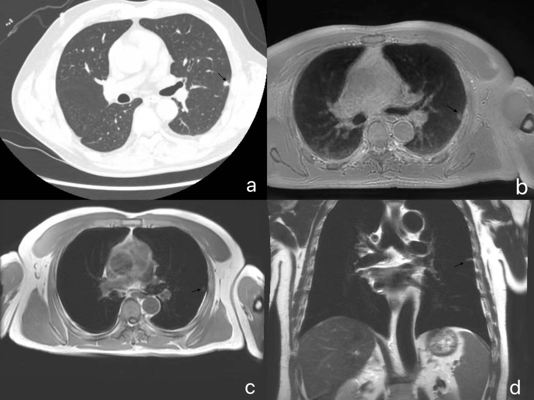
Figure 1. A 54-year-old male with a nodule (arrow) in the upper lobe of the left lung. (a)The CT showed an 8 mm circular nodule. (b) Axial ZTE sequences showed an 8 mm circular hyperintensity. (c)T1WI showed an 8 mm circular isointensity. (d)The coronal sequence showed an 8mm ellipsoidal hyperintensity.
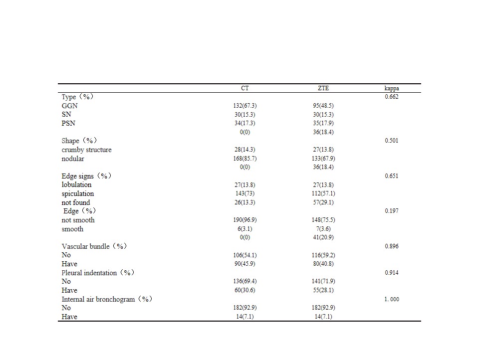
Table 1 Consistency comparison of CT and ZTE detection results
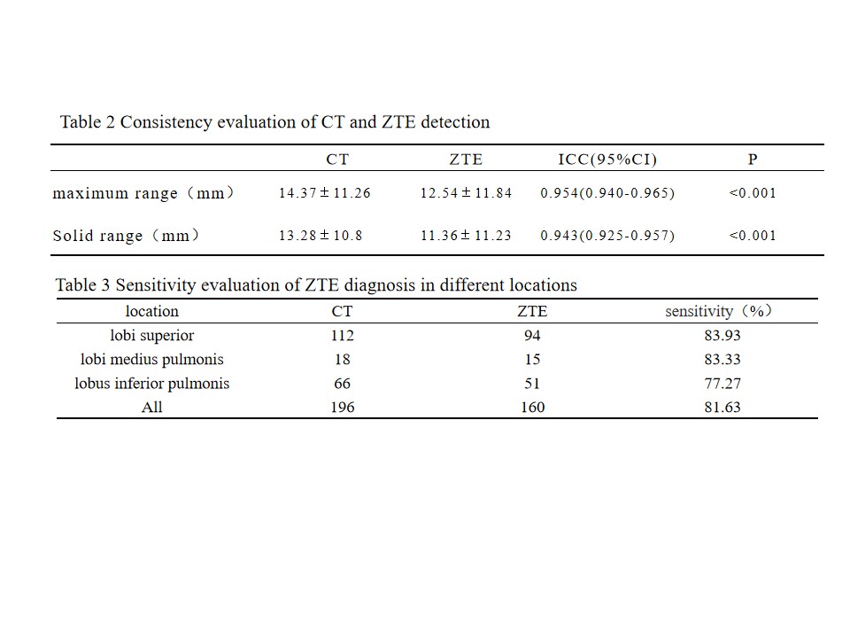
Table 2 Consistency evaluation of CT and ZTE detection
Table 3 Sensitivity evaluation of ZTE diagnosis in different locations
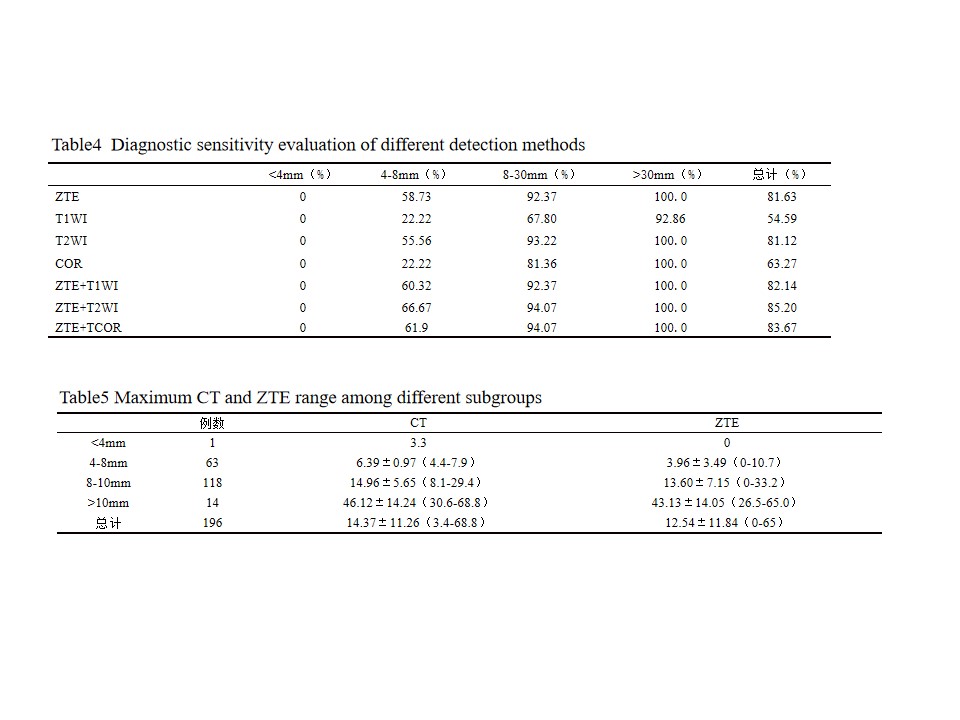
Table4 Diagnostic sensitivity evaluation of different detection methods
Table5 Maximum CT and ZTE range among different subgroups
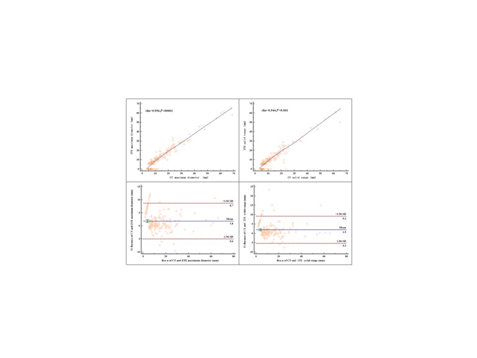
Figure 2. Pearson correlation and Bland-Altman analyses between CT and ZTE.