4800
T1 Relaxation-Enhanced Steady-State (T1RESS) Imaging with Radial Sampling for Robust Brain Examination at 3T and 0.55T1The Bernard and Irene Schwartz Center for Biomedical Imaging, Department of Radiology, New York University Grossman School of Medicine, New York, NY, United States, 2Radiology, Northshore University HealthSystem, Evanston, IL, United States, 3Siemens Medical Solutions, New York, NY, United States, 4Application Development, Siemens Healthcare GmbH, Erlangen, Germany
Synopsis
Keywords: Data Acquisition, Brain
A new class of sequences, named T1 relaxation-enhanced steady-state (T1RESS), has been recently proposed and showed improved lesion conspicuity in contrast-enhanced exams. T1RESS sequences combine steady-state acquisition, either balanced or unbalanced with weak gradient spoiling, with periodic contrast-modifying preparation pulses to create T1 weighting of the contrast. Here, we present radial versions of T1RESS sequences using the stack-of-stars trajectory, which offer improved motion robustness and enable dynamic imaging through use of advanced reconstructions such as GRASP. Volunteer scans acquired at 3T are shown for both sequences. Moreover, the balanced version is demonstrated at 0.55T to highlight its SNR efficiency.Introduction
Contrast-enhanced T1-weighted acquisition is an important component of many clinical protocols, such as for detection of brain tumors and metastases. Failure to identify small lesions can potentially have significant impact on the prognosis of patients. Recently, a new class of pulse sequences has been proposed, named T1 relaxation-enhanced steady-state (T1RESS), which offers improved conspicuity of enhancing lesions1,2. T1RESS uses a steady-state-based rapid acquisition combined with periodic contrast-modifying preparation to modulate the image contrast with the T1 relaxation time. T1RESS sequences can be used either with balanced steady state (bSSFP) or unbalanced steady state with weak gradient spoiling, which suppresses intravascular signal. Here, we describe radial T1RESS variants based on the stack-of-stars 3D trajectory, which offer improved motion robustness and enable advanced reconstruction for dynamic imaging such as the GRASP technique3. Both radial sequences are demonstrated for brain imaging in volunteers at 3T. The balanced version is furthermore shown at low field strength where the SNR efficiency of the underlying bSSFP mechanism and absence of banding artifacts make it a promising option to counter SNR losses.Methods
Sequence DesignA stack-of-stars 3D bSSFP sequence with golden-angle ordering has been implemented following the description for radial GRE acquisition4. In the balanced T1RESS (bT1RESS) version (Figure 1a), a non-selective contrast-modifying (CM) RF pulse is generated prior to acquiring stacks of projections for each angle. The amount of T1 weighting can be altered by varying the CM flip angle and waiting period (TI) before the readout train. An α/2 preparation and multiple dummy pulses are generated to establish steady state. After the readout train (before repeating the CM pulse), residual magnetization is stored in the longitudinal plane using an α/2 flip-back pulse5.
For the unbalanced T1RESS (uT1RESS) version (Figure 1b), the CM pulse is generated before the dummy pulses for the next angle (without α/2 preparation and flip-back pulses). After rewinding frequency-encoding and phase-encoding gradients at the end of each TR, a weak gradient spoiler (Figure 1b.i) is applied in constant frequency-encoding direction throughout the entire acquisition, which creates a controllable amount of flow-induced dephasing.
Experiments
Exemplary datasets were acquired in healthy volunteers (after obtaining informed consent) using clinical 3T systems (MAGNETOM Skyra/Prisma, Siemens Healthineers) equipped with 20-channel head-coil arrays. For bT1RESS, one 3D slab with 64 slices was acquired in sagittal orientation. Relevant parameters included: FOV 300x300x192mm3, resolution 1.17x1.17x3mm3, 500 projections, CM FA 90°, TI 300ms, FA 32°, BW 890Hz/px, TR/TE 3.75/1.87ms, duration 4:50min. Identical parameters were used for uT1RESS but TR was extended to 4.55ms to fit the weak spoilers, resulting in total duration of 5:15min. For comparison, bSSFP was acquired without CM pulses and otherwise identical parameters (duration 2:10min). Moreover, a radial spoiled 3D GRE scan was performed in axial orientation using following parameters: FOV 256x256x176mm3, resolution 1x1x1mm3, 500 projections, FA 12°, BW 700Hz/px, TR/TE 4.14/1.94ms, duration 2:47min.
Volunteer scans were also performed on a prototypic 0.55T scanner (modified MAGNETOM Aera, Siemens Healthineers). Only the bT1RESS version was acquired due to the higher SNR of the bSSFP acquisition. Parameters similar to the 3T scans were used, except for FA 70°, TI 200ms, and TR/TE 4.84/2.42ms. Durations were 4:06min and 2:16min for bT1RESS and bSSFP.
Additional isotropic scans in coronal orientation were performed with uT1RESS in a volunteer at 3T. Parameters included: FOV 320x320x180mm3, resolution 1.25x1.25x1.25mm3, FA 43°, TR 3.80ms, 512 projections, BW 1030Hz/px, slice oversampling 22.2%, PF 6/8, duration 4:28min.
Results
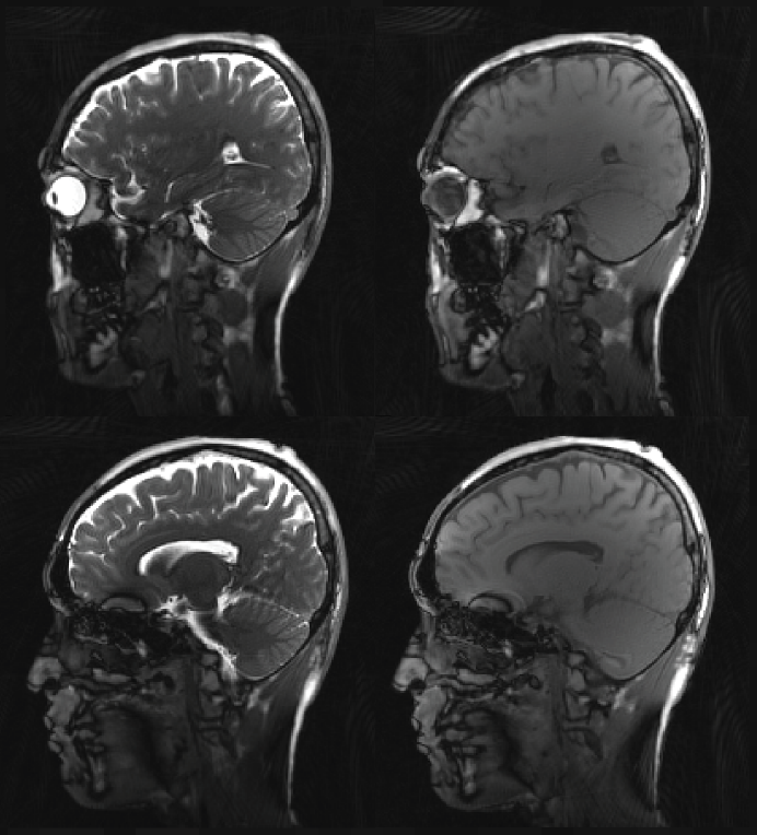
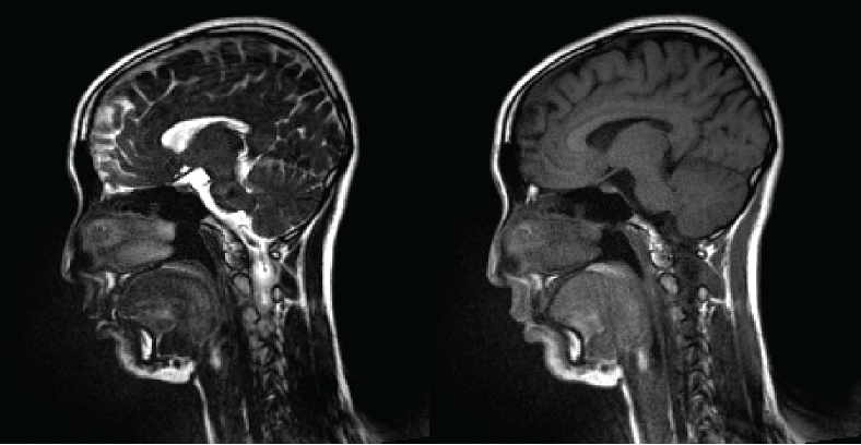
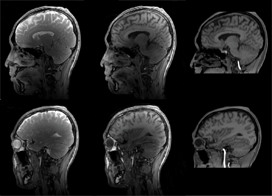
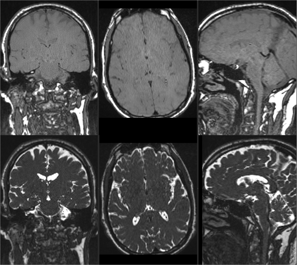
Figure 2 and 3 compare images acquired with bSSFP and bT1RESS at 3T and 0.55T. The signal intensity of the CSF is suppressed using bT1RESS through periodic application of CM pulses. Banding artifacts appear at 3T near air-tissue boundaries and are absent at 0.55T. Figure 4 shows uT1RESS images acquired at 3T for CM flip angles of 0° and 90°. As in bT1RESS, the CM pulses generate T1 weighting and suppress CSF. The weak spoilers render the blood vessels dark, which appear bright in the spoiled 3D GRE images and may impact lesion visibility. Banding artifacts are absent with uT1RESS compared to bT1RESS at the same field strength. Figure 5 shows the corresponding isotropic uT1RESS scans in coronal orientation.Discussion
Here, we present radial versions of T1RESS sequences that generate T1 weighting and suppress CSF signal by repeatedly applying contrast-modifying pulses, which improves lesion visibility in contrast-enhanced exams. The unbalanced variant renders blood vessels dark by applying weak spoilers along frequency-encoding directions, which induces flow-related phase dispersion. Due to the unbalanced gradients, the original uT1RESS sequence is more motion sensitive than bT1RESS. We anticipate that radial k-space sampling, as demonstrated, will help to overcome this limitation and outperform the original Cartesian version. bT1RESS, on the other hand, is often affected by banding artifacts when used at high field. At low field strength, however, banding artifacts are negligible. This makes the proposed bT1RESS version promising for T1-weighted imaging with high SNR on novel low-field MRI systems. Future work will include comparing T1RESS to established contrast-enhanced sequences in patients with brain tumors. Moreover, we will investigate the combination with GRASP reconstruction, which is compatible with the radial acquisition scheme and enables DCE-MRI at high temporal resolution.Acknowledgements
This work was supported in part by NIH R01 CA263091 and performed under the rubric of the Center for Advanced Imaging Innovation and Research (CAI2R, www.cai2r.net), an NIBIB National Center for Biomedical Imaging and Bioengineering (NIH P41 EB017183).References
1. Edelman R, Leloudas N, Pang J, Bailes J, Merrell R, Koktzoglou I. Twofold improved tumor-to-brain contrast using a novel T1 relaxation-enhanced steady-state (T1RESS) MRI technique. Science advances. 2020 Oct 28;6(44):eabd1635.
2. Edelman RR, Koktzoglou I, Leloudas N, Pang J. T1 Relaxation-Enhanced Steady-State (T1RESS): An Improved Three-Dimensional Method for Contrast-Enhanced Imaging of Brain Tumors.
3. Feng L, Grimm R, Block KT, Chandarana H, Kim S, Xu J, Axel L, Sodickson DK, Otazo R. Golden-angle radial sparse parallel MRI: combination of compressed sensing, parallel imaging, and golden-angle radial sampling for fast and flexible dynamic volumetric MRI. Magn Reson Med. 2014 Sep;72(3):707-17.
4. Block KT, Chandarana H, Milla S, Bruno M, Mulholland T, Fatterpekar G, et al. Towards Routine Clinical Use of Radial Stack-of-Stars 3D Gradient-Echo Sequences for Reducing Motion Sensitivity. Vol. 18, Journal of the Korean Society of Magnetic Resonance in Medicine. 2014;p.87.
5. Scheffler K, Heid O, Hennig J. Magnetization preparation during the steady state: fat-saturated 3D TrueFISP. Magn Reson Med. 2001;45(6):1075-1080.
Figures
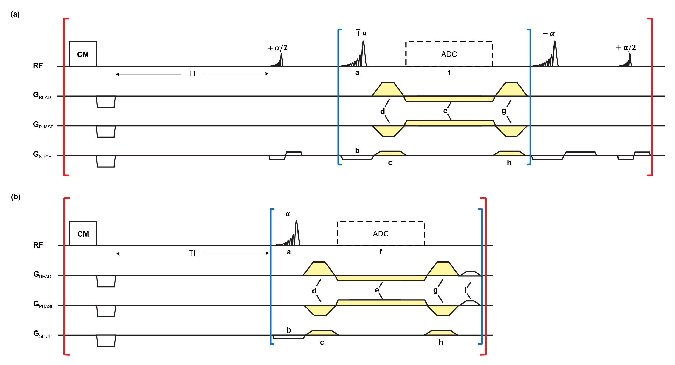
Fig 1: Radial T1RESS sequences. Red indicates outer loop over projection angles, blue shows inner loop over partitions.
bT1RESS: a) excitation pulse with FA ±α, b) slab-selection, c) slab-selection rewinder, d) prephase, e) readout, f) ADC, g) rewinder, and h) slab-selection prephase gradients.
uT1RESS: a) excitation pulse with FA α, b) slab-selection, c) slab-selection rewinder, d) prephase, e) readout, f) ADC, g) rewinder, h) slab-selection prephase, and i) weak readout-spoiler gradients.
d, e, g are modulated by GREAD= sin(φ), GPHASE= cos(φ). c, h are modulated by partition #.