4796
Radial Magnetic Resonance Fingerprinting for quantification of T1 in metastatic breast tumor.1Cancer Systems Imaging, MD Anderson Cancer Center, Houston, TX, United States, 2Department of Radiology, Case Western Reserve University, Cleveland, OH, United States, 3Imaging Physics, MD Anderson Cancer Center, Houston, TX, United States
Synopsis
Keywords: MR Fingerprinting/Synthetic MR, Cancer
Quantitative Magnetic Resonance Imaging (MRI) involves pixel-wise mapping of longitudinal relaxation time T1, transverse relaxation time T2 and proton density M0 and other relevant parameters at each location in the tissue to be characterized. The goal of this study is to investigate potential benefits of Magnetic Resonance Fingerprinting in quantifying T1 relaxation times in metastatic breast tumor using radial acquisition in tumor with high temporal and spatial resolution.Introduction
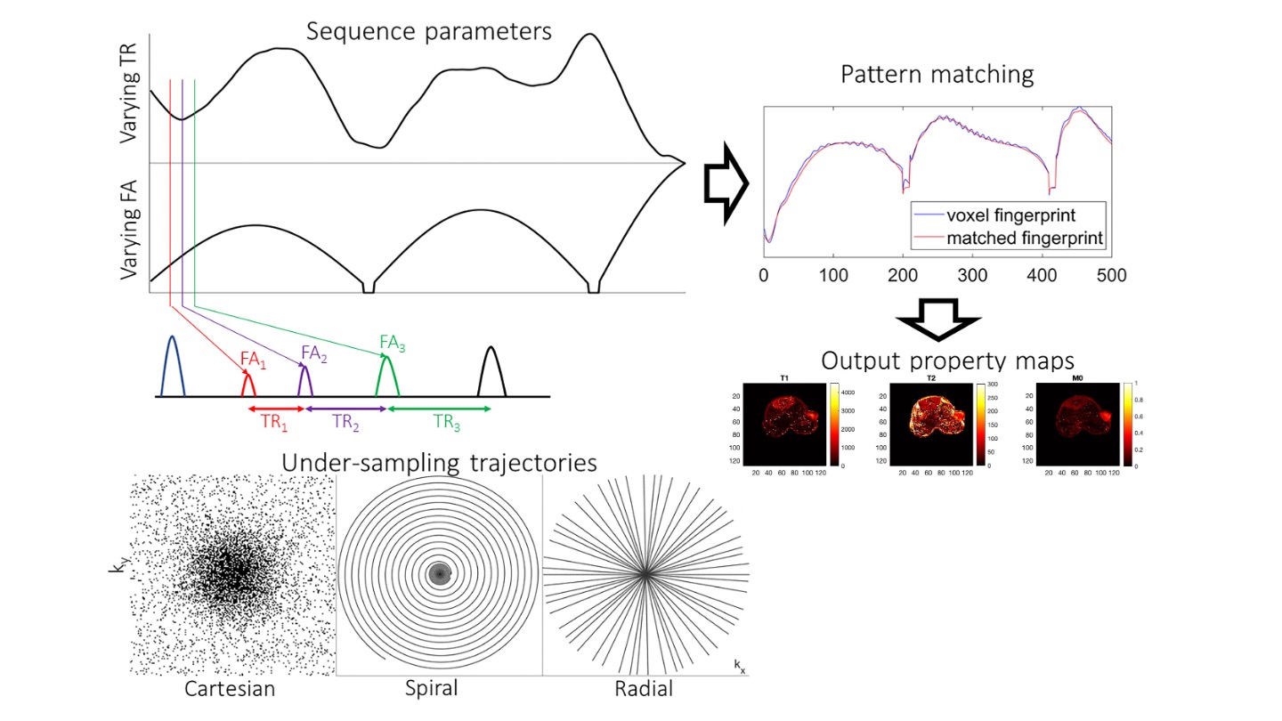
Quantitative Magnetic Resonance Imaging (MRI) involves pixel-wise mapping of longitudinal relaxation time T1, transverse relaxation time T2 and proton density M0 and other relevant parameters at each location in the tissue to be characterized. Magnetic Resonance Fingerprinting (MRF) is a promising new approach for rapid quantitative imaging. [1,2] Magnetic Resonance Fingerprinting (MRF), inspired by compressed sensing, consists of, fundamentally changing the acquisition and the magnetic parameters estimation process. Figure 1 illustrates an overview of how MRF works. In summary: A pseudo-random acquisition protocol - randomly varying radiofrequency (RF) pulses and repetition time (TR) - causes unique signal evolutions in different materials (i.e. a “fingerprint” of the material) that depends on the MR signal model and acquisition parameters. The response is measured in (k, t)-space and can be reconstructed using a non-uniform Fourier transform. Undersampling can accelerate the acquisition but results in noisier images. A pattern matching algorithm matches a predefined dictionary, which is built using the well-established Bloch equation formalism to “fingerprint” and recovers magnet parameters. [3-5] The radial trajectory design presents incoherent undersampling artifacts and is less sensitive to errors due to systemic imperfections as compared with spiral trajectory.The goal of this study is to investigate potential benefits of radial sampling in quantifying T1 relaxation times in metastatic breast tumor using MRF with a single-shot acquisition in tumor with high temporal and spatial resolution.
Methods
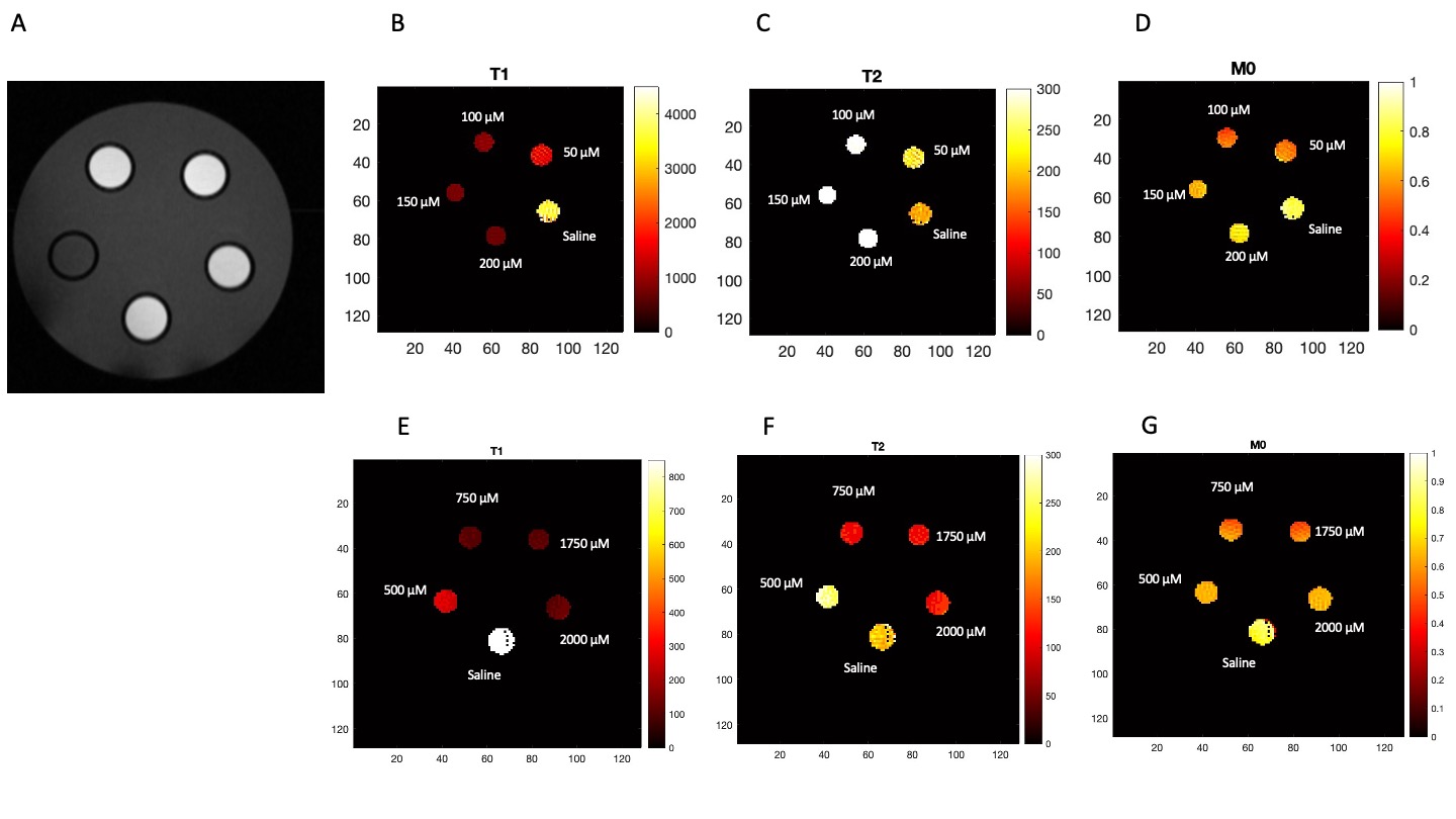
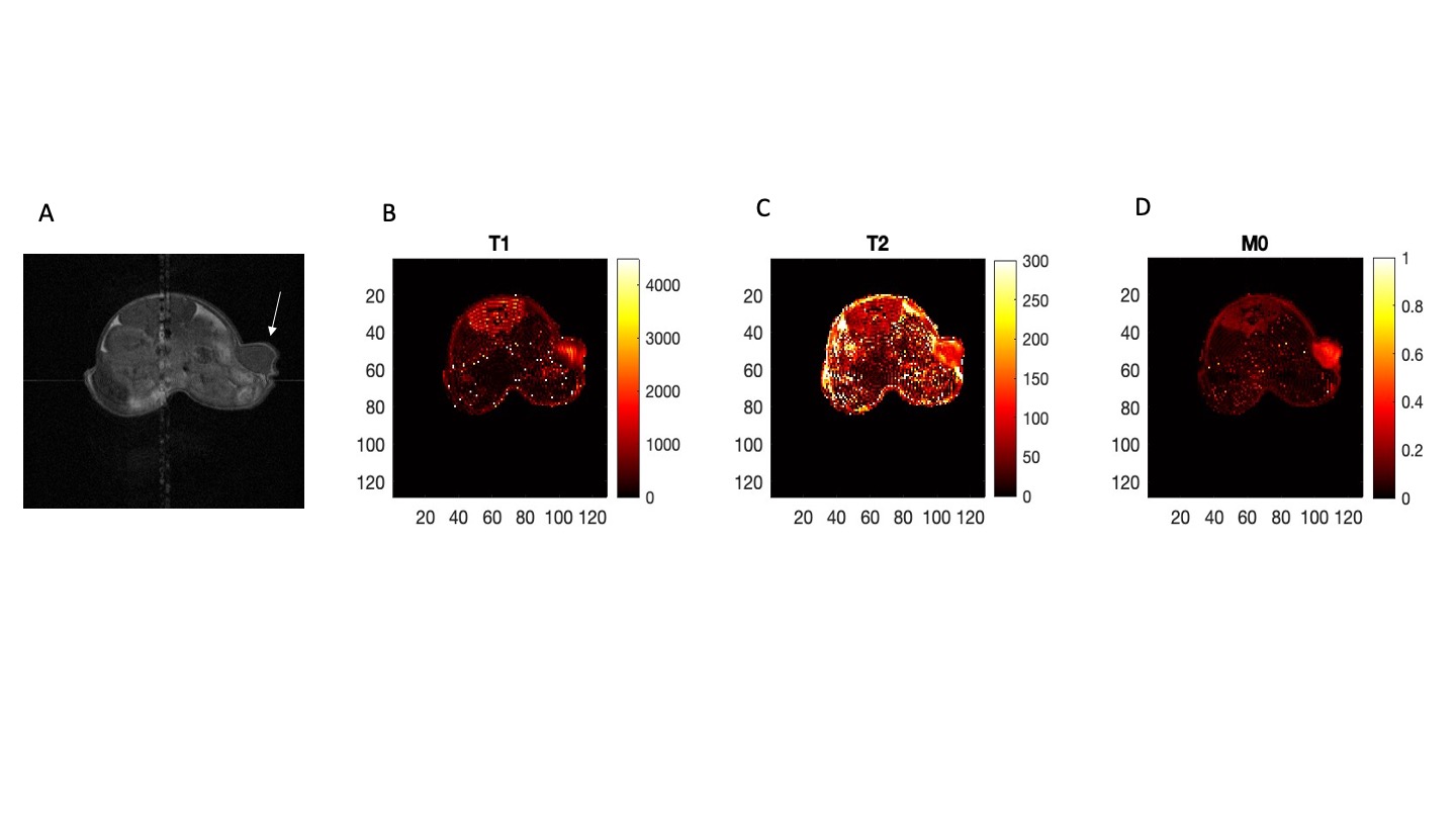
The accuracy of the proposed technique was first evaluated in vitro to determine the accuracy of the MRF method in comparison to saturation recovery (SR) spin echo. Gadovist phantoms at 0, 50, 100, 150, 200, 500, 1000, 1750, 2500 µM in saline were prepared that have T1 times of 0.1 – 3.5 sec. T1 values were determined by nonlinear least squares fitting using a three-parameter fit of a region-of-interest (ROI).In vivo MRI: 6-8 weeks old female athymic nude mice (n=3) were subcutaneously implanted with 2.0 X 10⁶ 4T1 cells in left flank with 27-gauge insulin syringe. Mice were imaged with MRF after showing signs of palpable tumor (~5 mm), injected with gadovist (0.1mmol Gd/kg), acquisition parameters were – TR = 1000 ms, 8 times undersampling data, acquisition matrix -256X25, matrix – 128X128 for a FOV of 37.5X37.5 mm2 with a slice thickness of 1.5 mm. The total acquisition time was 7 minutes.
Results
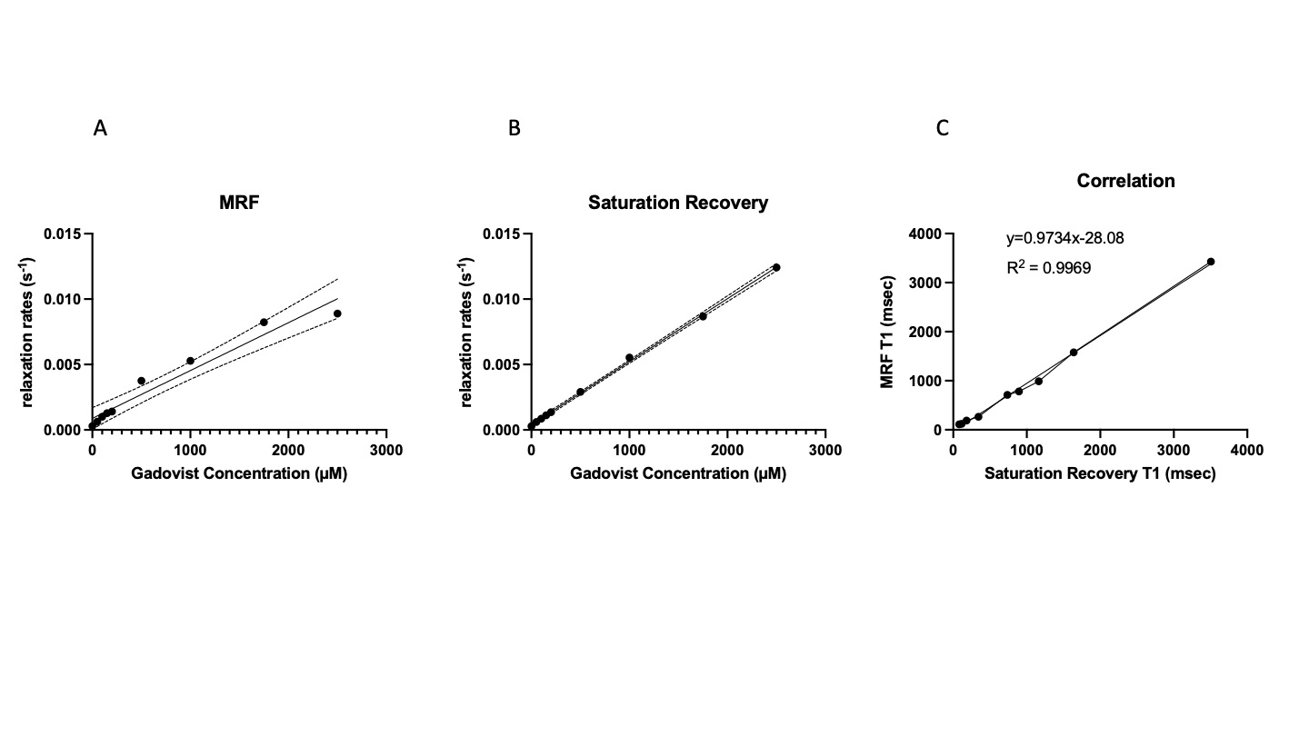
Gadovist phantoms at different concentrations were measured with MRF and compared with SR. Correlation was found to be R2 = 0.9969; Figure 3. Quantification of MRF gadovist phantoms (T1, T2, M0) parameters are shown in Figure 2.Mean T1 maps of tumor were 1118.8 ± 52.5 msec which was in accordance with SR measurements (1003 msec). Figure 4 represents quantification of MRF T1, T2 and M0 parameters. MRF was in good agreement with SR in phantoms and in vivo metastatic breast tumor.
Conclusion
MRF is a relatively new approach in the field of quantitative MRI that aims to suppress system imperfections and jointly quantify different tissue parameters like T1, T2 relaxation rates and proton density. This work has showcased the potential of radial MRF to quantify T1 relaxation times with high spatial and temporal resolution. It is the first preclinical in vivo contrast enhanced measurement and is under development to decrease scan duration.Future MRF work
Radial trajectory has lower k‐space sampling efficiency compared with a spiral trajectory; thus, it requires multiple repetitions of the MRF acquisition cycle or temporal averaging through k‐space view sharing. We will develop and validate dual-contrast MRF with spiral trajectories to measure T1 and T2 accurately and precisely by co-administering Gd-based and Dy-based contrast agent.Acknowledgements
No acknowledgement found.References
1. Ma, D., Gulani, V., Seiberlich, N., Liu, K., Sunshine, J. L., Duerk, J. L., & Griswold, M. A. (2013). Magnetic resonance fingerprinting. Nature, 495(7440), 187–192.
2. Panda, A., Mehta, B. B., Coppo, S., Jiang, Y., Ma, D., Seiberlich, N., Griswold, M. A., & Gulani, V. (2017). Magnetic Resonance Fingerprinting-An Overview. Current opinion in biomedical engineering, 3, 56–66.
3. Bipin Mehta, B., Coppo, S., Frances McGivney, D., Ian Hamilton, J., Chen, Y., Jiang, Y., Ma, D., Seiberlich, N., Gulani, V., & Alan Griswold, M. (2019). Magnetic resonance fingerprinting: a technical review. Magnetic resonance in medicine, 81(1), 25–46.
4. Dikaios, N., Protonotarios, N. E., Fokas, A. S., & Kastis, G. A. (2021). Quantification of T1, T2 relaxation times from Magnetic Resonance Fingerprinting radially undersampled data using analytical transformations. Magnetic resonance imaging, 80, 81–89.
5. Cloos, M. A., Assländer, J., Abbas, B., Fishbaugh, J., Babb, J. S., Gerig, G., & Lattanzi, R. (2019). Rapid Radial T1 and T2 Mapping of the Hip Articular Cartilage With Magnetic Resonance Fingerprinting. Journal of magnetic resonance imaging : JMRI, 50(3), 810–815.
Figures