4794
Navistar: Golden-Angle Stack-of-Stars Sampling with Embedded 2D Navigators for Highly-Accelerated 4D Dynamic MRI1BioMedical Engineering and Imaging Institute (BMEII), Icahn School of Medicine at Mount Sinai, New York, NY, United States, 2Center for Advanced Imaging Innovation and Research (CAI2R), NYU Grossman School of Medicine, New York, NY, United States
Synopsis
Keywords: Data Acquisition, Motion Correction
This work proposes a new radial sampling scheme called Navistar for highly-accelerated 4D dynamic MRI. Navistar periodically inserts 2D navigators into a golden-angle radial stack-of-stars acquisition. The embedded 2D navigators provide spatially-resolved information that can be used for different purposes, such as improved temporal-basis estimation for image reconstruction or respiratory-motion estimation. In our experiments, we have shown that Navistar sampling can be combined with low-rank subspace-constrained image reconstruction for highly-accelerated free-breathing 4D DCE-MRI of the liver with sub-second temporal resolution, which helps reduce intraframe respiratory blurring and eliminates the need for explicit respiratory-motion compensation.Introduction
The low-rank subspace model has been shown to be a powerful and effective method for high-accelerated dynamic MRI (1). Construction of a subspace by exploiting the low-rank condition of underlying dynamic images can lead to reduced degrees of freedom in image reconstruction, so that higher acceleration rates and improved reconstruction quality can be achieved compared to standard compressed-sensing or low-rank reconstruction with generic sparsity constraints. In low-rank subspace reconstruction, the subspace is typically generated with a pre-estimated temporal basis, which can be estimated from an analytic signal model (2, 3) or additionally acquired training data (4). Image reconstruction quality is highly dependent on the accuracy of temporal basis.Based on the low-rank subspace model, GRASP-Pro (Golden-angle RAdial Sparse Parallel imaging with imProved performance) has recently been developed for highly-accelerated free-breathing DCE-MRI using standard golden-angle stack-of-stars sampling, and its performance substantially outperforms standard compressed-sensing reconstruction (5, 6). In the current GRASP-Pro implementation, however, the temporal basis is estimated from the centers of k-space in acquired stack-of-stars data (6), which provide limited spatial information and are sensitive to various factors such as gradient delay and/or eddy currents.
To address this limitation and to further improve the performance of GRASP-Pro, we introduce a new radial sampling scheme called Navistar, which periodically inserts 2D navigators into golden-angle radial stack-of-stars acquisition. The embedded 2D navigators allow for improved estimation of a temporal basis for low-rank subspace reconstruction and enable more reliable respiratory-motion detection if needed. In combination with GRASP-Pro, the performance of Navistar sampling has been demonstrated for highly-accelerated free-breathing 4D DCE-MRI of the liver with sub-second temporal resolution, which helps reducing intraframe respiratory blurring and eliminates the need for additional compensation of respiratory motion.
Methods
Based on the low-rank property, dynamic MR images $$$\mathbf{x}$$$ can be decomposed as $$$\mathbf{x}=\mathbf{UV}$$$, where $$$\mathbf{U}$$$ denotes the temporal basis that is typically estimated before iterative reconstruction, and $$$\mathbf{V}$$$ denotes the spatial basis, which provides spatial characteristics for the dynamic images under $$$\mathbf{U}$$$ (1). Due to the low-rank condition of most dynamic MR images, only a few dominant components in $$$\mathbf{U}$$$ are sufficient to approximate $$$\mathbf{x}$$$, which leads to $$$\mathbf{x}\approx \mathbf{UV}$$$, where $$$\mathbf{k}$$$ represents the first $$$\mathbf{k}$$$ major basis components in $$$\mathbf{U}$$$. Based on this model, GRASP-Pro reconstruction is implemented by solving for $$$\mathbf{V}_{\mathbf{k}}$$$:$$\tilde{\mathbf{V}}_{\mathbf{K}}=arg\min_{\mathbf{V}_{\mathbf{K}}}\frac{1}{2}\parallel{\mathbf{EU}_{\mathbf{K}}\mathbf{V}_{\mathbf{K}}-\mathbf{y}}\parallel_{2}^{2}+\lambda _{1}R_{1}(\mathbf{U}_{\mathbf{K}}\mathbf{V}_{\mathbf{K}})+\lambda_{2}R_{2}(\mathbf{V}_{\mathbf{K}})\quad\quad\quad$$
Here, $$$\mathbf{E}$$$ denotes a multicoil-encoding operator, $$$\mathbf{y}$$$ represents undersampled dynamic k-space data, and $$$\mathit{R}$$$ is a regularizer that can be enforced on spatial and/or temporal dimensions. Once $$$\mathbf{V}_{\mathbf{k}}$$$ is reconstructed, $$$\mathbf{x}$$$ can then be recovered as $$$\tilde{\mathbf{x}}=\mathbf{U}_{\mathbf{k}} \mathbf{V}_{\mathbf{k}}$$$.
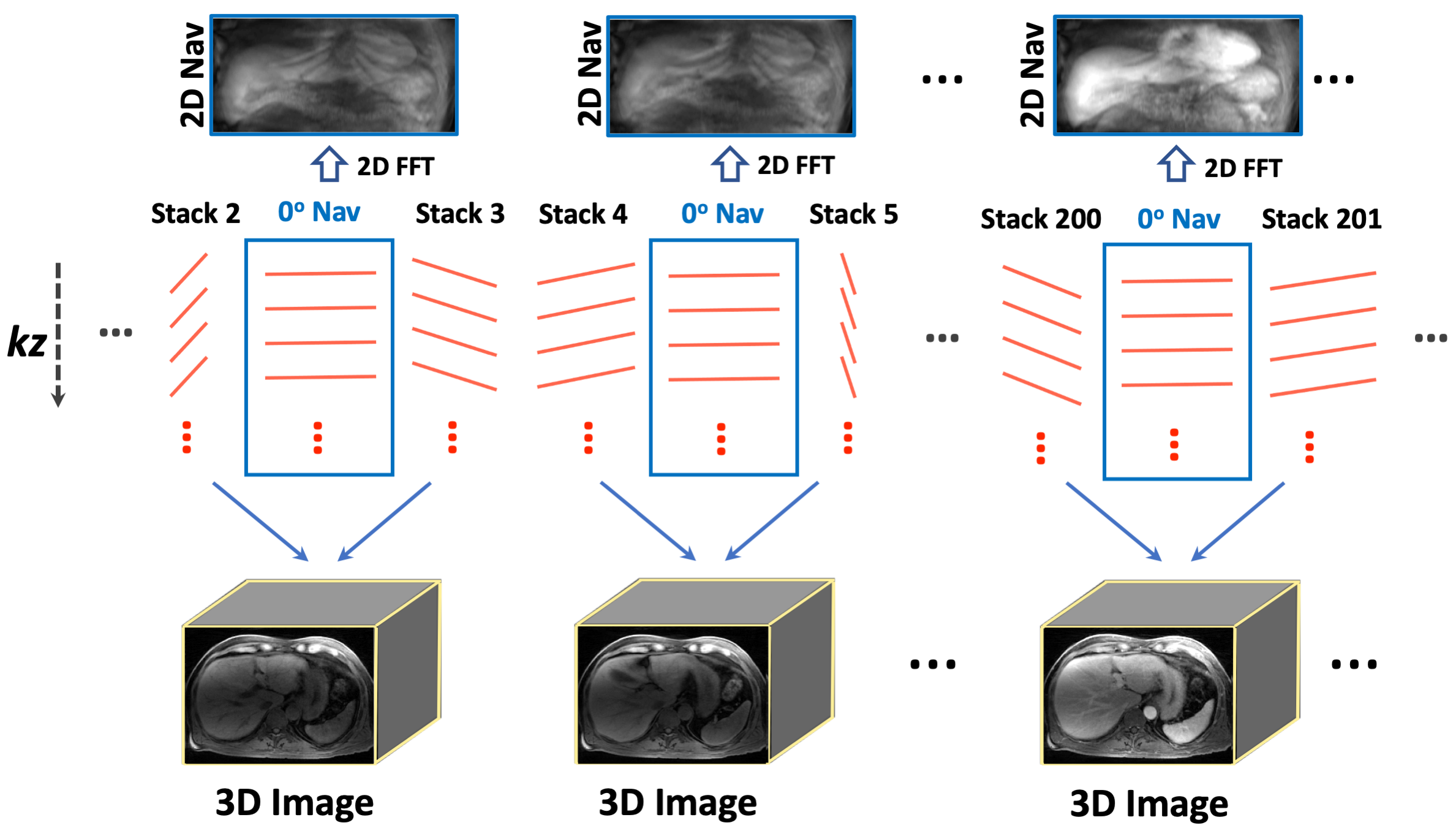
The new Navistar sampling scheme is implemented as shown in Figure 1. A 2D projection with zero-degree acquisition angle is periodically inserted into the standard golden-angle stack-of-stars sampling. In our current implementation, one 2D navigator is acquired every two radial stacks. During image reconstruction, those 2D navigators are first transformed to 2D projections with a 2D FFT, and they are used to generate a temporal basis using principal component analysis (PCA). Dynamic image reconstruction is then performed by combining every two radial stacks as one image volume. For the example shown in Figure 1, three 3D images are reconstructed from stacks 2&3, stacks 4&5 and stacks 200&201, respectively. This reconstruction scheme leads to highly-accelerated data acquisition and generation of hundreds of dynamic volumes with a sub-second temporal resolution to reduce intra-frame blurring.
The proposed method was tested in one volunteer and three patients with confirmed hepatocellular carcinoma (HCC) for free-breathing DCE-MRI of the liver. Relevant imaging parameters included: FOV=360x360mm2, matrix size=256x256, spatial resolution=1.4x1.4mm2, slice thickness=5mm, number of slices=40, 75% slice partial Fourier, TR/TE=2.75/1.21ms, flip angle=10o, total acquisition time= 270s. A total of 2400 radial stacks were acquired, including 1600 regular stacks with golden-angle rotation and 800 navigator stacks (rotation angle=zero). Image reconstruction was performed using GRASP-Pro (6), in which the temporal basis for each dataset was estimated from the acquired 2D navigators. Standard GRASP reconstruction (7) was performed for comparison.
Results
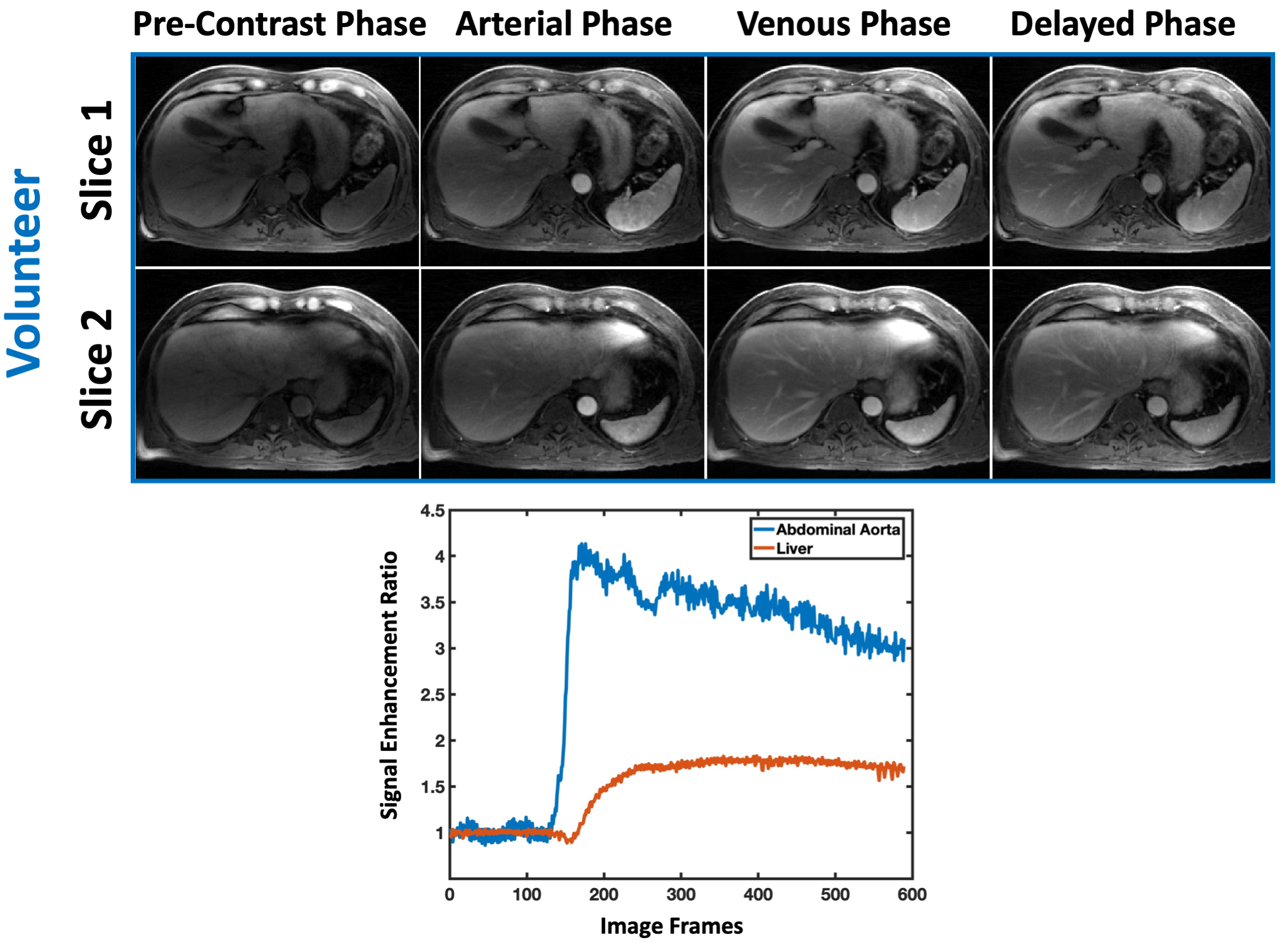
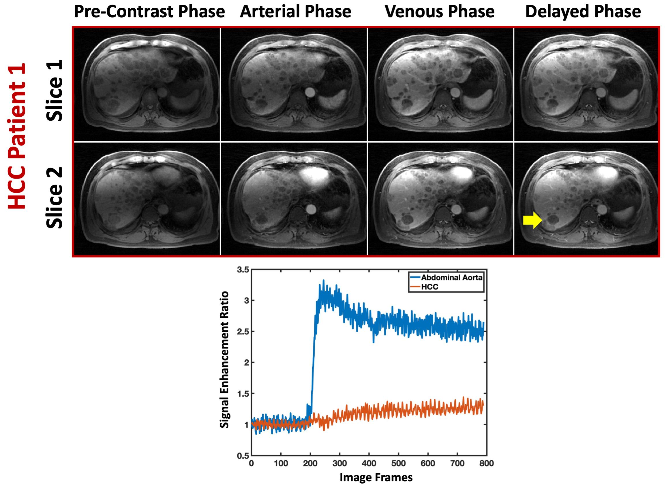
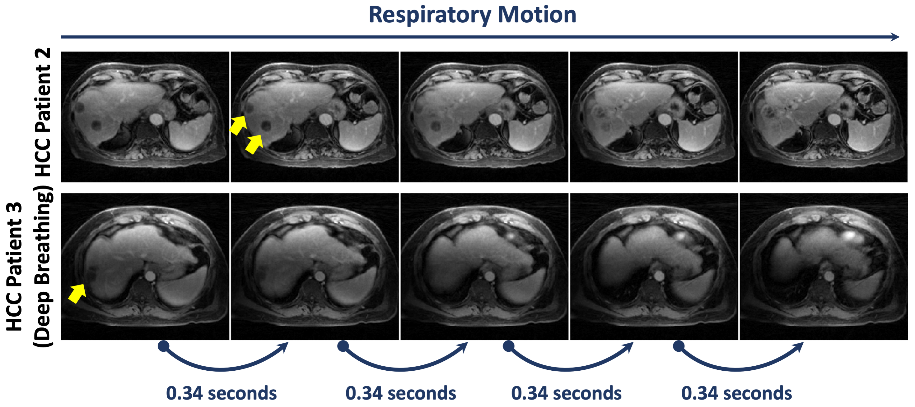
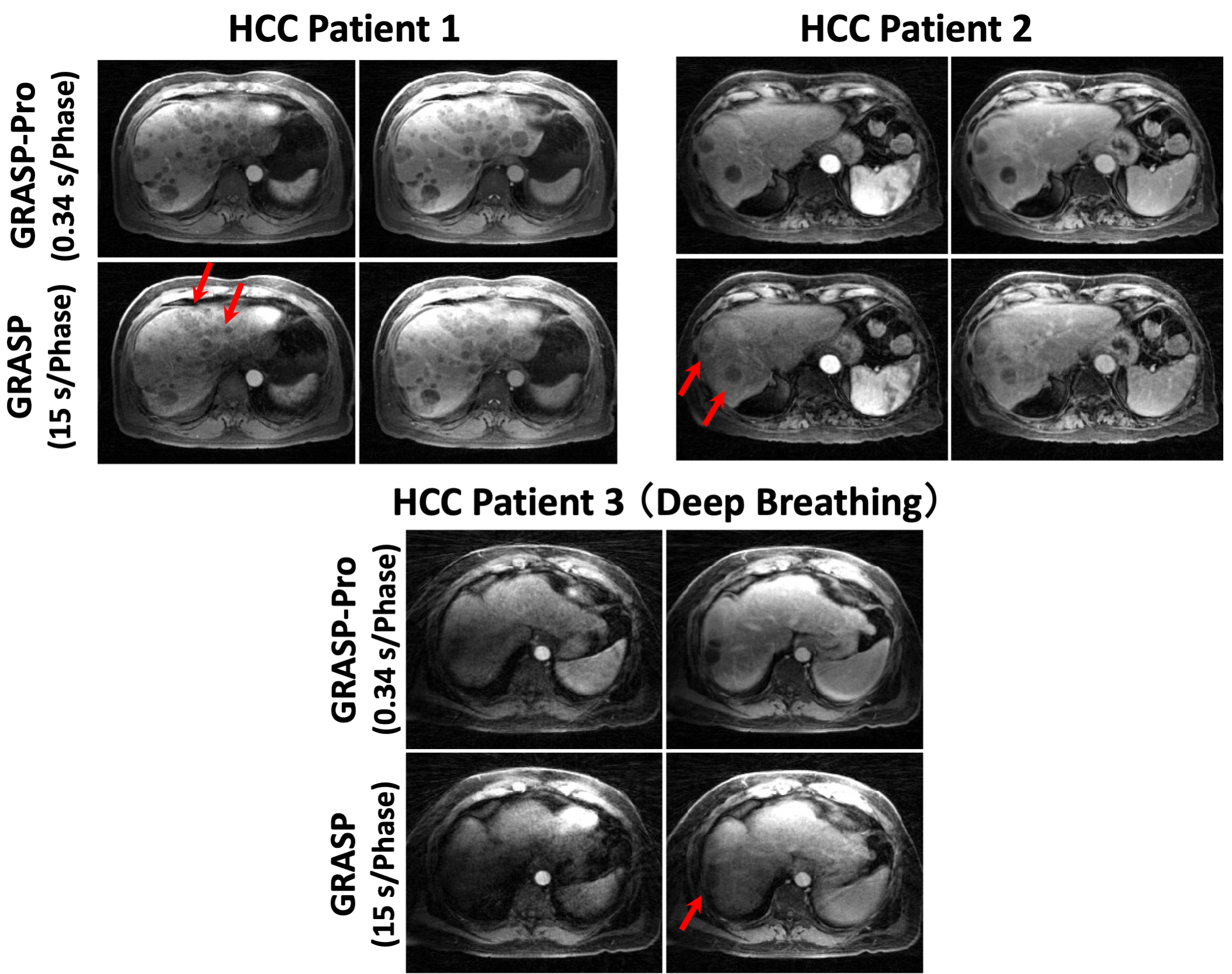
Figure 2 shows different contrast phases of liver images from the volunteer. With only 2 radial stacks in each dynamic volume, a temporal resolution of 0.34 seconds/volume was achieved, and no explicit respiratory-motion compensation was needed. Corresponding contrast-enhancement curves the abdominal aorta and liver parenchyma are also shown. Figure 3 shows the same results from an HCC patient, together with contrast-enhancement curves from the abdominal aorta and one HCC. Note that the HCCs do not show significant contrast enhancement because they are post-treatment lesions.Figure 4 shows five consecutive dynamic frames from two HCC patients (one slice is displayed for each patient), which indicate that respiratory motion can be resolved even in deep breathing. Figure 5 compares the GRASP-Pro reconstruction with standard GRASP reconstruction with low temporal resolution. Despite substantially higher acceleration, GRASP-Pro outperforms GRASP with better delineation of the HCCs and vessel-tissue contrast, while the GRASP reconstruction suffers from respiratory blurring (red arrows).
Conclusion
This work describes Navistar sampling, a new radial acquisition strategy that can be combined with low-rank subspace reconstruction for real-time 4D MRI. It permits highly-accelerated dynamic image acquisition to achieve sub-second temporal resolution, which eliminates the need for explicit respiratory motion detection and motion compensation, which is often challenging in free-breathing DCE-MRI.Acknowledgements
This work was supported in part by the NIH R01EB030549 and R01EB018308.References
1. Liang ZP: Spatiotemporal imaging with partially separable functions. 2007 4th IEEE Int Symp Biomed Imaging From Nano to Macro - Proc 2007:988–991.
2. Tamir JI, Uecker M, Chen W, et al.: T 2 shuffling: Sharp, multicontrast, volumetric fast spin‐echo imaging. Magn Reson Med 2017; 77:180–195.
3. Feng L, Liu F, Soultanidis G, et al.: Magnetization-prepared GRASP MRI for rapid 3D T1 mapping and fat/water-separated T1 mapping. Magn Reson Med 2021; 86:97–114.
4. Zhao B, Haldar JP, Christodoulou AG, Liang ZP: Image reconstruction from highly undersampled (k, t)-space data with joint partial separability and sparsity constraints. IEEE Trans Med Imaging 2012; 31:1809–1820.
5. Feng L, Wen Q, Huang C, Tong A, Liu F, Chandarana H: GRASP-Pro: imProving GRASP DCE-MRI through self-calibrating subspace-modeling and contrast phase automation. Magn Reson Med 2020; 83:94–108.
6. Feng L: 4D GRASP MRI at Sub-Second Temporal Resolution. NMR Biomed. 2022 Oct 19;e4844. doi: 10.1002/nbm.4844. Online ahead of print.
7. Feng L, Grimm R, Block KT obia., et al.: Golden-angle radial sparse parallel MRI: combination of compressed sensing, parallel imaging, and golden-angle radial sampling for fast and flexible dynamic volumetric MRI. Magn Reson Med 2014; 72:707–717.
Figures