4762
An analysis of longitudinal changes in dynamic brain activity with MHE patients treated initially with artificial livers: A follow-up fMRI study1Ningxia Medical University, Ningxia, China, 2General Hospital of Ningxia Medical University, Ningxia, China, 3GE Healthcare MR Research, Beijing, China, Beijing, China
Synopsis
Keywords: Psychiatric Disorders, Brain, Minimal hepatic encephalopathy cognition
Artificial liver support is an increasingly important intermediate step in the treatment of patients with liver failure. Yet there is still a lack of imaging studies to evaluate the efficacy of artificial liver support (especially on cognitive function of MHE). This study sought to evaluate the temporal variability of ReHo in the brain spontaneous activity after artificial liver support and correlated it with the PHES.We found that the dReHo in the right superior frontal gyrus decreased and were negatively correlated with NCT-A and positively correlated with DST.Dynamic regional indexes might be a novel neuro-imaging biomarker for artificial liver efficacy evaluation.Introduction
Patients with advanced liver cirrhosis have a higher risk of developing hepatic encephalopathy (HE) [1], and as the disease progresses, some patients also suffer from central nervous system compromise, which may manifest as cognitive dysfunction and behavioral abnormalities [2-4], signaling a progressive disease state [5]. Artificial liver support has been shown to be an effective treatment and is able to improve HE in acute and acute-on-chronic liver failure [6], can improve clinical symptoms and reduce the damage of toxins on body functions.Early detection and appropriate management of minimal hepatic encephalopathy (MHE) is of profound significance. In fact, researchers have conducted many resting-state functional magnetic resonance imaging (fMRI) studies on early recognition and mechanism research for MHE [7-12]. However, the clinical lack of cognitive function change objective imaging indicators to evaluate the efficacy of patients with liver failure with MHE, especially in dynamic regional homogeneity. Studies in both animals and humans have demonstrated that rest brain is a highly dynamic system, which could be characterized by non-stationary spatial temporal functional organization [13]. It is important to explore whether the observed aberrant associations between dynamic regional homogeneity and cognitive change. Therefore, our study based on the dynamic index of brain spontaneous activity of the longitudinal changes and related clinical indicators.Materials and Methods
This study was a prospective longitudinal study. All study procedures were conducted in accordance with the Declaration of Helsinki. After explaining the aims and steps in the procedure of the current study, all the participants and their guardians signed written informed consent. A total of 10 patients with MHE were included in this pretest. Resting-state fMRI and Psychometric Hepatic Encephalopathy Score (PHES) were performed before and after 6 months of artificial liver support. The GE Architect 3.0T MR scanner and 48-channel head coil were performed on conventional MRI and resting-state blood oxygenation level-dependent functional magnetic resonance imaging before and 6 months after BOLD-fMRI was performed using the Ax BOLD rest36sl sequence, TR=2000ms, TE=30 ms, Flip angle 90°, F0V250mm×250mm, rectangular area 64X64, number of layers: 35, layer thickness 3.6mm, scan dynamic 180 times, scan time was 6min. Artificial liver rs-BOLD image data before and 6 months after treatment. The collected raw images were preprocessed in Matlab2012a using the resting-state brain imaging data processing and analysis tool (DPARSF_V4.5, http: /wrestler.Net) and SPM12. The time-domain dynamic analysis module of DPABI software was utilized to calculate the dynamic ReHo (dReHo) variability using the sliding window method to reflect the temporal dynamics of regional neural activity. Using the "Statistical analysis module" in the DPABI software, the statistical min value of P<0.05 was obtained by Paired t-tests before and after artificial liver treatment, the statistical threshold was P<0.05, corrected by Gaussian Random Field (GRF)theory (P<0.001 at the voxel-level and P<0.05 at mass level ).Then, the dReHo variability values of the brain regions with statistically significant differences were extracted before liver versus 6 months after treatment for each subject separately. The correlation between the amount of dReHo variability and the improvement of PHES was analyzed using Spearman correlation.Results
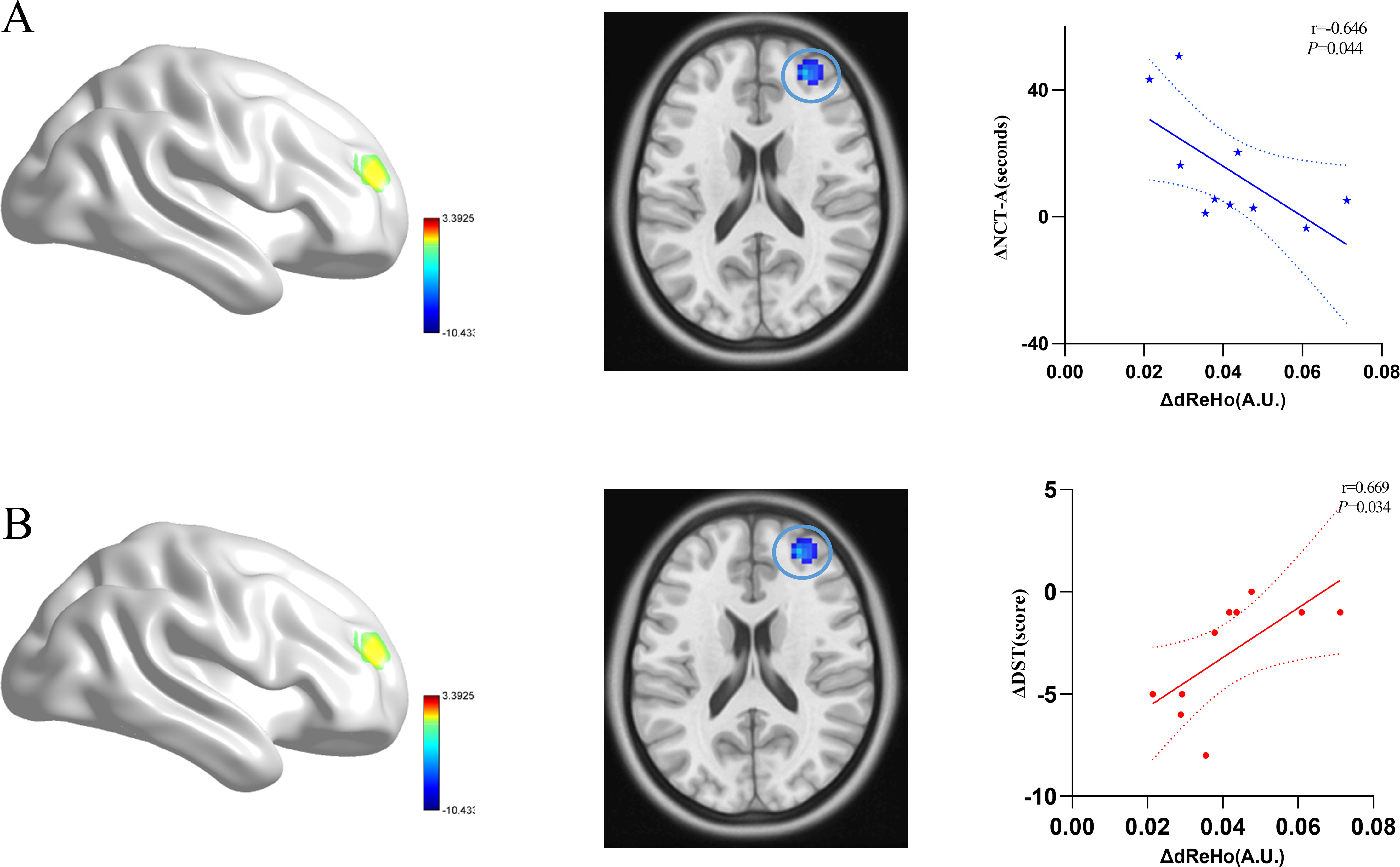
Basic clinical information (gender, age, education level, etc) of MHE patients was summarized in Table1, the spatial coordinate information of the brain regions with significant differences in dReHo variability was presented in Table2. T plot distribution pattern obtained by dReHo variability paired sample t-test before and after artificial liver support (Fig. 1A), and the dReHo in the right middle frontal gyrus and superior frontal gyrus were decreased after artificial liver support (with Gaussian airport correction, P<0.001, P<0.05) (Fig. 1B). Significant difference in test scores before and after artificial liver (P<0.05) (Fig. 2), the amount of pre- and post-dReHo reduction was negatively correlated with the improvement of NCT-A score (r=-0.646, P=0.044) and positively correlated with DST (r=0.669, P=0.034) (Fig. 3).Discussion and Conclusion
In this study, we found that dReHo in the right middle frontal gyrus was reduced after 6 months of artificial liver support in the treatment of MHE and correlated with PHES, suggesting that the improvement in cognitive function after treatment may be related to the recovery of function in this brain region. The middle prefrontal cortex is thought to be related to be basic functions of the subconscious mind such as emotion in the brain. The first changes in this brain region after artificial liver support indicate that this brain region is more sensitive to the response to treatment. The current findings about the dynamic neural characteristics of MHE patients treated initially with artificial liver support may expand our understanding of cognition related to neural mechanisms.Acknowledgements
J.R.Z thanks X.D.W for guidance as his advisor during his dissertation work. All of authors thank the Science and Technology Department of Ningxia for its financial support.References
[1]. Zhang, G., et al., The short-term effect of liver transplantation on the low-frequency fluctuation of brain activity in cirrhotic patients with and without overt hepatic encephalopathy. Brain Imaging Behav, 2017. 11(6): p. 1849-1861.
[2]. Shawcross, D.L., Diagnosis and management of hepatic encephalopathy. Br J Nurs, 2018. 27(Sup3): p. S7-S13.
[3]. Hasan, L.Z. and G.Y. Wu, Novel Agents in the Management of Hepatic Encephalopathy: A Review. J Clin Transl Hepatol, 2021. 9(5): p. 749-759.
[4]. Rudler, M., et al., Diagnosis and Management of Hepatic Encephalopathy. Clin Liver Dis, 2021. 25(2): p. 393-417.
[5]. Rose, C.F., et al., Hepatic encephalopathy: Novel insights into classification, pathophysiology and therapy. J Hepatol, 2020. 73(6): p. 1526-1547.
[6]. Stadlbauer, V., G.A. Wright and R. Jalan, Role of artificial liver support in hepatic encephalopathy. Metab Brain Dis, 2009. 24(1): p. 15-26.
[7]. Zafiris, O., et al., Neural mechanism underlying impaired visual judgement in the dysmetabolic brain: an fMRI study. Neuroimage, 2004. 22(2): p. 541-52.
[8]. Qi, R., et al., Altered resting-state brain activity at functional MR imaging during the progression of hepatic encephalopathy. Radiology, 2012. 264(1): p. 187-95.
[9]. Ni, L., et al., Altered regional homogeneity in the development of minimal hepatic encephalopathy: a resting-state functional MRI study. PLoS One, 2012. 7(7): p. e42016.
[10]. Chen, H., et al., Identifying minimal hepatic encephalopathy in cirrhotic patients by measuring spontaneous brain activity. Metabolic brain disease, 2016. 31(4).
[11]. Yue, C., et al., Identification of minimal hepatic encephalopathy based on dynamic functional connectivity. Brain Imaging and Behavior, 2021. 15(5).
[12]. Guo, J., et al., Altered dynamic spontaneous neural activity in minimal hepatic encephalopathy. Frontiers in Neurology, 2022. 13.
[13]. Fu, Z., et al., Characterizing dynamic amplitude of low-frequency fluctuation and its relationship with dynamic functional connectivity: An application to schizophrenia. Neuroimage, 2018. 180(Pt B): p. 619-631.
Figures
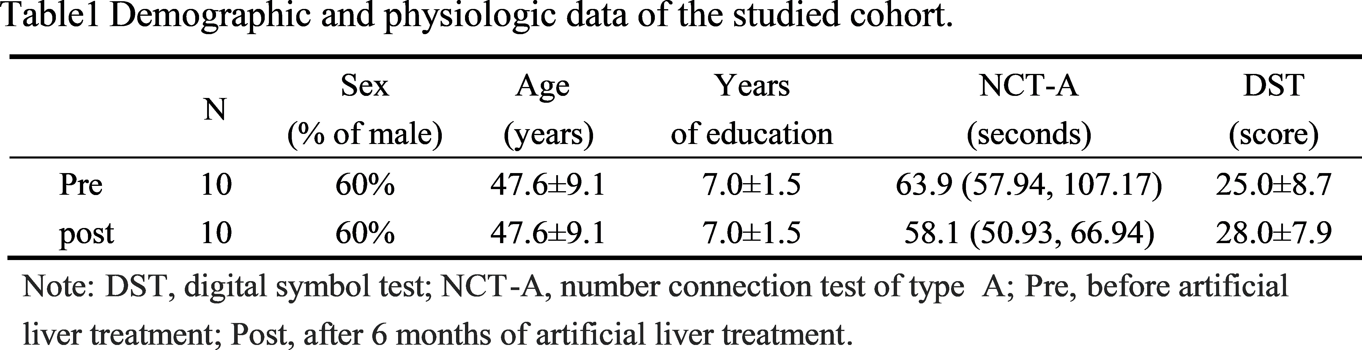
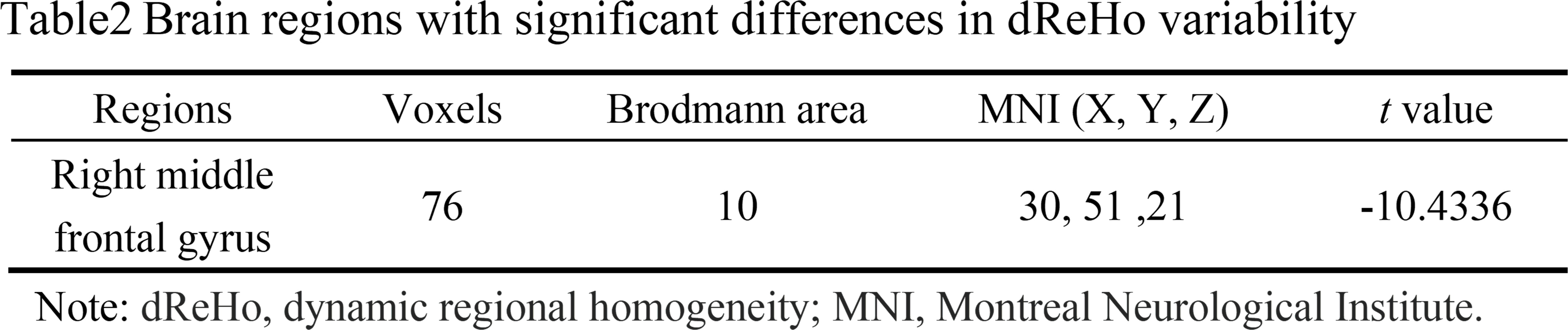
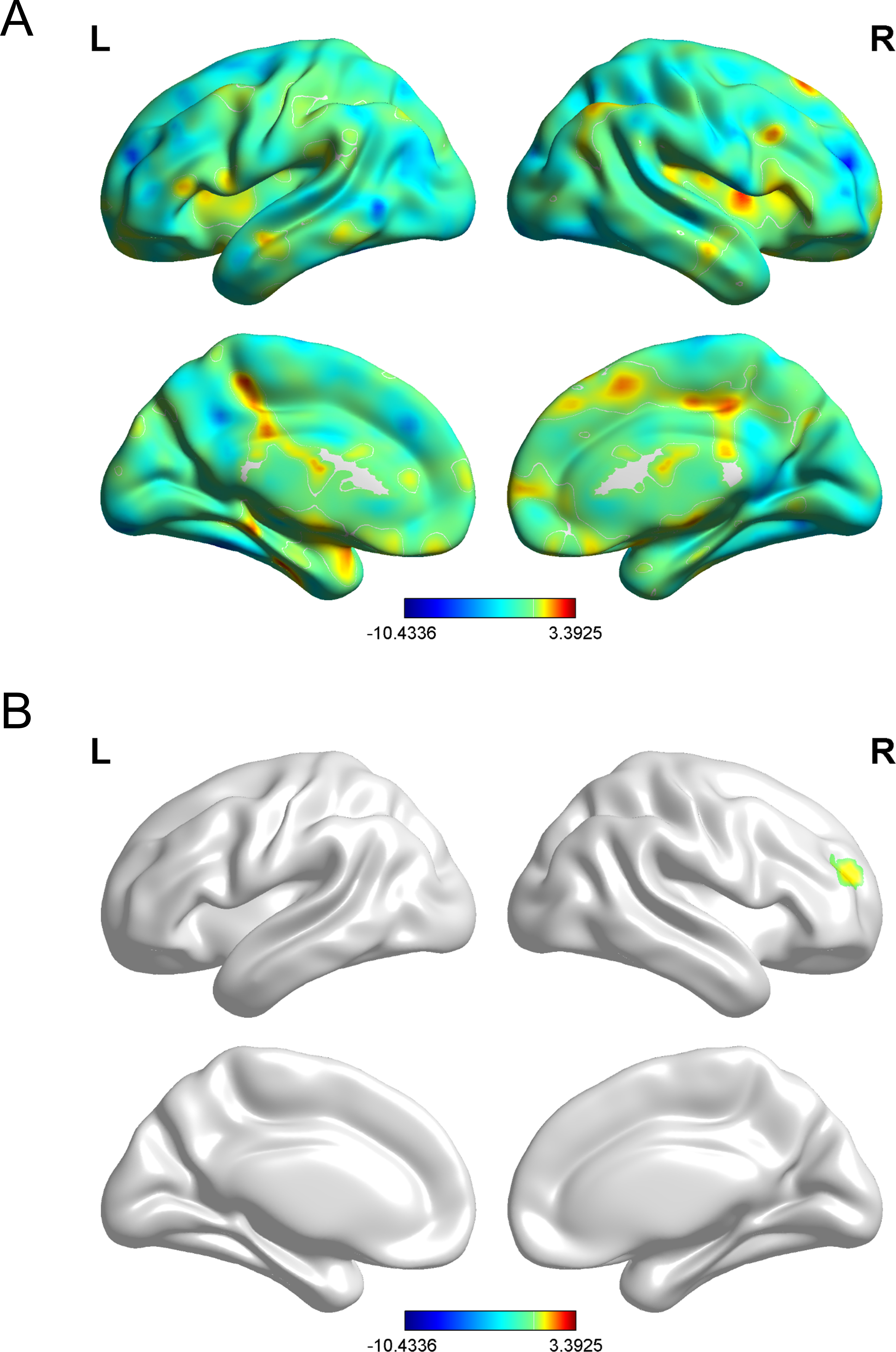
Fig.1 A. The distribution pattern of dReHo variability in the patients with MHE. The red and blue colors, respectively, indicate high and low dReHo variability. “L” denotes the left hemisphere and “R” denotes the right hemisphere.
B. Brain regions where significant differences in dReHo variability were placed across the two groups (before and after artificial liver support).
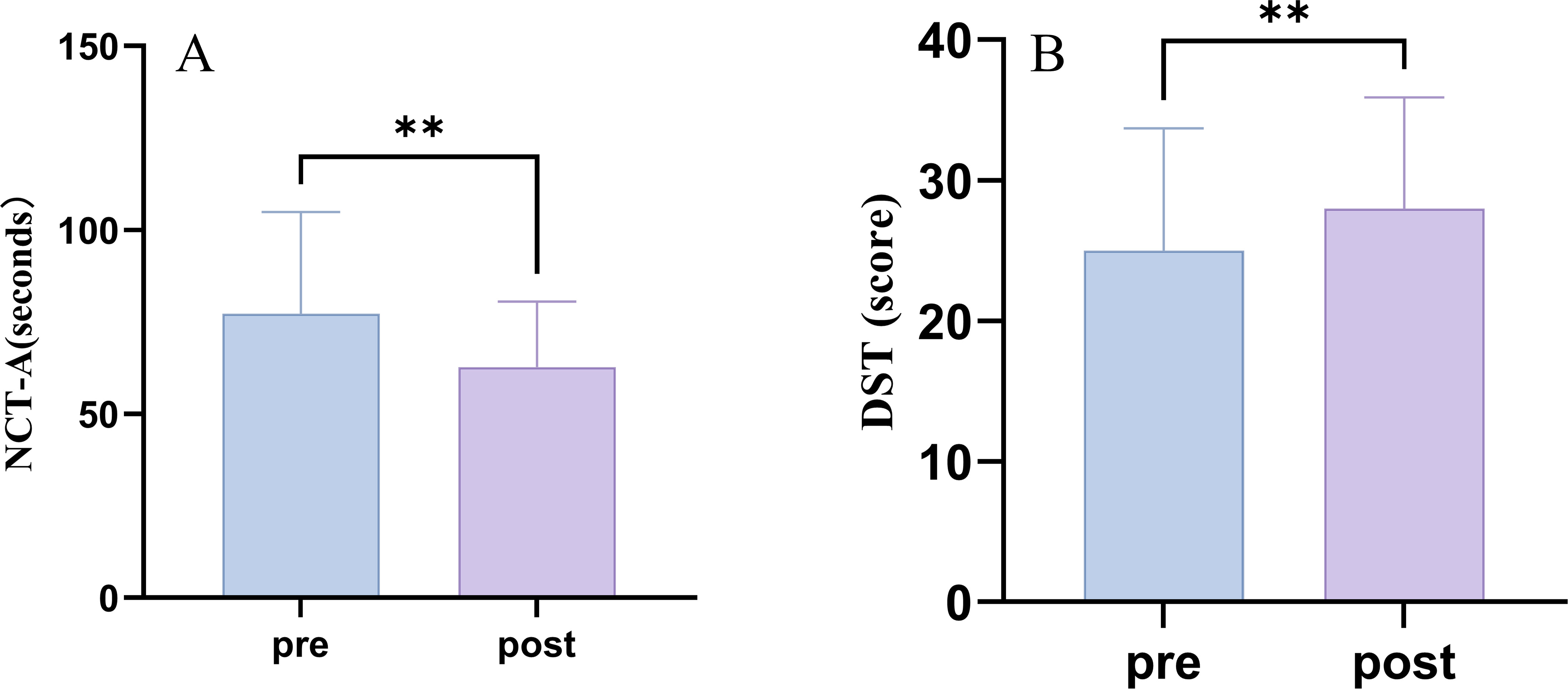
Fig.2 Changes in NCT-A and DST scores with two group. For the number connection test A(NCT-A), the Mann-Whitney U-test was used to compare the differences between the groups (pre-post), for the digit symbol test (DST),the paired t-test was used to compare the differences, the difference was significant(P<0.05).