4760
Altered cerebellar GABA-mediated cerebral functional networks in patients with schizophrenia: A transcranial magnetic stimulation study1Huaxi MR Research Center (HMRRC), Department of Radiology, West China Hospital of Sichuan University, Chengdu, China, 2Research Unit of Psychoradiology, Chinese Academy of Medical Sciences, Chengdu, China, 3Philips Healthcare, Xi'an, China, 4Mental Health Center, West China Hospital of Sichuan University, Chengdu, China, 5Functional and Molecular Imaging Key Laboratory of Sichuan Province, Chengdu, China
Synopsis
Keywords: Psychiatric Disorders, Neuroscience, schizophrenia, cerebellum, gamma-aminobutyric acid (GABA)
To investigate the cerebellar GABA-mediated cerebral functional networks, we measured cerebellar basal and stimulus-activated GABA content in schizophrenia patients (SCZ) and healthy controls (HC) with cerebellar transcranial magnetic stimulation (TMS). We utilized graph theory to calculate global and regional property changes of cerebral functional networks and their correlations with cerebellar GABA at baseline and after TMS. The results show significant increase of the global efficiency, nodal clustering coefficient of left ventral attention network (VAN) after TMS in SCZ. This provides new molecular evidence for neuropathology of cerebellum dysfunction in SCZ and reveal potential interaction with cerebral functional networks.Introduction
Gamma-aminobutyric acidergic (GABAergic) neuronal deficit has been proposed as central to the neuropathology of schizophrenia (SCZ). Previous studies have found that the GABAergic neurons in cerebellar cortex exert both basal and stimulus-activated inhibition on cerebellar nuclei[1], and cerebellar GABAergic deficits in SCZ could decrease cerebellar inhibition[2]. The abovementioned works elicit cerebral dysfunction by cerebello-thalamo-cortical loop[3], theorizing the so-called “cerebello-cortical inhibition” (CBI) hypothesis[4]. However, the potential pathophysiological mechanism of cerebello-cerebral dysfunction in schizophrenia and its correlation with GABA content remains unclear. High frequency transcranial magnetic stimulation (TMS) and intermittent theta burst stimulation (iTBS) of the midline cerebellum can relieve the negative symptoms of schizophrenia, whose potential physiological mechanism could be related to increasing the sum of excitatory postsynaptic potentials leading to neuronal excitation of the stimulating spot[5] and regulating the neurotransmitter secretion[6]. Combining TMS and magnetic resonance spectroscopy (MRS) enables measurement of GABA content. This study aimed to explore the alteration of cerebellar GABA content and its interaction with cortical networks. We utilized graph theory to calculate the global and regional property changes in individual functional networks and their correlations with cerebellar GABA at baseline and after TMS.Material and Methods
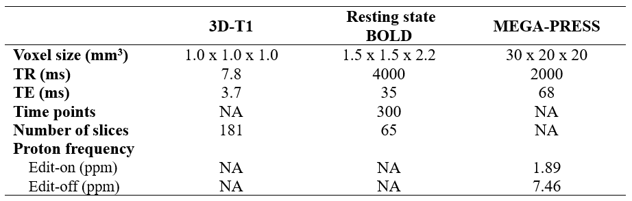
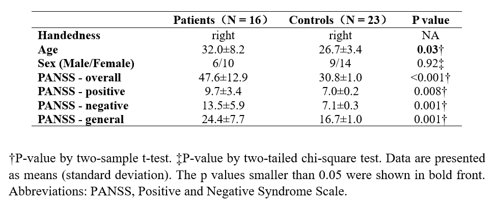
Sixteen patients with stable schizophrenia were recruited. All the patients were on regular antipsychotic medication and stable as prescribed. The diagnosis of SCZ was determined by structured clinical interviews conducted by experienced psychiatrists according to the diagnostic and statistical manual of mental disorders-IV (DSM-IV). Twenty-three demographically matched healthy controls (HC) were recruited through posters and website advertisements and were filtrated using a non-patient version of the clinical definitive interview (SCID-NP). The severity of psychotic symptoms in subjects was assessed with the Positive and Negative Syndrome Scale (PANSS) by an experienced assessor before the MRI scanning. All subjects underwent two MRI scans immediately before and after TMS using a 3.0T scanner (Elition, Philips Healthcare) with a 32-channel head coil. The examination protocols include high resolution three-dimensional T1-weighted (3D-T1) imaging, high resolution resting-state BOLD imaging and MEscher-GarwOod point resolved spectroscopy (MEGA-PRESS). The detailed imaging parameters were shown in table 1. After the baseline MR scanning, subjects were guided to treatment room to receive TMS. The TMS coil was pointed to the middle (i.e., along the midline) of the bilateral Crus I/II of the posterior cerebellum. All subjects received two TMS sequences (iTBS and 15 Hz TMS). MEGA-PRESS data were processed using the Gannet 3.0 toolbox (http://www.gabamrs.com/). To further ensure the robustness of our results, only edited spectra with GABA fitting error of less than 15% were included in final analyses. Functional MRI data were processed using the CBIG toolbox (https://github.com/ThomasYeoLab/CBIG). Graph theory analysis was calculated using the DPABI toolbox (http://rfmri.org/dpabi). We applied the individual-specific cortical functional network parcellation according to Kong’s work[7]. Two sample t-tests were used to compare the altered GABA content between SCZ and HC. Paired t-tests were then performed to detect significant cerebellar GABA content changes and global and regional topological properties before and after TMS in each group, respectively. We further tested for correlations to determine the relationships between the GABA change and the above topological properties changes with significant differences in SCZ. Significance was determined as P < 0.05.Results
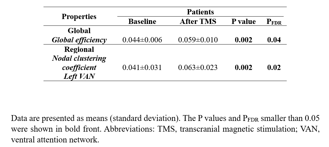
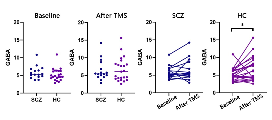
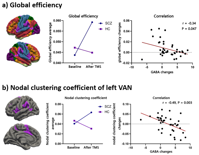
Demographic and clinical characteristics of SCZ and HC were shown in table 2. For GABA content, the left cerebellar GABA at baseline and after TMS between SCZ and HC showed no significant difference (P > 0.05). Within group analysis revealed that GABA content in the left cerebellar voxel of interest (VOI) was significantly increased after TMS in HC (P = 0.03), but no statistically significant difference in SCZ (P = 0.25), figure 1. After TMS, global efficiency increased in SCZ (PFDR = 0.04, table 3). There was a negative correlation between the global efficiency change and the left cerebellar GABA change (r = -0.34, P = 0.047, figure 2a), which indicated the more increase of cerebellar GABA after TMS, the less change of global efficiency of cortical network. In SCZ group, the nodal clustering coefficient of left VAN was increased after TMS (PFDR = 0.02, table 3). The changes of the nodal clustering coefficient of left VAN (r = -0.49, P = 0.003, figure 2b) were negatively correlated with the change of the cerebellar GABA.Disscussion
In this study, we found basal level of cerebellar GABA in SCZ did not differ significantly from that of HC, but the stimulus-activated cerebellar GABAergic neuronal ability is reduced in SCZ. Furthermore, we found that the global efficiency of the cortical networks increased after cerebellar TMS in SCZ, but not in HC. Then the node clustering coefficient of left VAN increased in SCZ after TMS. Moreover, the changes of global efficiency and regional topological properties in left VAN were negatively correlated with the change of cerebellar GABA content after TMS. These findings suggest that stimulus-activated impairment of cerebellar GABAergic neurons exists in SCZ and the impairment affects cortical functional network.Conclusion
In conclusion, we observed that the changes of GABA content were significantly associated with topological properties of individual cortical networks in SCZ. The findings may provide new molecular evidence for the neuropathology of cerebellum dysfunction in SCZ and a potential theoretical basis for the schizophrenic treatment.Acknowledgements
No acknowledgement found.References
[1] Bao, S., et al., Cerebellar cortical inhibition and classical eyeblink conditioning. Proc Natl Acad Sci U S A, 2002. 99(3): p. 1592-7.
[2] van Rootselaar, A.-F., et al., Decreased cortical inhibition and yet cerebellar pathology in 'familial cortical myoclonic tremor with epilepsy'. Movement Disorders: Official Journal of the Movement Disorder Society, 2007. 22(16): p. 2378-2385.
[3] Cao, H., et al., Cerebello-Thalamo-Cortical Hyperconnectivity Classifies Patients and Predicts Long-Term Treatment Outcome in First-Episode Schizophrenia. Schizophr Bull, 2022. 48(2): p. 505-513.
[4] Fernandez, L., et al., Assessing cerebellar brain inhibition (CBI) via transcranial magnetic stimulation (TMS): A systematic review. Neurosci Biobehav Rev, 2018. 86: p. 176-206.
[5] Croarkin, P.E., et al., High-frequency repetitive TMS for suicidal ideation in adolescents with depression. J Affect Disord, 2018. 239: p. 282-290.
[6] Cuypers, K. and A. Marsman, Transcranial magnetic stimulation and magnetic resonance spectroscopy: Opportunities for a bimodal approach in human neuroscience. Neuroimage, 2021. 224: p. 117394.
[7] Kong, R., et al., Spatial Topography of Individual-Specific Cortical Networks Predicts Human Cognition, Personality, and Emotion. Cereb Cortex, 2019. 29(6): p. 2533-2551.
Figures