4759
Disrupted striatum-centered tracts and positive symptoms in schizophrenia patients
Yuanqiang Zhu1, Chen Wang1, Fan Guo1, and Yingjuan Chang1
1Air Force Medical University, Xi'an, China
1Air Force Medical University, Xi'an, China
Synopsis
Keywords: Psychiatric Disorders, Neuroscience
Stronger tract strengths of the bilateral striatum with dlPFC were detected in schizophrenia patients. Our result provided system-level insights into abnormal connectivity strength of striatal circuits in schizophrenia patients. And the strength might have potential as neuroimaging biomarkers for positive symptoms.Introduction:
Striatum is one central processing hub, receiving input from the cortex, and striatal dysfunction is a fundamental element in schizophrenia [1]. The cortical-subcortical basal ganglia network is also of interest in neuropsychiatric conditions including schizophrenia [2]. Dysfunction in the striatum could plausibly lead to a range of heterogeneous symptoms observed in schizophrenia, including positive, negative, and cognitive symptoms. Although impaired striatal connectivity has been frequently described in schizophrenia and the correlation between negative symptoms, its link with positive symptoms has not been carefully studied. We aim to identify the striatum-centered structural networks based on diffusion tensor imaging (DTI) that contribute to the positive symptom in schizophrenia patients.
Material and Method: Tract strengths of striatal circuits were compared in 73 schizophrenia patients and 77 healthy controls, using seed-based classification by DTI probabilistic tractography with 10 a priori target masks (mOFC, dlPFC, vlPFC, IFG, posterior cingulate cortex (PCC), ACC, dorsal ACC (dACC), supplementary motor area (SMA), hippocampus and amygdala). Linear regression was conducted to test whether the striatal-centered tract strength was associated with the schizophrenia Positive and Negative Syndrome Scale (PANSS) scores or cognitive assessment. Canonical correlation analysis (CCA) was further used to examine the multivariate relationship between tract strength of striatal circuits and clinical measurements.
Results: Compared with the healthy controls, schizophrenia patients showed stronger tract strength in the bilateral striatum- dorsolateral prefrontal cortex (dlPFC), left striatum-amygdala and left striatum- supplementary motor area (SMA), but weaker tract strength in the striatum-dACC (dorsal anterior cingulate cortex) in the right hemisphere. Pearson correlation analysis showed positive correlation between positive PANSS scores in the bilateral striatum-dlPFC tract strength in schizophrenia patients. CCA revealed schizophrenia patients with stronger tract strength of bilateral striatum-dlPFC, left striatum-medial orbitofrontal cortex, left striatum-hippocampus, right striatum-dACC, right striatum-ACC and weaker tract strength of right striatum-SMA, right striatum-ventrolateral prefrontal cortex (vlPFC) showed more serious positive symptoms.
Conclusion: The findings provided insights into the relationship between striatal-centered tracts and positive symptoms in schizophrenia patients. Our study may provide a potential circuitry-level biomarker to study the neurobiological mechanisms of positive symptoms.
Acknowledgements
This study was supported by the Key R&D Program Projects, National Science Foundation of China (Grant No.2016YFC1306900), the National Natural Science Foundation of China under Grant Nos. 81974215 and 81801772, Key R&D Program Projects of Shaanxi, China (No.2021SF-287 and 2022JM-575), Boost Program of Xijing Hospital (XJZT18ML84 and XJZT19ML56), China Postdoctoral Science Foundation (2019M653963), and Military Medical Science and Technology Youth Training Program (20QNPY049).References
1, Sorg, C., et al., Increased intrinsic brain activity in the striatum reflects symptom dimensions in schizophrenia. Schizophr Bull, 2013. 39(2): p. 387-95. 2.
2, Levitt, J.J., et al., Miswiring of Frontostriatal Projections in Schizophrenia. Schizophr Bull, 2020. 46(4): p. 990-998.
Figures
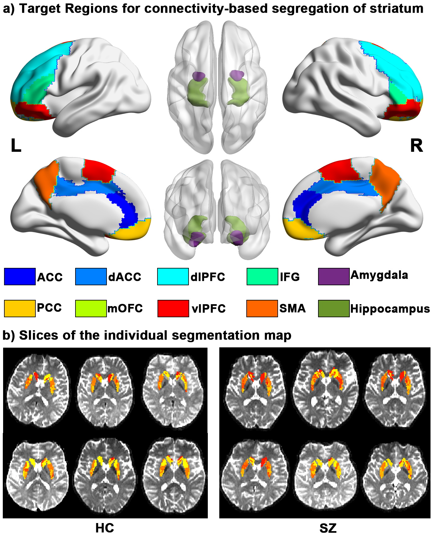
Figure 1. Target regions for seed-based tractography. (a) The target brain masks (8 cortical cortex and 2 subcortical regions) for striatum tractography. (b) Slices of the individual segmentation map of striatum (color theme: red–yellow in FSL 5.0.9).
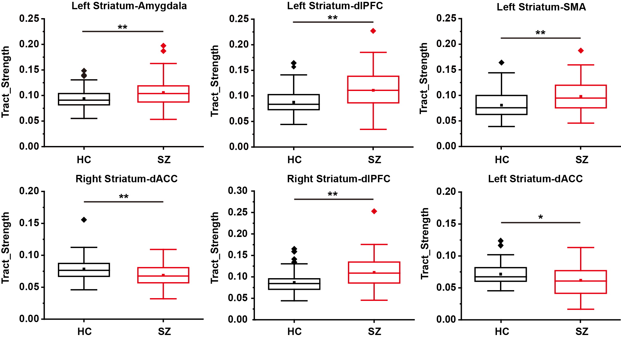
Figure 2. Differences in the tract strength of striatal tracts between HC and SZ (**p < 0.0025, Bonferroni correction, *p < 0.005, no correction).
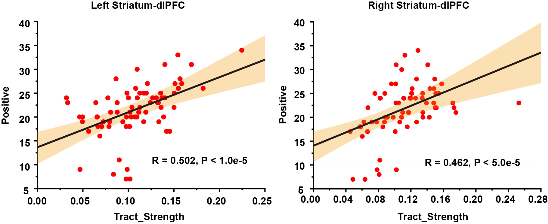
Figure 3. Relationship between the tract strength of the bilateral striatum-dlPFC and positive symptoms (all p < 0.05/160 = 0.0003, Bonferroni correction).
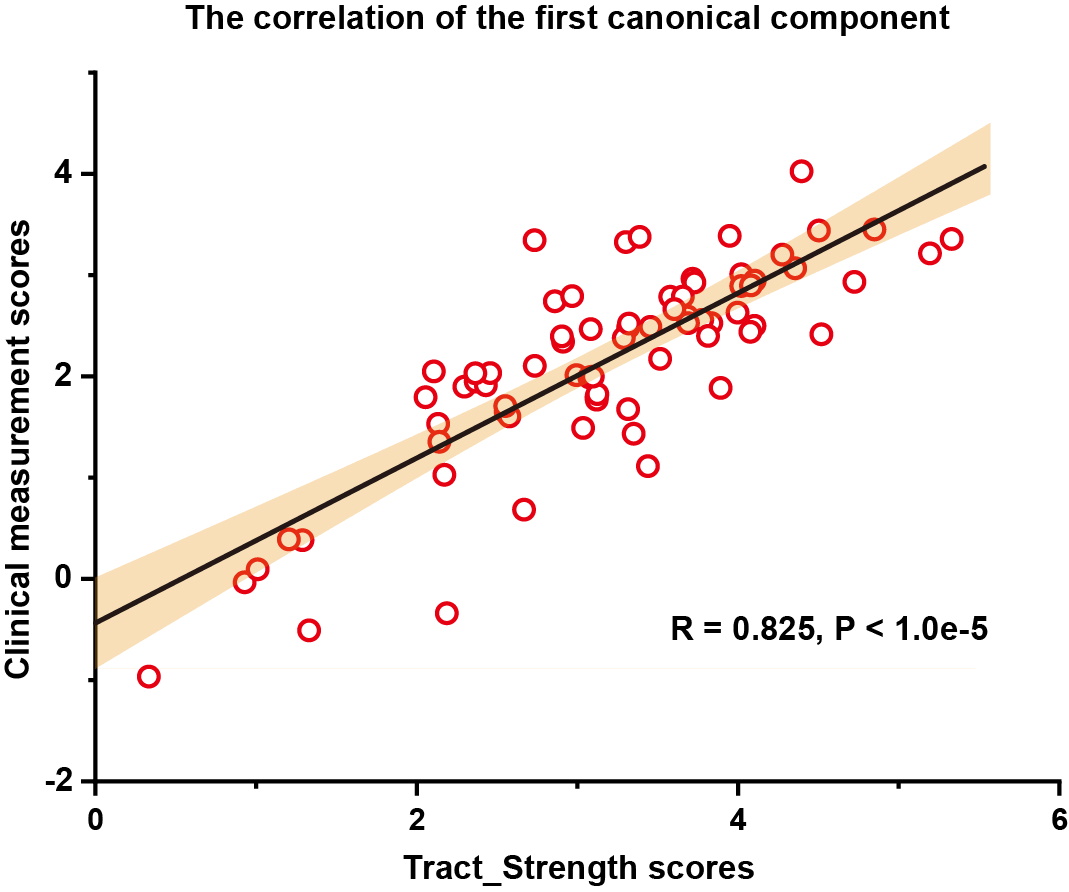
Figure 4. The scatterplot for the canonical variates of the clinical measurements and the tract strength.
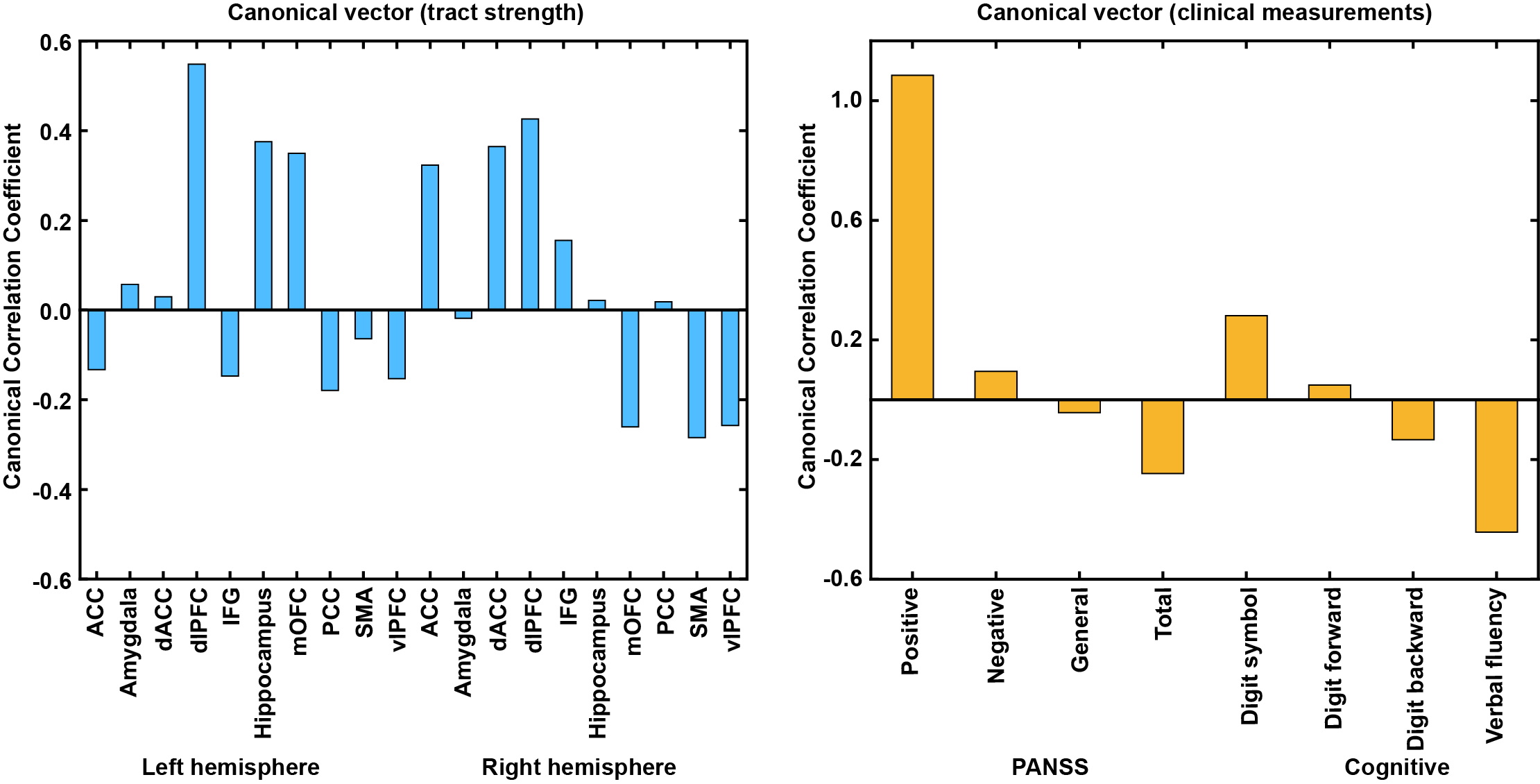
Figure 5. The standardized canonical correlation coefficients for the significant canonical component across tract strength of striatal circuits and clinical measurements.
DOI: https://doi.org/10.58530/2023/4759