4739
A Fat-suppression, Image-subtraction Method Using Deep Learning for the Detection of Knee Abnormalities on MRI
Tsutomu Inaoka1, Akihiko Wada2, Tomoya Nakatsuka1, Masayuki Sugeta1, Akinori Yamamoto1, Hisanori Tomobe1, Ryousuke Sakai1, Hiroyuki Nakazawa1, Masaru Sonoda3, Rumiko Ishikawa1, Shusuke Kasuya1, and Hitoshi Terada1
1Radiology, Toho University Sakura Medical Center, Sakura, Japan, 2Radiology, Juntendo University, Tokyo, Japan, 3Radiology, Seirei Sakura Citizen Hospital, Sakura, Japan
1Radiology, Toho University Sakura Medical Center, Sakura, Japan, 2Radiology, Juntendo University, Tokyo, Japan, 3Radiology, Seirei Sakura Citizen Hospital, Sakura, Japan
Synopsis
Keywords: Machine Learning/Artificial Intelligence, Joints
This fat-suppression subtraction-image method using a DL model with 2D CNNs may be useful for the detection and classification of abnormalities on knee MRI.Introduction
The development of deep learning (DL), which is an emerging field of artificial intelligence (AI), has facilitated clinical decision support for interpreting echocardiograms, chest radiographs, and magnetic resonance images (MRI). DL-based MRI diagnosis of internal joint derangement offers many exciting possibilities. Many investigational DL algorithms for internal joint derangement have been developed to detect tears of the anterior cruciate ligament (ACL), meniscus, and articular cartilage in the knee, rotator cuff tears in the shoulder, and Achilles tendon tears in the ankle. We hypothesized that abnormal findings could more easily be detected by subtracting normal knee fat-suppression images from those with abnormal findings using DL. We devised a DL model with 2D CNNs to generate fat-suppression images from original non-fat-suppression images acquired with two different sequences, to detect and classify abnormalities (fat-suppression image-subtraction method). In this study, we assessed the feasibility of using this model.Materials and methods
All images were obtained in our institution using a 3 T MR scanner (Magnetom Skyra, Siemens Healthcare, Erlangen, Germany) with an 8-channel knee coil. All studies consisted of 2D-FSE T1-weighted images (T1WI) and T2-weighted images (T2WI) with and without fat suppression in the sagittal plane. Forty-five knee studies in 45 consecutive symptomatic patients (mean age, 54.6 ± 20.3 years; 16 males; 21 right) were included. In addition, 12 knee MR studies in six healthy volunteers who had neither symptoms nor history of trauma in the knee (mean age, 34.2 ± 9.5 years; 4 males; 6 right) were included.Deep learning (DL) model
The DL model uses 2D CNNs on the open-source Neural Network Console ver.2.1 deep learning library, which was commercially developed (Sony Network Communications, Tokyo, Japan, https://dl.sony.com) and was based on the Python programming language (version 3.6.3; Python Software Foundation, Wilmington, DE, USA). Our DL algorithm consisted of two consecutive processes: generation of fat-suppression images and detection and classification of abnormalities (Figure 1). In these processes, FS-T2WI with only normal finding, FS-T2WI with normal and abnormal findings, and subtraction images between FS-T2WI with only normal finding and FS-T2WI with normal and abnormal findings were synthesized from acquired T1WI and T2WI.
Image assessments
A total of 2,472 image datasets, including the acquired T1WI, acquired T2WI, synthesized FS-T2WI, and subtraction images, were created. The image quality of 2,472 image datasets was assessed. The presence or absence of overall abnormalities on image sets including the acquired T1WI, acquired T2WI, synthesized FS-T2WI, and subtraction images was determined by the radiologist. The presence or absence of abnormalities of the ACL, bone marrow, articular cartilage, menisci, and joint effusion with capsular distention, soft-tissue edema, and other fluid collections was also determined.
Results
Generation of fat-suppression imagesOf the 2,472 image datasets, 2,203 (89.1%) were judged to be of adequate image quality and 269 (10.9%) were judged to be of inadequate image quality. The reasons for inadequate image quality were blurring and misregistration at the anatomical edges, particularly in the medial and lateral aspects of the knee on the subtraction images.
Detection and classification of abnormalities on knee MRI
Accuracy, average precision, average recall, F-measure, and sensitivity of our DL model for determining whether presence or absence of overall abnormalities on knee MRI were 0.895, 0.894, 0.894, 0.894, and 0.905, respectively. AUROC (95% confidence interval) was 0.931 (0.899–0.963). Accuracies, average precisions, average recalls, F-measures, sensitivities, and AUROCs to detect each abnormality are shown in Table 1. Representative cases are shown in Figures 2 and 3.
Discussion
We present a fat-suppression subtraction-image method using 2D CNN DL algorithms for the detection and classification of abnormal findings on knee MRI. We devised a DL model with 2D CNNs for the synthesis of fat-suppression images from two different non-fat-suppressed 2D-FSE imaging sequences. The accuracy, sensitivity, and AUROC of our DL algorithms for the detection of overall abnormalities (normal or abnormal) were 89.5%, 90.5%, and 0.931, respectively. These findings indicate that this fat-suppression subtraction-image method using DL will be useful in cases of poor image quality or in the absence of original fat-suppression images.There are some limitations in this study. First, we included a small amount of image data acquired in patients and healthy volunteers in this study. The protocols and parameters of knee MRI were fixed. External cross-validation is needed. Larger studies in an uncontrolled environment to confirm the clinical usefulness of our preliminary observation are warranted. Next, we did not correlate with a surgical standard of reference. Finally, the diagnostic performance of human readers assisted by our DL model was not evaluated. While our initial results are promising, correlation with surgical findings as the gold standard and further technical development will be required before this method can be fully implemented in clinical practice.
Conclusion
This fat-suppression subtraction-image method using a DL model with 2D CNNs may be useful for the detection and classification of abnormalities on knee MRI.Acknowledgements
No acknowledgement found.References
1. Astuto B, Flament I, Namiri NK, Shah R, Bharadwaj U, Link TM, et al. Automatic deep learnig-assisted detection and grading of abnormalities in knee MRI studies. Radiology AI 2021; 3(3):e200165.
2. Pedoia V, Norman B, Mehany SN, Bucknor MD, Link TM, Majumdar S. 3D convolutional neural networks for detection and severity staging of meniscus and PFJ cartilage morphological degenerative changes in osteoarthritis and anterior cruciate ligament subjects. J Magn Reson Imaging 2019;49(2):400–410.
3. Liu F, Zhou Z, Samsonov A, et al. Deep learning approach for evaluating knee MR images: achieving high diagnostic performance for cartilage lesion detection. Radiology 2018;289(1):160–169.
Figures
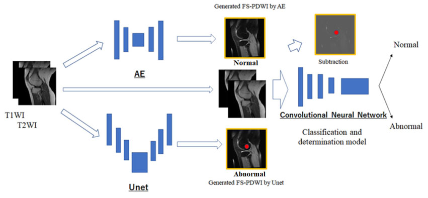
Figure 1. The generation of fat-suppression images without abnormal findings (normal
fat-suppression images) using convolutional encoder-decoder network (AE). The generation of fat-suppression images with normal and/or abnormal
findings using U-Net network. The detection and classification of
abnormalities on knee MR images using two-dimensional convolutional neural
network.
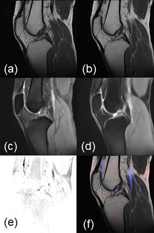
Figure 2. Normal fat-suppression
image. (a) acquired sagittal T1WI, (b) acquired sagittal T2WI, (c) synthesized FS-T2WI by U-Net, (d) synthesized FS-T2WI by AE, (e) subtraction image result after
subtraction of synthesized fat-suppression images by U-Net and by AE, (f)
attention map image.
(c, d) FS-T2WIs generated by U-Net and AE are both of
adequate quality. (e, f) There is no abnormal finding on the subtraction image
and attention map image.
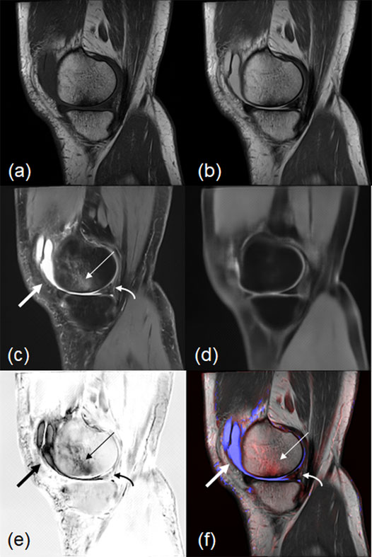
Figure 3. Fat-suppression image
with abnormal findings. (a) acquired sagittal T1WI, (b) acquired
sagittal T2WI, (c) FS-T2WI synthesized by U-Net, (d) FS-T2WI synthesized
by AE, (e) subtraction image between fat-suppression images synthesized by
U-Net and synthesized by AE, (f) attention map image.
(c, d) FS-T2WIs generated by U-Net and AE both are of
adequate image quality. (c, e, f) Intraarticular fluid collection (arrow), bone
marrow abnormality (thin arrow), and meniscus abnormality (curved arrow) are shown
on the subtraction and attention map images.
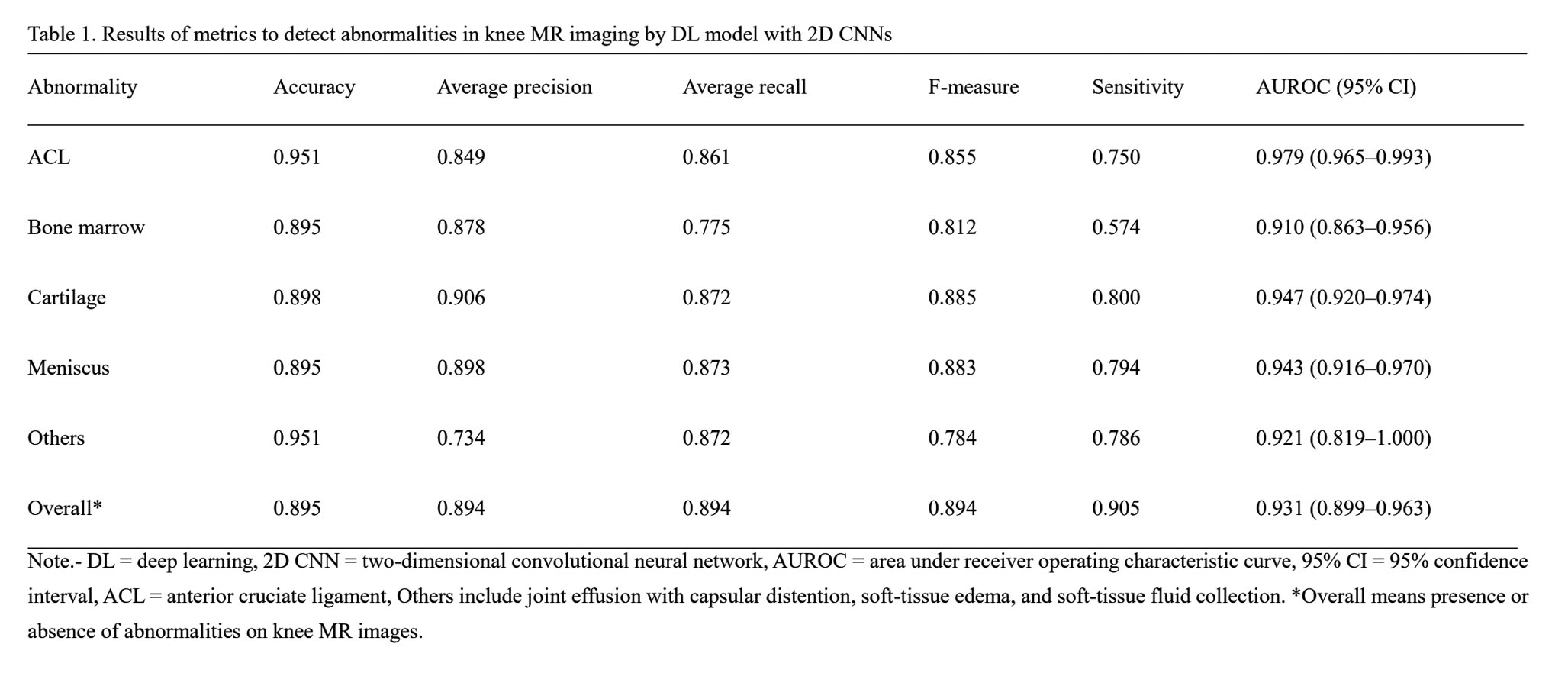
Table 1. Results of metrics
to detect abnormalities in knee MR imaging by DL model with 2D CNNs.
DOI: https://doi.org/10.58530/2023/4739