4717
Dual responsive MR probe based on oxidation-triggered chemical shift1Department of Diagnostic and Interventional Radiology, Institute for Clinical and Experimental Medicine, Prague, Czech Republic, 2First Faculty of Medicine, Institute of Biophysics and Informatics, Charles University, Prague, Czech Republic, 3Department of Polymer and Colloid Immunotherapeutics, Institute of Macromolecular Chemistry, Czech Academy of Sciences, Prague, Czech Republic
Synopsis
Keywords: Non-Proton, Tumor
Presented novel phosphorus- and fluorine-containing polymer possess high sensitivity at 31P/19F-MR and a large chemical shift from biological phosphorus signal (∆δ=60 ppm) due to phosphorothioate P=S group in its structure. The probe represents a conceptually new approach for phosphorus MR, as it undergoes oxidation-induced structural changes in the presence of ROS, represented in greater amounts in cancer tissue. The additional 19F-MR signal serve as an on-site information on the polymer distribution in the organism.
Introduction
31P-MR is used mainly for assessing various metabolites in vivo. Its further potential for imaging is tested, but due to high physiological background signal, it requires a specific exogenous probe1. Presented polymer is a metal-free phosphorus-containing probe for a specific 31P-MR, as phosphorothioate group (P=S) in its structure creates a high chemical shift from biological signal (P=O). Moreover, the P=S groups are prone to metabolic oxidation to phosphoester groups P=O, when exposed to reactive oxygen species (ROS), represented in greater amounts in cancer tissue. This feature can serve as a promising functional in situ sensor of oxidative stress. Here, we provide the results confirming ROS production in the cell line using fluorescent imaging and the polymer measurement using MR in both in vitro (31P-MRS) and in vivo (31P-MRS/19F-MRI) testing of the oxidation-triggered chemical shift.Methods
The polymer (Poly[O-(2-(methacryloyloxy)ethyl)O-(2(trimethylamoniumyl)ethyl)phosphorothioate]: pTMPC) was synthesized by controlled radical polymerization technique of the corresponding zwitterionic monomer. MR spectroscopy and imaging were performed on 4.7T scanner using custom dual 1H/31P solenoid (in vitro) and 1H/31P/19F surface (in vivo) coils. The fluorescence measurement was used to test 4T1 cell line (96-well plate; 0.1x10⁶ mL−1) ROS production and was performed on optical imaging system using OxyBurst fluorescent dye (OB) and with additional 20 and 60 µL of H2O2, as a control. The OB is a non-fluorescent dye, which is oxidised by ROS to fluorescein derivate. In the first step of MR measurements, the in vitro oxidation (conversion from P=S to P=O) was tested using singlepulse 31P-MRS (TR=500 ms, ST=5 min, BW=200 ppm) of the pTMPC in two conditions: i) the polymer (cP=10 mmol L−1) mixed with the hydrogen peroxide (c=20 mmol L−1; mimicking hypoxic tumor tissue), ii) the polymer (cP=2 mmol L−1 ) added to 4T1 cancer cell line suspension (40×106 mL−1 cells). Finally, 4T1 cells suspension (0.3×10⁶ in PBS) was injected subcutaneously to Balb/C mouse, which, at tumor volume ~0.3 mL, underwent intratumoral injection of the dual 31P/19F pTMPC (cP=100 mmol L−1; cF=112.5 mmol L−1; 0.2 mL in PBS). The animal was monitored using several MR methods: 1H-MRI (localization), 31P/19F-MRS single pulse (TR=200/500 ms, ST=10/5 min, BW=200/40 ppm) and 19F-MRI (CSI; TR=200 ms, ST=1h). Isoflurane was used during the in vivo MR. The 31P-MRS signal quantification and 1H/19F-MR image overlay were carried out in ImageJ and Matlab software. The 31P-MRS signal amplitude was averaged from each hour and is presented as a percentage change from the experiment onset. For quantification the P=S signal was used, because when the phosphorus signal is converted by oxidation it overlay with the signal originating from the organism. The 19F-MRI was used as an on-site signal to track the polymer in vivo.Results/Discussion
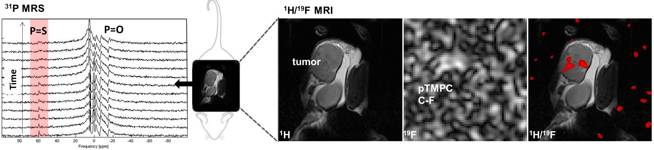
The fluorescent measurement confirm that 4T1 cells alone can produce ROS. In the first phase of the experiment, the fluorescence signal from the cells was comparable to the control (with additional H2O2; Fig.1). The MR signal conversion from P=S to P=O accompanied by a significant frequency shift in the 31P-MR spectrum was observed in vitro. The polymer underwent oxidation and the P=S amplitude was reduced by 91.1% (H2O2) and 36.0% (4T1 cells; Fig.2). The structural modifications ensured chemical shift in in vivo 31P and 19F-MR, making it easily distinguishable from the biological background (31P-MRS: ∆δ=60 ppm) and isoflurane-based anaesthesia (19F-MRS: ∆δ=20 ppm). The in vivo oxidation of the polymer in tumor resulted in 43% signal conversion of the 31P-MRS signal in first 6 hours and the 1H/19F-MRI measured 24h post-injection confirm that the dual pTMPC is localized in the tumor, assuring that the 31P-MR signal conversion was triggered by hypoxic tumor tissue (Fig.3).Conclusion
Phosphorus-containing polymer showed both a high sensitivity for 31P-MR and a chemical shift from biological phosphorus signal. Furthermore, it can serve as a sensitive sensor of pathological conditions, as it undergoes oxidation-induced structural changes in the presence of ROS. With favourable MR properties and biocompatibility, the probe represents a conceptually new approach for 31P-MR. The 19F-MR signal can provide an on-site information on the polymer distribution in the organism during the P=S to P=O conversion in the 31P-MRS.Acknowledgements
The project was supported by the Ministry of Health of the Czech Republic [NU20-08-00095], SGS project no. 2022/3002 of the Technical University of Liberec and by MH CR-DRO (Institute for Clinical and Experimental Medicine IKEM, IN00023001).References
1. Kracíková, L., Ziółkowska, N., Androvič, L., Klimánková, I., Červený, D., Vít, M., Jirak, D. Phosphorus‐containing Polymeric Zwitterion: A pioneering bioresponsive probe for 31P‐magnetic resonance imaging. Macromolecular Bioscience. 2022; 2100523. doi:10.1002/mabi.202100523.Figures
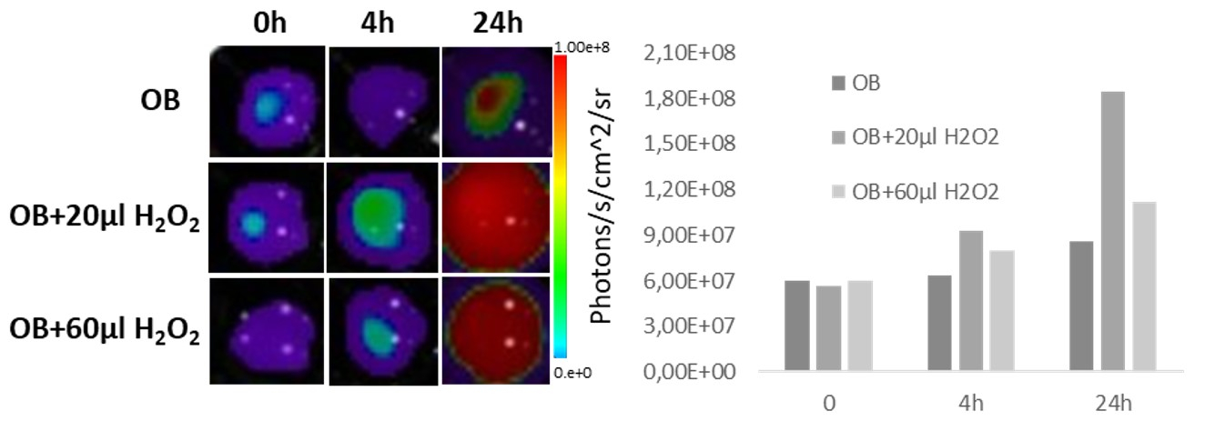
The in vitro fluorescent measurement of reactive oxygen species (ROS) production in 4T1 cancer cell line tested by non-fluorescent dye OxyBurst (OB), which is oxidised by ROS to fluorescein derivate. The results are presented as maximum radiation (Photons/s/cm^2/sr) in the region of interest (ROI) in time (0, 4, 24 hours).
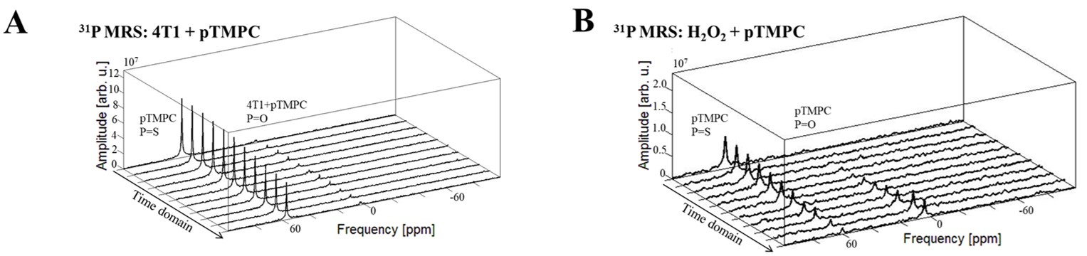
MR results of the in vitro measurement. (A) 31P MR spectra of the polymer (cP=2 mmol L−1) added to 4T1 cancer cell line suspension (40×106 mL−1 cells) and (B) the polymer (cP=10 mmol L−1) mixed with the hydrogen peroxide (c=20 mmol L−1; mimicking hypoxic tumor tissue).