4700
Global Longitudinal Strain Associates with Adverse Outcomes in Nonischemic Dilated Cardiomyopathy without Myocardial Scar1Fuwai Hospital and National Center for Cardiovascular Diseases, Beijing, China
Synopsis
Keywords: Cardiomyopathy, Cardiomyopathy
Despite late gadolinium enhancement (LGE) have already proved a strong association with adverse outcomes in non-ischemic dilated cardiomyopathy (NIDCM) patients, for those who with negative LGE, mortality remains a clinical issue. This study therefore aimed to investigate the prognostic impact of cardiac magnetic resonance (CMR) feature tracking derived strain in NIDCM patients without myocardial scar.
Purpose
Despite late gadolinium enhancement (LGE) have already proved a strong association with adverse outcomes in non-ischemic dilated cardiomyopathy (NIDCM) patients, for those who with negative LGE, mortality remains a clinical issue. This study therefore aimed to investigate the prognostic impact of cardiac magnetic resonance (CMR) feature tracking derived strain in NIDCM patients without myocardial scar.Methods
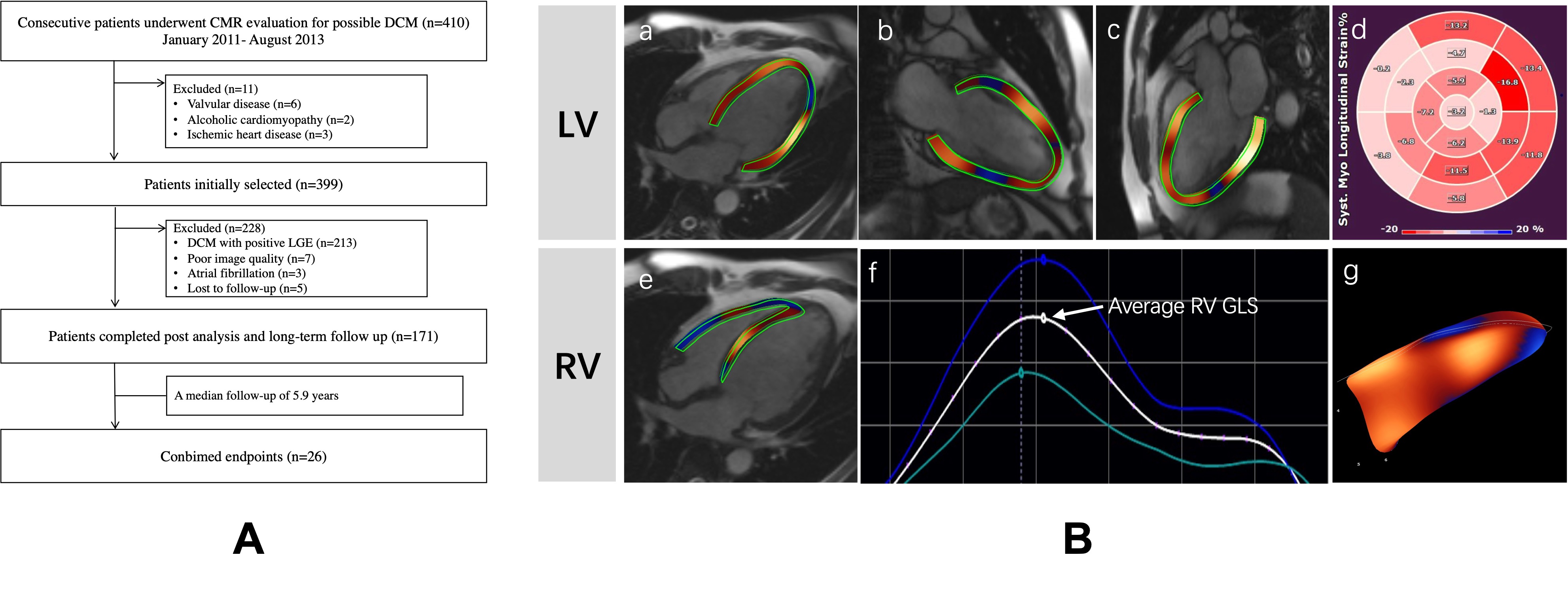
Consecutive patients with NIDCM who underwent CMR between 2012 and 2014 were retrospectively evaluated, and those with positive LGE were excluded. Ventricular systolic dysfunction was measured as LV and RV global longitudinal strain (GLS) by CMR feature tracking. All patients were followed up for the combined endpoints (CE) including all-cause mortality and heart transplantation.Results
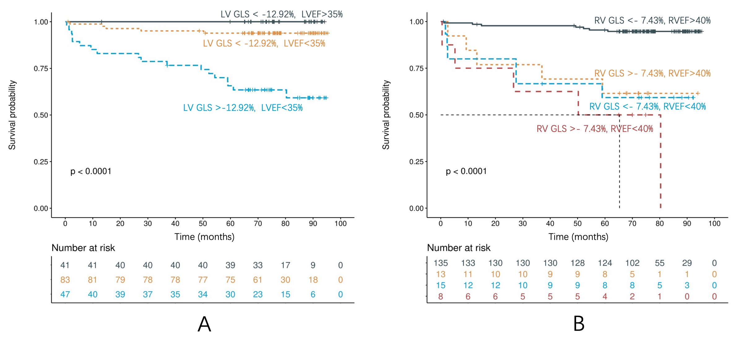
A total of 171 patients with NIDCM were followed up for a median of 5.9 years, and 26 patients reached CE. Both LV and RV GLS were significantly associated with endpoints. Using separate multivariate COX analysis models, LV-GLS offered incremental prognostic value for endpoints beyond NYHA class and NT-proBNP(HR, 1.377; P<0.001). RV-GLS also exhibited an independent prognostic value in addition to NYHA class, NT-proBNP, and anti-arrhythmic medication(HR, 1.142; P=0.002). Furthermore, among patients of LVEF less than 35%, patients with a LVGLS>-12.92% had a higher rate of CE(log-rank, P<0.001).Conclusion
Biventricular GLS associated with adverse outcomes in NIDCM patients without myocardial scar, and provided risk stratification value in the subgroup of reduced EF.Acknowledgements
This study was made possible by the wonderful work of colleagues in department of Magnetic Resonance Imaging, Fuwai hospital, Beijing, China.References
[1] Weintraub RG, Semsarian C, Macdonald P. Dilated cardiomyopathy[J]. Lancet, 2017,390(10092):400-414. DOI: 10.1016/S0140-6736(16)31713-5.
[2] Japp AG, Gulati A, Cook SA, et al. The diagnosis and evaluation of dilated cardiomyopathy[J]. J Am Coll Cardiol, 2016,67(25):2996-3010. DOI: 10.1016/j.jacc.2016.03.590.
[3] Halliday BP, Cleland JGF, Goldberger JJ, et al. Personalizing risk stratification for sudden death in dilated cardiomyopathy: The past, present, and future[J]. Circulation, 2017,136(2):215-231. DOI: 10.1161/CIRCULATIONAHA.116.027134.
[4] Smiseth OA, Torp H, Opdahl A, et al. Myocardial strain imaging: How useful is it in clinical decision making?[J]. Eur Heart J, 2016,37(15):1196-1207. DOI: 10.1093/eurheartj/ehv529.
[5] Romano S, Judd RM, Kim RJ, et al. Feature-tracking global longitudinal strain predicts death in a multicenter population of patients with ischemic and nonischemic dilated cardiomyopathy incremental to ejection fraction and late gadolinium enhancement[J]. JACC Cardiovasc Imaging, 2018,11(10):1419-1429. DOI: 10.1016/j.jcmg.2017.10.024.
[6] Chimura M, Onishi T, Tsukishiro Y, et al. Longitudinal strain combined with delayed-enhancement magnetic resonance improves risk stratification in patients with dilated cardiomyopathy[J]. Heart, 2017,103(9):679-686. DOI: 10.1136/heartjnl-2016-309746.
[7] Liu T, Gao Y, Wang H, et al. Association between right ventricular strain and outcomes in patients with dilated cardiomyopathy[J]. Heart, 2020, DOI: 10.1136/heartjnl-2020-317949.
Figures